Review
. 2021;4(1):231-247.
doi: 10.1007/s42247-021-00175-9. Epub 2021 Mar 8.

Advances in emergent biological recognition elements and bioelectronics for diagnosing COVID-19

Affiliations
Review

Advances in emergent biological recognition elements and bioelectronics for diagnosing COVID-19

Praopim Limsakul et al. Emergent Mater. 2021.

Abstract

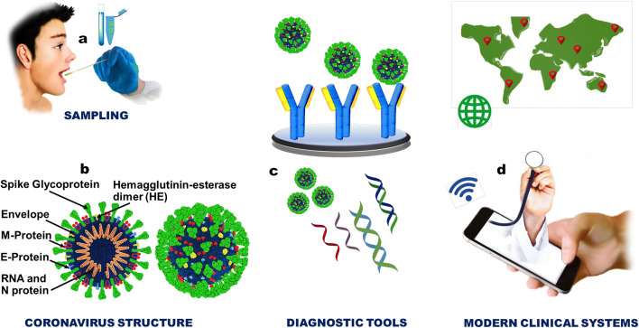
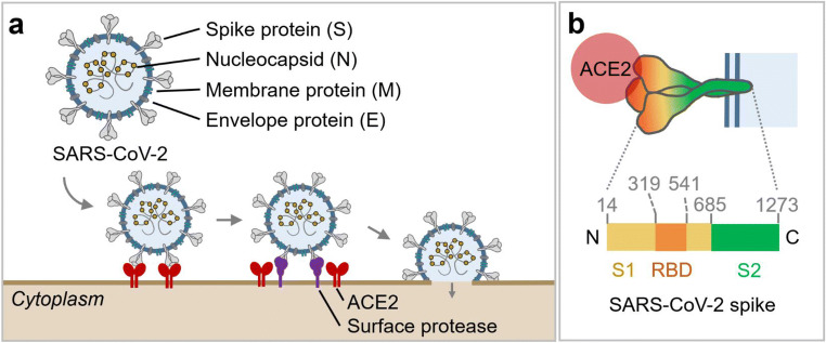
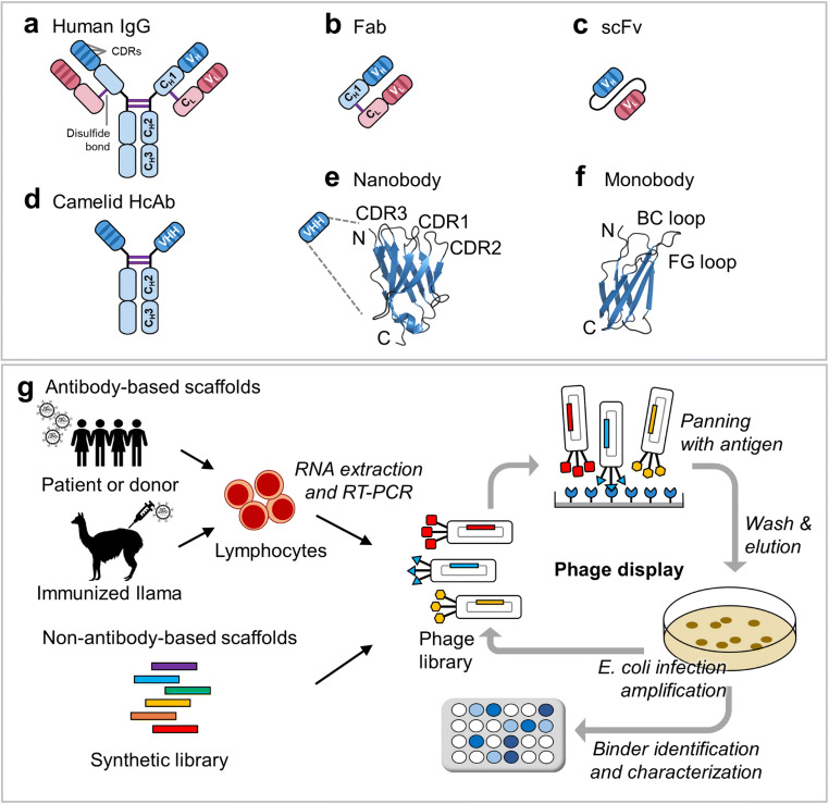
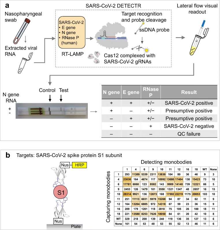
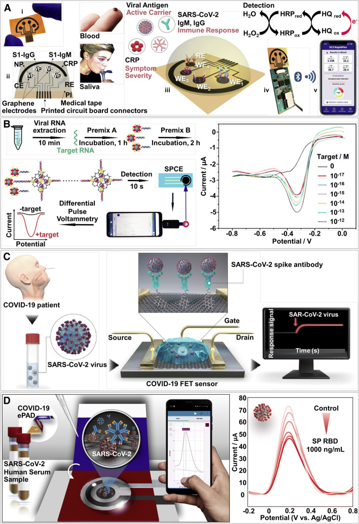
Coronaviruses pose a serious threat to public health. Tremendous efforts are dedicated to advance reliable and effective detection of coronaviruses. Currently, the coronavirus disease 2019 (COVID-19) diagnosis mainly relies on the detection of the severe acute respiratory syndrome coronavirus 2 (SARS-CoV-2) genetic materials by using reverse transcription-polymerase chain reaction (RT-PCR) assay. However, simpler and more rapid and reliable alternatives are needed to meet high demand during the pandemic. Biosensor-based diagnosis approaches become alternatives for selectively and rapidly detecting virus particles because of their biorecognition elements consisting of biomaterials that are specific to virus biomarkers. Here, we summarize biorecognition materials, including antibodies and antibody-like molecules, that are designed to recognize SARS-CoV-2 biomarkers and the advances of recently developed biosensors for COVID-19 diagnosis. The design of biorecognition materials or layers is crucial to maximize biosensing performances, such as high selectivity and sensitivity of virus detection. Additionally, the recent representative achievements in developing bioelectronics for sensing coronavirus are included. This review includes scholarly articles, mainly published in 2020 and early 2021. In addition to capturing the fast development in the fields of applied materials and biodiagnosis, the outlook of this rapidly evolving technology is summarized. Early diagnosis of COVID-19 could help prevent the spread of this contagious disease and provide significant information to medical teams to treat patients.

Keywords: Antibody; Antibody-like molecule; Biorecognition material; COVID-19; SARS-CoV-2; Virus.


Conflict of interest statement

Conflict of interestThe authors declare that they have no conflict of interest.

Figures

Fig. 1
Fig. 1
Illustrations of the SARS-CoV-2 and the concept of using biosensing technologies for effective COVID-19 diagnosis, including a sampling, b coronavirus structure, c biodiagnostic tools, and d modern clinical systems, which seeks to deliver important data and allows fast and efficient managements of the individual and public health
Fig. 2
Fig. 2
The structure and cell entry mechanism of SARS-CoV-2. a Four structural proteins of SARS-CoV-2 include spike (S), nucleocapsid (N), membrane (M), and envelop (E) proteins. To enter host cells, the SARS-CoV-2 S protein binds to host angiotensin-converting enzyme 2 (ACE2) receptors (shown in red). Subsequently, the surface protease, e.g., transmembrane protease serine 2 (TMPRSS2, shown in purple), cleaves at the S1/S2 boundary which triggers a series of conformational changes that lead to fusion between the viral envelope and the target host cell membrane. b Schematic of the SARS-CoV-2 S protein. The total length is 1273 amino acids which consist of a signal peptide (residue: 1–13) located at the N-terminus, the S1 subunit (residue: 14–685, shown in yellow), and the S2 subunit (residue: 686–1273, shown in green). In the S1 subunit, there are an N-terminal domain (residue: 14–305) and a receptor-binding domain (RBD; residue: 319–541, highlighted in orange). In the S2 subunit, there are the fusion peptide (FP; residue: 788–806), heptapeptide repeat sequence 1 and 2 (residue: 912–984 for HR1, and 1163–1213 for HR2), transmembrane domain (TM; residue: 1213–1237), and cytoplasm domain (residue: 1237–1273). Using the RBD, the trimeric spike molecule binds to ACE2
Fig. 3
Fig. 3
Antibodies and antibody-like molecules and schematic overview of their selection pipeline. The structure and size of selected biorecognition molecules. a Human immunoglobulin G (IgG, ~150 kDa) antibody consists of two heavy (blue) and two light (pink) chains connected by disulfide bonds (S–S). Both chains consist of several constant domains (CL and CH1–CH3) and the variable domains (VL and VH). The variable domains contain the complementarity-determining regions (CDRs), which determine antibody-binding specificity. b The antigen-binding fragment (Fab, ~55 kDa) is composed of each VH, CH1, VL, and CL domains. c The single-chain variable fragment (scFv, ~28 kDa) is composed of each VH and VL domains connected via a linker. d The camelid heavy-chain antibody (HcAbs, ~96 kDa) is made up of only two heavy chains; each of which consists of two constant (CH2 and CH3) domains and a single variable (VHH, ~12–15 kDa) domain. e Structure of the nanobody (e.g., vhhGFP4, PDB ID:3OGO; [51]). f Structure of the monobody (~10 kDa) (e.g., PDB ID: 1TTG; [52]). g Selection of biorecognition molecules using the phage display technology. (Top) For antibody-based scaffolds, an antibody library is generated using lymphocytes (B cells) isolated from either human or camelids (immunized by viral proteins). Subsequently, lymphocyte RNA encoding recognition protein binders is transcribed into cDNA by RT-PCR. A binder phage library is then created in the M13 phage that allows protein binders to be presented on the phage surface. The protein binder library undergoes a panning process [53]. Several cycles of panning are conducted for the selection of high-affinity binders. After that, all selected binders will be verified. (Bottom) For non-antibody-based scaffolds, synthetic peptide libraries are used, and the same procedure is repeated. In addition to the phage display, the yeast surface display can be applied to screen desired molecules [54]. ad and g are adapted with permission from [55]. Copyright 2018, The Company of Biologists
Fig. 4
Fig. 4
Applications of biorecognition elements in detecting SARS-CoV-2. a Schematic of SARS-CoV-2 DETECTR workflow. Viral RNA is extracted from COVID-19 patient samples and used as an input to DETECTR (LAMP preamplification and Cas12-based detection). The readout of this DETECTR can be visualized by a lateral flow strip. A positive result requires detection of at least one of the two SARS-CoV-2 viral gene targets (N gene or E gene). RNase P gene is used as a control, and QC represents quality control. Adapted with permission from [80], Copyright 2020, Springer Nature, b A schematic representation of the sandwich ELISA using the monobody. The monobody can replace antibody for the sandwich ELISA. The S1 subunit of SARS-CoV-2 S protein is added to the monobody-immobilized microplate. After the S1 subunit is bound with the monobody and unbound molecules are washed away, the binding between the S1 subunit and the monobody is identified by detecting monobody-HRP. All possible combinations (nine S1-binding monobodies) are tested as shown in the inset table. Nus represents Nus-Tag fused at the C terminus of the monobody. HRP is horseradish peroxidase enzyme that is used to amplify signal in photometric assays by catalyzing the conversion chemiluminescent substrates. Adapted from [63], Copyright, The Authors, some rights reserved; exclusive licensee AAAS. Distributed under a Creative Commons Attribution NonCommercial License 4.0 (CC BY-NC)
Fig. 5
Fig. 5
Examples of electrochemical sensors for COVID-19 diagnosis. a A wireless graphene-based telemedicine platform (i) an image of a disposable and flexible graphene array, (ii) an illustration of the graphene sensor array layout, (iii) schematic illustrations of the detection of SARS-CoV-2 viral proteins, antibodies (IgG and IgM), and inflammatory biomarker C-reactive protein (CRP) in blood and saliva (also see Table 2), (iv) an image of the electrochemical sensing system connected to a printed circuit board for signal processing and wireless communication, and (v) data transmission via wireless to a user interface. WE, CE, and RE: stand for working electrode, counter electrode, and reference electrode. Adopted with permission from [91], Copyright 2020, Elsevier, b A schematic illustration of SARS-CoV-2 detection using the screen-printed electrode; the electrochemical detection process using a smartphone along with differential pulse voltammograms for different concentrations of artificial target for the SARS-CoV-2 biosensor. Adopted with permission from [92], Copyright 2020, Elsevier, c A schematic diagram of the COVID-19 field-effect transistor (FET) based biosensor. Adapted with permission from [90, 93], Copyright 2020, American Chemical Society. This permission is granted for the duration of the World Health Organization (WHO) declaration of COVID-19 as a global pandemic, d The detection principle of the COVID-19 electrochemical paper-based analytical device in human serum sample along with square wave voltammetric responses tested with different concentrations of the SARS-CoV-2 spike protein. Adopted with permission from [94], Copyright 2020, Elsevier

Similar articles

Cited by

References

    1. COVID-19 Coronavirus Pandemic. 2021 [cited Access January 18, 2021]; Available from: https://www.worldometers.info/coronavirus/
    1. Oran DP, Topol EJ. Prevalence of Asymptomatic SARS-CoV-2 infection. Ann. Intern. Med. 2020;173:362–367. doi: 10.7326/M20-3012. - DOI - PMC - PubMed
    1. Lai C-C, Shih T-P, Ko W-C, Tang H-J, Hsueh P-R. Severe acute respiratory syndrome coronavirus 2 (SARS-CoV-2) and coronavirus disease-2019 (COVID-19): the epidemic and the challenges. Int. J. Antimicrob. Agents. 2020;55:105924. doi: 10.1016/j.ijantimicag.2020.105924. - DOI - PMC - PubMed
    1. Liang K-H, Chang T-J, Wang M-L, Tsai P-H, Lin T-H, Wang C-T, Yang D-M. Novel biosensor platforms for the detection of coronavirus infection and severe acute respiratory syndrome coronavirus 2. J. Chin. Med. Assoc. 2020;83:701–703. doi: 10.1097/JCMA.0000000000000337. - DOI - PMC - PubMed
    1. Gandhi M, Yokoe DS, Havlir DV. Asymptomatic transmission, the Achilles’ heel of current strategies to control Covid-19. N. Engl. J. Med. 2020;382:2158–2160. doi: 10.1056/NEJMe2009758. - DOI - PMC - PubMed

LinkOut - more resources