Review
. 2021;4(1):187-209.
doi: 10.1007/s42247-021-00195-5. Epub 2021 Mar 10.

Affinity biosensors developed with quantum dots in microfluidic systems

Affiliations
Review

Affinity biosensors developed with quantum dots in microfluidic systems

Sultan Şahin et al. Emergent Mater. 2021.

Abstract

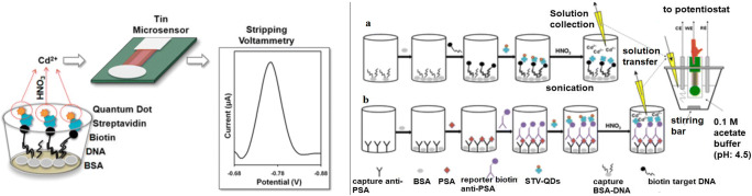
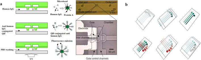
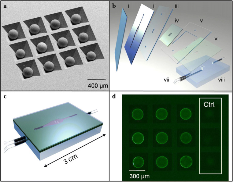
Quantum dots (QDs) are synthetic semiconductor nanocrystals with unique optical and electronic properties due to their size (2-10 nm) such as high molar absorption coefficient (10-100 times higher than organic dyes), resistance to chemical degradation, and unique optoelectronic properties due to quantum confinement (high quantum yield, emission color change with size). Compared to organic fluorophores, the narrower emission band and wider absorption bands of QDs offer great advantages in cell imaging and biosensor applications. The optoelectronic features of QDs have prompted their intensive use in bioanalytical, biophysical, and biomedical research. As the nanomaterials have been integrated into microfluidic systems, microfluidic technology has accelerated the adaptation of nanomaterials to clinical evaluation together with the advantages such as being more economical, more reproducible, and more susceptible to modification and integration with other technologies. Microfluidic systems serve an important role by being a platform in which QDs are integrated for biosensing applications. As we combine the advantages of QDs and microfluidic technology for biosensing technology, QD-based biosensor integrated with microfluidic systems can be used as an advanced and versatile diagnostic technology in case of pandemic. Specifically, there is an urgent necessity to have reliable and fast detection systems for COVID-19 virus. In this review, affinity-based biosensing mechanisms which are developed with QDs are examined in the domain of microfluidic approach. The combination of microfluidic technology and QD-based affinity biosensors are presented with examples in order to develop a better technological framework of diagnostic for COVID-19 virus.

Keywords: Biosensors; Microfluidic systems; Quantum dots.


Figures

Fig. 1
Fig. 1
Fluorescence photographs of probes incubated with different concentrations (a) MCF-7 cells (from a to e: 180, 2 × 10³, 2 × 10⁴, 105, and 2 × 10⁶ cells/mL, respectively), (b) HL-60 cells (from a to e: 210, 3 × 10³, 3 × 10⁴, 3 × 10⁵, and 3 × 10⁶ cells/mL, respectively), and (c) K562 cells (from a to e: 200, 103, 2 × 10⁴, 2 × 10⁵, and 2 × 10⁶ cells/mL, respectively). Fluorescence emission change after quenching QD fluorescence with GO structure at the end of hybridization of aptamer and cells at different concentrations, (d) MCF-7 cells (from a to h: 0, 180, 500, 2 × 10³, 2 × 10⁴, 105, 2 × 10⁶, and 2 × 10⁷ cells/mL, respectively), (e) HL-60 cells (from a to h: 0, 210, 500, 3 × 10³, 3 × 10⁴, 3 × 10⁵, 3 × 10⁶, and 3 × 10⁷ cells/mL, respectively) and (f) K562 cells (from a to h: 0, 200, 500, 103, 2 × 10⁴, 2 × 10⁵, 2 × 10⁶, and 2 × 10⁷ cells/mL, respectively). Logarithmic calibration curve for (g) MCF-7 cell, (h) HL-60 cells and (i) K562 cells. Reprinted with permission from Ref. [63]. Copyright © 2016 Elsevier
Fig. 2
Fig. 2
PEC signals from F-doped SnO2 conducting glass modified with hybrid structures (a) g-C3N4, (b) CdS, (c) g-C3N4@CdS. Reprinted with permission from Ref. [94]. Copyright © 2016, American Chemical Society
Fig. 3
Fig. 3
Schematic presentation of the electrochemical immunoassay procedure. (a) Electrochemical affinity assay for the detection of C533G mutation of RET gene and (b) electrochemical immunoassay for the detection of PSA in human serum. Reprinted with permission from Ref. [105]. Copyright © 2013, American Chemical Society
Fig. 4
Fig. 4
(a) hIgG injection from the inlet of the microchannel, anti-hIgG conjugated QD injection and washing step (left), microscopy image of microchannels (right) Ref. [54]. (b) Protein chip designed by immobilizing antibody to the surface of the PDMS channels. Reprinted with permission from Ref. [57]. Copyright © 2010, American Chemical Society
Fig. 5
Fig. 5
Fiber optic system integrated to microfluidic system, light source, and syringe pump from Ref. [111]
Fig. 6
Fig. 6
Design of a QD–GO quenching biosensing system in microfluidic system. Reprinted with permission from Ref. [64]. Copyright © 2016 Elsevier
Fig. 7
Fig. 7
(left) Schematic diagram of in situ synthesis of PDMS–GNP composite microreactors and immunoassay analytical procedure based on QD label. (right) Schematic representation of the electrochemical analysis via the flow injection mode on the microchip Ref. [14]
Fig. 8
Fig. 8
A microfluidic chip design with magnetic carrier supports which are collected in the detection zone by help of external magnetic force. Reprinted with permission from Ref. [62]. Copyright © 2014 Elsevier
Fig. 9
Fig. 9
A design that allows Salmonella bacteria to be bound with magnetic particles and combined with MnO2-QD complex structure to detect GSH and QD in the separation chamber of the microfluidic system. Ref. [67]
Fig. 10
Fig. 10
Microfluidic chip with microbead array for virus DNA analysis. (a) An overview of the microfluidic chip design. (b) A cross section of the reaction and detection area. (c) Photograph of the microchip. (d) A microscope image of the reaction area. Reprinted with permission from Ref. [58]. Copyright © 2010 Elsevier
Fig. 11
Fig. 11
(a) SEM images of beads in anisotropically etched silicon chip. (b) Chip (iv) is fitted between double-sided adhesive layer (ii) and cover slip (i) with laminate layers (iii, v, vi) included to direct fluid flow through PMMA base (viii) and inlet and outlet ports (vii). (c) Sealed LoC assembly. (d) Fluorescent image of beads after immunoassay including negative controls as imaged with one second of CCD camera integration (exposure) time. Reprinted with permission from Ref. [56]. Copyright © 2009 Elsevier
Fig. 12
Fig. 12
Schematic representation of QD based μ-PAD design and fabrication allowing multiple detection. Reprinted with permission from Ref. [63]. Copyright © 2016 Elsevier

Similar articles

Cited by

References

    1. Zhu S, Zhang J, Qiao C, Tang S, Li Y, Yuan W, Li B, Tian L, Liu F, Hu R, Gao H, Wei H, Zhang H, Sun H, Yang B. Strongly green-photoluminescent graphene quantum dots for bioimaging applications. Chem. Commun. 2011;47(24):6858–6860. doi: 10.1039/C1CC11122A. - DOI - PubMed
    1. Ruan J, Song H, Qian Q, Li C, Wang K, Bao C, Cui D. HER2 Monoclonal antibody conjugated RNase-A-associated CdTe quantum dots for targeted imaging and therapy of gastric cancer. Biomaterials. 2012;33(29):7093–7102. doi: 10.1016/j.biomaterials.2012.06.053. - DOI - PubMed
    1. Frasco MF, Chaniotakis N. Semiconductor quantum dots in chemical sensors and biosensors. Sensors. 2009;9(9):7266–7286. doi: 10.3390/s90907266. - DOI - PMC - PubMed
    1. Yang C, Hu L, Zhu H-Y, Ling Y, Tao J-H, Xu C-X. RGO Quantum dots/ZnO hybrid nanofibers fabricated using electrospun polymer templates and applications in drug screening involving an intracellular H2O2 sensor. J. Mater. Chem. B. 2015;3(13):2651–2659. doi: 10.1039/C4TB02134G. - DOI - PubMed
    1. Zhang L, Sun Y, Liang Y-Y, He J-P, Zhao W-W, Xu J-J, Chen H-Y. Ag Nanoclusters could efficiently quench the photoresponse of CdS quantum dots for novel energy transfer-based photoelectrochemical bioanalysis. Biosens. Bioelectron. 2016;85:930–934. doi: 10.1016/j.bios.2016.06.018. - DOI - PubMed

LinkOut - more resources