Case Reports
. 2020 Nov 6;5(1):204-209.
doi: 10.1016/j.mayocpiqo.2020.08.004. eCollection 2021 Feb.

Progressive Monoarticular Inflammatory Arthritis Following Influenza Vaccination

Affiliations
Case Reports

Progressive Monoarticular Inflammatory Arthritis Following Influenza Vaccination

Laurel A Littrell et al. Mayo Clin Proc Innov Qual Outcomes. .

Abstract

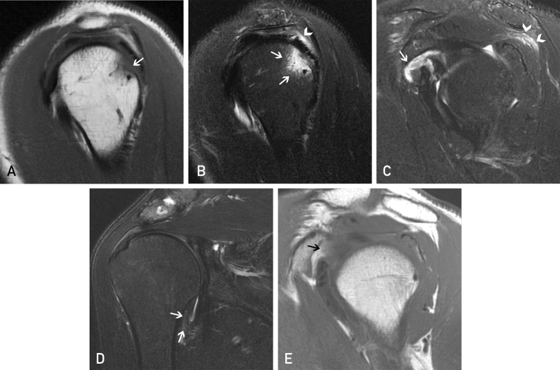
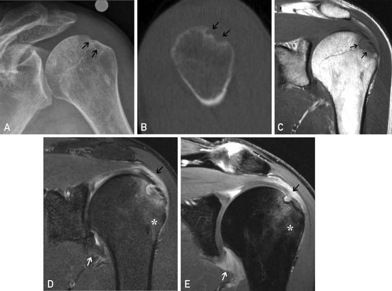
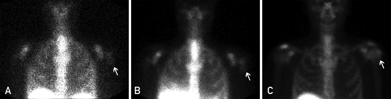
Musculoskeletal injury is an uncommon but usually self-limited complication of vaccine administration. We present a case of progressive inflammatory monoarthritis of the shoulder characterized by bone erosion, bursitis, and severe synovitis caused by an influenza vaccine administered to the ipsilateral deltoid region. Clinical symptoms began within 2 hours of vaccination, with progressive decline in function over 6 weeks. Magnetic resonance imaging examinations performed 5 months apart demonstrated progressive erosive changes of the greater tuberosity, rotator cuff injury, and extensive enhancing synovitis of the glenohumeral joint and subacromial/subdeltoid bursa. After the exclusion of septic arthritis and osteomyelitis, the patient underwent nonoperative treatment and experienced near-complete recovery at 32 months. Although inflammatory arthritis of the shoulder following vaccination is rare, there have been previous reports of it. Clinicians and radiologists need to be aware of this potential complication to ensure an accurate diagnosis.

Keywords: CT, computed tomography; MRI, magnetic resonance imaging.


Figures

Figure 1
Figure 1
Magnetic resonance imaging of the left shoulder obtained 6 weeks after vaccine administration and onset of symptoms. (A) Sagittal T1 and (B, C) sagittal fat-suppressed T2-weighted images demonstrate focal cortical irregularity and indistinctness of posterior margin of the greater tuberosity (arrow in A) with surrounding bone marrow edema within the posterolateral humeral head superiorly (arrows in B). There is also focal fluid and edema in the overlying subacromial/subdeltoid bursa and superficial fibers of the infraspinatus tendon (arrowhead in B). The edema within the infraspinatus tracks medially within the superficial and superior fibers of the muscle belly (arrowheads in C). There is evidence of synovitis in the rotator interval and subscapularis recess (arrow in C). (D) Coronal T2-weighted image demonstrates minimally increased T2 signal within the inferior glenohumeral ligament (arrows in D), and (E) sagittal T1 image demonstrates obliteration of the retrocoracoid fat and thickening of the coracohumeral ligament (arrow in E), findings that are suggestive of adhesive capsulitis in the appropriate clinical setting.
Figure 2
Figure 2
Radiographic, computed tomographic, and magnetic resonance images from 6 months after vaccine administration and onset of symptoms and 5 months after the magnetic resonance imaging examination shown in Figure 1. (A) Anteroposterior radiograph of the left shoulder in internal rotation suggests progression of cortical erosion along the posterior greater tuberosity (arrows in A, B, and C). (B) Sagittal reformatted computed tomographic image and (C) coronal T1 magnetic resonance imaging confirm the progression of the erosive changes. (D) Coronal T2 and (E) post–gadolinium-enhanced coronal spoiled gradient recalled acquisition (SPGR) (E) demonstrate the enhancing bone marrow edema underlying the erosions (asterisks in D and E) and the enhancing synovitis and capsulitis (white arrows in D and E) and subacromial/subdeltoid bursitis (black arrows in D and E).
Figure 3
Figure 3
Planar anteroposterior images of the shoulders from (A) indium-111 white blood cell scan 24 hours after injection, (B) Tc-99m sulfur colloid bone marrow scan approximately 1 hour after radiotracer injection, and (C) delayed phase planar anteroposterior image from 3-phase Tc-99m-methyl diphosphonate bone scan 3 hours after injection demonstrating a focus of increased white blood cell activity near the junction of the left humeral head and neck (arrow in A), which is matched by sulfur colloid uptake (arrow in B), indicating that the white blood cell activity is due to activated bone marrow rather than infection or osteomyelitis. In addition, the mildly increased uptake on the delayed phase images (arrow in C) is less intense than would be expected in the presence of osteomyelitis.

References

    1. Historical Reference of Seasonal Influenza Vaccine Doses Distributed. https://www.cdc.gov/flu/prevent/vaccinesupply.htm Available at:
    1. 2017-2018 Estimated Influenza Illnesses, Medical visits, and Hospitalizations Averted by Vaccination in the United States. https://www.cdc.gov/flu/vaccines-work/averted-estimates.htm Available at:
    1. The Vaccine Adverse Event Reporting System (VAERS) https://wonder.cdc.gov/vaers.html Available at: - PubMed
    1. Bodor M., Montalvo E. Vaccination-related shoulder dysfunction. Vaccine. 2007;25(4):585–587. - PubMed
    1. Atanasoff S., Ryan T., Lightfoot R., Johann-Liang R. Shoulder injury related to vaccine administration (SIRVA) Vaccine. 2010;28(51):8049–8052. - PubMed

Publication types

LinkOut - more resources