. 2021 Jan 6;4(1):217-240.
doi: 10.1016/j.matt.2020.10.022. Epub 2020 Nov 18.

Symbiotic Photosynthetic Oxygenation within 3D-Bioprinted Vascularized Tissues

Affiliations

Symbiotic Photosynthetic Oxygenation within 3D-Bioprinted Vascularized Tissues

Sushila Maharjan et al. Matter. .

Abstract

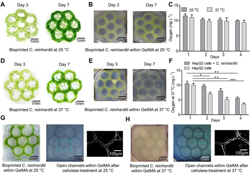
In this study, we present the photosynthetic oxygen (O2) supply to mammalian cells within a volumetric extracellular matrix-like construct, whereby a three-dimensional (3D)-bioprinted fugitive pattern encapsulating unicellular green algae, Chlamydomonas reinhardtii (C. reinhardtii), served as a natural photosynthetic O2-generator. The presence of bioprinted C. reinhardtii enhanced the viability and functionality of mammalian cells while reducing the hypoxic conditions within the tissues. We were able to subsequently endothelialize the hollow perfusable microchannels formed after enzymatic removal of the bioprinted C. reinhardtii-laden patterns from the matrices following the initial oxygenation period, to obtain biologically relevant vascularized mammalian tissue constructs. The feasibility of co-culture of C. reinhardtii with human cells, the printability and the enzymatic degradability of the fugitive bioink, as well as the exploration of C. reinhardtii as a natural, eco-friendly, cost-effective, and sustainable source of O2 would likely promote the development of engineered tissues, tissue models, and food for various applications.


Conflict of interest statement

AUTHOR CONTRIBUTIONS S.M. designed and performed the experiments, collected and analyzed the data, and prepared the manuscript; J.A., C.C., A.G.R., D.H, C.D., E.C., M.D.R., D.B., M.L.S, W.L., F.C., and G.Y. performed the experiments and revised the manuscript; Y.S.Z. conceptualized, designed, and supported the study, and prepared the manuscript. DECLARATION OF INTERESTS The authors declare no competing interest.

Figures

Figure 1.
Figure 1.. Formulation of the fugitive bioink and 3D bioprinting.
(A) Schematics of the bioink formulation. (B) Viscosity characterizations of the optimized bioink and the different components of the bioink. (C) Schematic illustration of the 3D bioprinting process. (D) Photographs of side and top views of the 3D-bioprinted honeycomb patterns with different layers.
Figure 2.
Figure 2.. 3D bioprinting of C. reinhardtii.
(A) Photographs showing bioprinted C. reinhardtii-laden honeycomb patterns with different layers at day 3. (B) Photographs of bioprinted C. reinhardtii-laden tubular, grid, and cross-grid patterns with different sizes. (C) Optical and fluorescence micrographs of bioprinted C. reinhardtii-laden honeycomb patterns. (D) Viability of C. reinhardtii at different days determined by live/dead assay using Sytox-orange. (E) Viability of the bioprinted C. reinhardtii within 4-layered honeycomb patterns at different days, as represented by OD750. Results are presented as means ± standard deviations.
Figure 3.
Figure 3.. Growth of bioprinted C. reinhardtii and measurements of dissolved O2 levels in medium.
(A) Photographs showing the bioprinted C. reinhardtii grown in TAP medium at 25 °C. (B) Photographs showing the bioprinted C. reinhardtii within a GelMA scaffold grown in TAP medium at 25 °C. (C) Levels of dissolved O2 released by bioprinted C. reinhardtii in TAP medium at 25 °C and 37 °C. Results are presented as means ± standard deviations. (D) Photographs showing the bioprinted C. reinhardtii grown in DMEM:TAP medium (1:1) at 37 °C. (E) Photographs showing the bioprinted C. reinhardtii within a GelMA scaffold grown in DMEM:TAP medium (1:1) at 37 °C. (F) Levels of dissolved O2 in DMEM:TAP medium (1:1) at 37 °C in the presence or absence of bioprinted C. reinhardtii embedded within GelMA/HepG2 matrices. Results are presented as means ± standard deviations. (G) Photographs showing the bioprinted C. reinhardtii pattern within a GelMA matrix at day 7 and the formation of open microchannels within the matrix after digestion of the C. reinhardtii pattern with 1 mg mL−1 of cellulase in citrate buffer (pH 6) at 25 °C for 12 h, followed by microscopic image showing the open microchannel. (H) Photographs showing the bioprinted C. reinhardtii pattern within a GelMA matrix at day 7 and the formation of open microchannels within the matrix after digestion of the C. reinhardtii pattern with 1 mg mL−1 of cellulase in citrate buffer (pH 6) at 37 °C for 4 h, followed by microscopic image showing the open microchannel.
Figure 4.
Figure 4.. Co-culture of bioprinted C. reinhardtii and HepG2 cells.
(A) Fluorescence micrographs showing CellTracker CMF2HC-labeled HepG2 cells (blue) in a GelMA construct and the embedded, bioprinted pattern of C. reinhardtii (red). (B) Fluorescence micrographs showing live (green)/dead (red)-stained HepG2 cells in GelMA constructs at day 4, in the absence (control) or presence of C. reinhardtii. (C) Viability of HepG2 cells in the absence (control) or presence of C. reinhardtii, at day 4 and day 7. Results are presented as means ± standard deviations. (D) Fluorescence micrographs showing the immunostaining of HIF-1α expression by HepG2 cells in the absence (control) or presence of C. reinhardtii at day 7. Nuclei were counter-stained with DAPI (blue). (E) Quantification of HIF-1α expression by HepG2 cells in the absence (control) or presence of C. reinhardtii at day 7. Results are presented as means ± standard deviations.
Figure 5.
Figure 5.. Functional studies of HepG2 cells.
(A) Fluorescence micrographs showing the expression of F-actin (red) by HepG2 cells within a GelMA construct at day 7 in the absence (control) and presence of C. reinhardtii. Nuclei were counter-stained with DAPI (blue). (B) Fluorescence micrographs showing the expression of CYP3A (green) by HepG2 cells within a GelMA construct at day 7 in the absence (control) and presence of C. reinhardtii. Nuclei were counter-stained with DAPI (blue). (C) Fluorescence micrographs showing the expression of CYP1A (red) by HepG2 cells within a GelMA construct at day 7 in the absence (control) and presence of C. reinhardtii. Nuclei were counter-stained with DAPI (blue). (D) Albumin secretion by HepG2 cells in GelMA constructs as determined by ELISA. Results are presented as means ± standard deviations. (E) Urea production by HepG2 cells in GelMA constructs as determined by ELISA. Results are presented as means ± standard deviations. In all these functional studies, the bioprinted C. reinhardtii-laden patterns within the HepG2/GelMA constructs were dissolve at day 4 by cellulase digestion.
Figure 6.
Figure 6.. Vascularization of microchannels.
(A) Fluorescence micrographs showing GFP-HUVECs (green) seeded in the channels surrounded by CM-Dil-stained HepG2 cells (red) in a GelMA construct, at day 5 of GFP-HUVEC seeding in the microchannels after removing the bioprinted C. reinhardtii-laden patterns by cellulase digestion. (B) Fluorescence micrographs showing live (green)/dead (red)-stained HepG2 cells in a GelMA construct and HUVECs in the microchannels at day 7 of HUVEC seeding. (C) Viability of HepG2 cells in the GelMA matrices at day 3 and day 7 of HUVEC seeding in the microchannels. The control group had no HUVECs post-seeded into the microchannels. Results are presented as means ± standard deviations. (D) Viability of HUVECs at day 3 and day 7 of seeding in the microchannels. Results are presented as means ± standard deviations. E) Fluorescence micrographs showing the immunostaining of CD31 expression (green) by the HUVECs at day 7 of HUVEC seeding in the microchannels. Nuclei were counter-stained with DAPI (blue).

References

    1. Matesanz R, Mahíllo B, Álvarez M, and Carmona M (2009). Global observatory and database on donation and transplantation: world overview on transplantation activities. Transplant Proc. 41, 2297–2301. - PubMed
    1. Giwa S, Lewis JK, Alvarez L, Langer R, Roth AE, Church GM, Markmann JF, Sachs DH, Chandraker A, and Wertheim JA (2017). The promise of organ and tissue preservation to transform medicine. Nat. Biotechnol. 35, 530. - PMC - PubMed
    1. Atala A, Kasper FK, and Mikos AG (2012). Engineering complex tissues. Sci. Transl. Med. 4, 160rv12–160rv12. - PubMed
    1. Zhang YS, Yue K, Aleman J, Mollazadeh-Moghaddam K, Bakht SM, Yang J, Jia W, Dell’Erba V, Assawes P, and Shin SR (2017). 3D bioprinting for tissue and organ fabrication. Ann Biomed Eng. 45, 148–163. - PMC - PubMed
    1. Ying GL, Jiang N, Maharjan S, Yin YX, Chai RR, Cao X, Yang JZ, Miri AK, Hassan S, and Zhang YS (2018). Aqueous Two-Phase Emulsion Bioink-Enabled 3D Bioprinting of Porous Hydrogels. Adv. Mater. 30, 1805460. - PMC - PubMed

LinkOut - more resources