. 2021 Nov 16;224(9):1509-1519.
doi: 10.1093/infdis/jiab139.

Serum and Cervicovaginal Fluid Antibody Profiling in Herpes Simplex Virus-Seronegative Recipients of the HSV529 Vaccine

Affiliations

Serum and Cervicovaginal Fluid Antibody Profiling in Herpes Simplex Virus-Seronegative Recipients of the HSV529 Vaccine

Kening Wang et al. J Infect Dis. .

Abstract

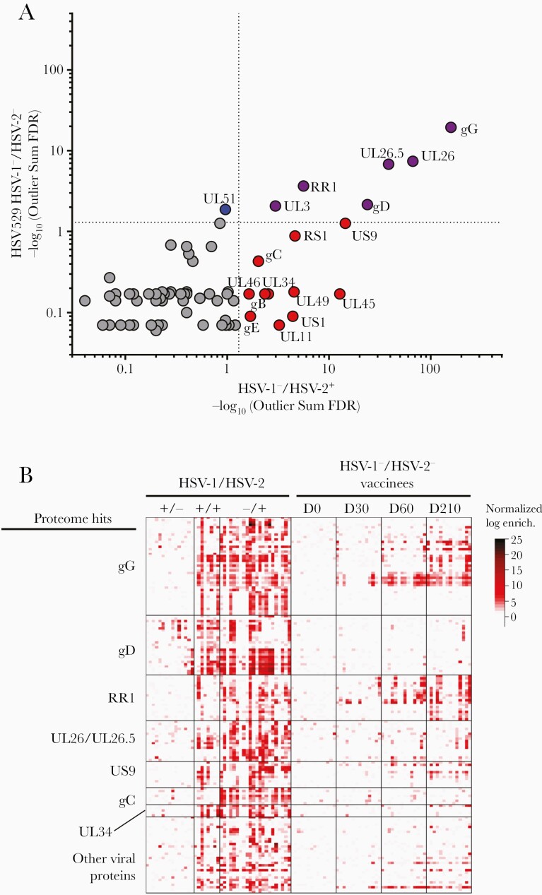
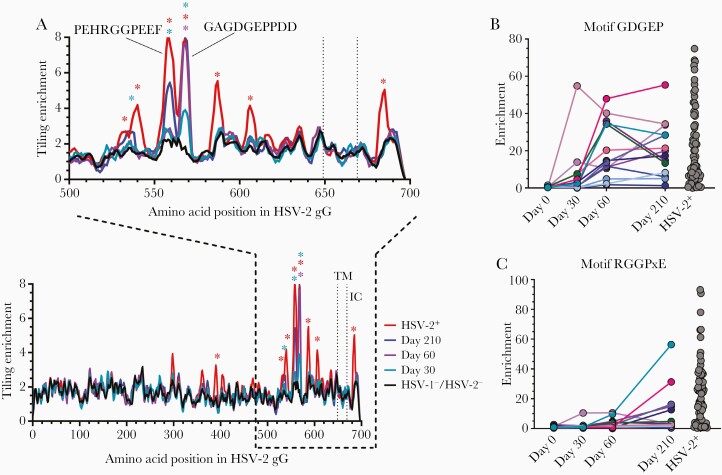
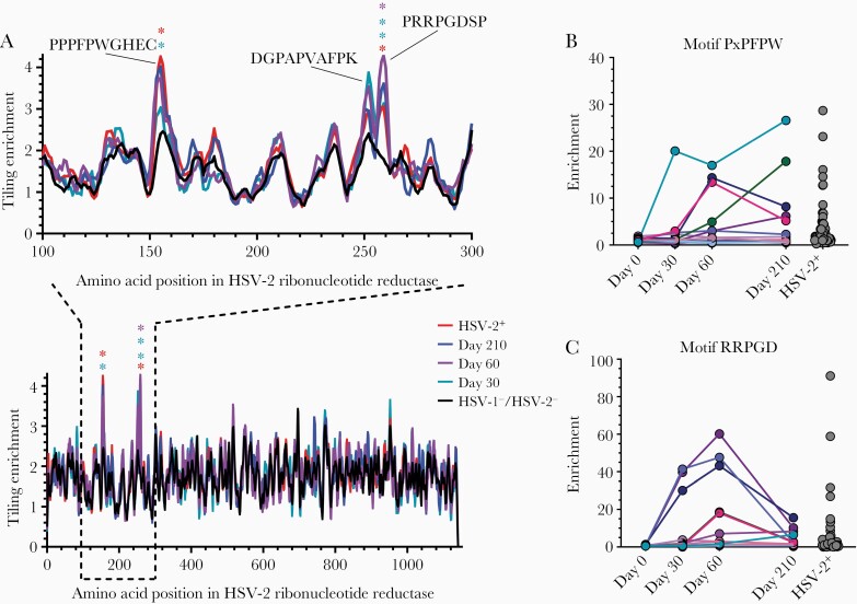
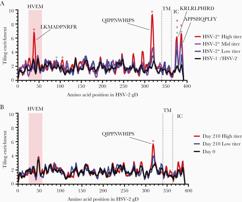
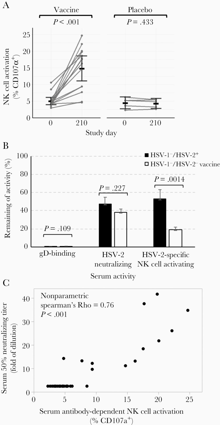
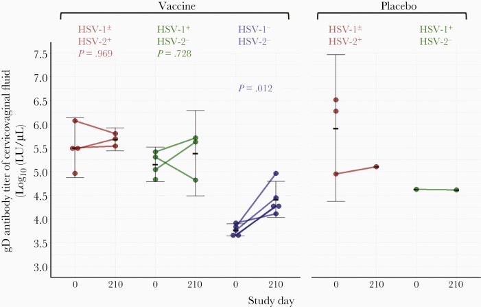
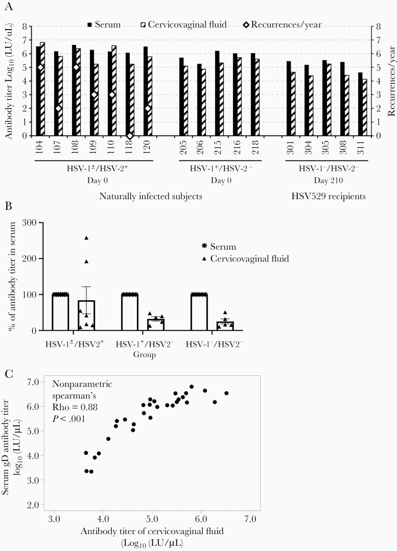
Previous herpes simplex virus type 2 (HSV-2) vaccines have not prevented genital herpes. Concerns have been raised about the choice of antigen, the type of antibody induced by the vaccine, and whether antibody is present in the genital tract where infection occurs. We reported results of a trial of an HSV-2 replication-defective vaccine, HSV529, that induced serum neutralizing antibody responses in 78% of HSV-1-/HSV-2- vaccine recipients. Here we show that HSV-1-/HSV-2- vaccine recipients developed antibodies to epitopes of several viral proteins; however, fewer antibody epitopes were detected in vaccine recipients compared with naturally infected persons. HSV529 induced antibodies that mediated HSV-2-specific natural killer (NK) cell activation. Depletion of glycoprotein D (gD)-binding antibody from sera reduced neutralizing titers by 62% and NK cell activation by 81%. HSV-2 gD antibody was detected in cervicovaginal fluid at about one-third the level of that in serum. A vaccine that induces potent serum antibodies transported to the genital tract might reduce HSV genital infection.

Keywords: HSV-2; antibody-dependent cellular cytotoxicity; genital herpes; glycoprotein D; herpes simplex; herpesvirus; replication-defective vaccine; vaccine.


Figures

Figure 1.
Figure 1.
Herpes simplex virus type 2 (HSV-2)–enriched epitope motifs identified using serum epitope repertoire analysis (SERA) recognized by antibodies in sera from HSV529 trial participants or herpes simplex virus type 1 (HSV-1) and/or HSV-2 naturally infected subjects. A, Statistically significant HSV-2 antigens identified by protein-based immunome-wide association study (PIWAS) in HSV-1/HSV-2 HSV529 vaccine recipients and HSV-1/HSV-2+ naturally infected subjects, ranked using the outlier sum false discovery rate (FDR). Six proteins were significant (FDR <.05) in both cohorts including gG, UL26.5/UL26 (the epitopes were all in the region that overlaps both UL26.5 and UL26), RR1, gD, and UL3 (purple), 11 significant only in naturally infected subjects (red), and 1 significant in only HSV529 subjects (blue). B, Heatmap of enrichments for epitope motifs from HSV-2 proteins. HSV-1/HSV-2 serostatus is indicated at the top of the figure. Each column of the heatmap represents data from 1 subject and each row represents an epitope motif. Colors indicate z scores for each epitope. Heatmap shows the 7 HSV-2 proteins identified as significant by PIWAS with >2 epitope motifs recognized in natural infection or vaccine recipients. The strongest responses in vaccine recipients is to epitope motifs in gG and RR1 with weaker responses to gD, UL26/UL26.5, US9, gC, and other viral proteins.
Figure 2.
Figure 2.
Herpes simplex virus type 2 (HSV-2) gG epitope motifs recognized by antibodies in herpes simplex virus type 1 (HSV-1)/HSV-2–negative HSV529 vaccine recipients and HSV-2 naturally infected subjects. A, Tiling analysis for HSV-2 gG shows epitope motifs from HSV-2+ (n = 72), HSV-1/HSV-2 HSV529 vaccine recipients (n = 14) at various days after vaccination, and HSV-1/HSV-2 controls (n = 48). Epitopes determined to be significantly enriched are denoted by asterisks (false discovery rate <.05), and transmembrane (TM) and intracellular (IC) domains are labeled. The kinetics of antibodies binding to motifs GDGEP (B) and RGGPxE (C) in serum for individual HSV-1/HSV-2 HSV529 vaccine recipients or HSV-2 naturally infected persons is shown.
Figure 3.
Figure 3.
Epitopes to the large subunit of herpes simplex virus type 2 (HSV-2) ribonucleotide reductase recognized by antibodies in sera. A, Tiling analysis for sera from HSV-2+ subjects (n = 72), HSV529 vaccine recipients (n = 14) at various days after vaccination, and herpes simplex virus type 1 (HSV-1)/HSV-2–negative controls (n = 48) to epitopes of the large subunit of the HSV-2 ribonucleotide reductase. Epitopes determined to be significantly enriched are denoted by asterisks (false discovery rate <.05). Sera for individual HSV529 vaccine recipients at various times after vaccination or HSV-2+ persons were analyzed for antibody to 2 motifs, PxPFPW (B) and RRPGD (C), in the large subunit of HSV-2 ribonucleotide reductase.
Figure 4.
Figure 4.
Herpes simplex virus type 2 (HSV-2) gD epitope motifs recognized by antibodies in sera of herpes simplex virus type 1 (HSV-1)/HSV-2–negative HSV529 vaccine recipients and HSV-2 naturally infected subjects. Tiling analysis of gD epitopes in sera from HSV-2+ naturally infected persons were separated into groups with high (n = 4), mid (n = 4), and low (n = 7) HSV-2 neutralizing titers compared to HSV-1/HSV-2 controls (n = 48) (A), and HSV-1/HSV-2 HSV529 vaccine recipients were separated into groups with high (n = 7) and low (n = 7) HSV-2 neutralizing titers at day 210 compared to day 0 samples (B). Epitopes determined to be significantly enriched are denoted by asterisks (false discovery rate <.01). Transmembrane (TM) and intracellular (IC) domains are labeled, and the herpesvirus entry mediator (HVEM) binding domain on gD is shaded.
Figure 5.
Figure 5.
HSV529 vaccination induces serum antibody that mediates herpes simplex virus type 2 (HSV-2)–specific antibody-dependent cell-mediated cytotoxicity (ADCC). A, Serum samples from herpes simplex virus type 1 (HSV-1)/HSV-2–negative subjects on days 0 and 210 were assayed for HSV-2–specific ADCC using a natural killer (NK) cell activation assay. NK cell activation was measured by detecting CD107a on the surface of NK92-CD16-GFP cells by fluorescence-activating cell sorting. The group mean of the percentage of NK cells positive for surface CD107a is shown. P values are based on paired t tests. B, The percentage of gD binding antibody, HSV-2 neutralizing antibody, and HSV-2–specific NK cell activating antibody after depletion of gD-binding antibody from sera of HSV-1/HSV-2+ naturally infected persons and HSV-1/HSV-2 persons vaccinated with HSV529. Error bars shows standard errors. C, Serum HSV-2–specific ADCC correlates positively with serum HSV-2 neutralizing titer. Sera from HSV-1/HSV-2 subjects on days 0 and 210 were assayed for HSV-2 neutralizing activity in Vero cells. The 50% inhibitory concentration was calculated by nonlinear regression in GraphPad Prism. ADCC activity significantly correlates with neutralizing titers (nonparametric Spearman ρ = 0.76, P < .001).
Figure 6.
Figure 6.
Comparison of gD antibody titers in cervicovaginal fluid before and after vaccination. Cervicovaginal fluid from each subject was collected using a vaginal cup before vaccination (study day 0) and 30 days after the third dose of vaccine (day 210) with HSV529. Herpes simplex virus type 2 (HSV-2) gD-specific antibody was measured by luciferase immunoprecipitation assay. All 5 herpes simplex virus type 1 (HSV-1)/HSV-2–negative subjects had significantly increased gD antibody titers in their cervicovaginal fluid after vaccination, but no significant change was seen in subjects in the HSV-1+ or HSV-2+ group. P values are based on paired t tests.
Figure 7.
Figure 7.
Correlation of the levels of herpes simplex virus type 2 (HSV-2) gD antibody in cervicovaginal fluid and in serum. A, Comparison of gD antibody titer in cervicovaginal fluid vs in serum on day 210 for each herpes simplex virus type 1 (HSV-1)/HSV-2–negative subject. gD antibody titers on day 0 of HSV-1±/HSV-2+ or HSV-1+/HSV-2 subjects were included as naturally infected controls. Recurrences of HSV per year are shown in open diamonds; the 3 subjects with the highest gD antibody titers in cervicovaginal fluid (104, 108, 110) also had the highest number of recurrences per year. B, Group mean gD antibody titer in cervicovaginal fluid expressed as the percentage of group mean gD antibody titer in serum (with the latter set at 100%). Error bars show standard errors. C, gD antibody titers of all available cervicovaginal fluid significantly correlate with their serum gD antibody titers (Spearman ρ = 0.88, P < .001).

Similar articles

Cited by

References

    1. Kohl S. Role of antibody-dependent cellular cytotoxicity in neonatal infection with herpes simplex virus. Rev Infect Dis 1991; 13:S950–2. - PubMed
    1. Sullender WM, Miller JL, Yasukawa LL, et al. . Humoral and cell-mediated immunity in neonates with herpes simplex virus infection. J Infect Dis 1987; 155:28–37. - PubMed
    1. Yeager AS, Arvin AM, Urbani LJ, Kemp JA 3rd. Relationship of antibody to outcome in neonatal herpes simplex virus infections. Infect Immun 1980; 29:532–8. - PMC - PubMed
    1. Prober CG, Sullender WM, Yasukawa LL, Au DS, Yeager AS, Arvin AM. Low risk of herpes simplex virus infections in neonates exposed to the virus at the time of vaginal delivery to mothers with recurrent genital herpes simplex virus infections. N Engl J Med 1987; 316:240–4. - PubMed
    1. Belshe RB, Heineman TC, Bernstein DI, et al. . Correlate of immune protection against HSV-1 genital disease in vaccinated women. J Infect Dis 2014; 209:828–36. - PMC - PubMed

Publication types

MeSH terms