. 2021 Mar 14;23(2):44.
doi: 10.1208/s12248-021-00576-y.

Anthracycline-Induced Cardiotoxicity: Molecular Insights Obtained from Human-Induced Pluripotent Stem Cell-Derived Cardiomyocytes (hiPSC-CMs)

Affiliations

Anthracycline-Induced Cardiotoxicity: Molecular Insights Obtained from Human-Induced Pluripotent Stem Cell-Derived Cardiomyocytes (hiPSC-CMs)

William P Bozza et al. AAPS J. .

Abstract

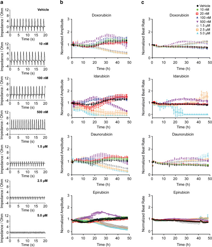
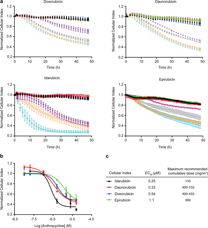
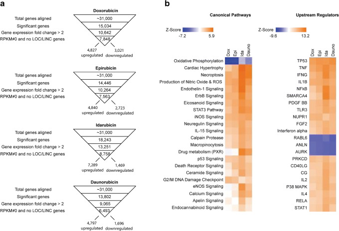
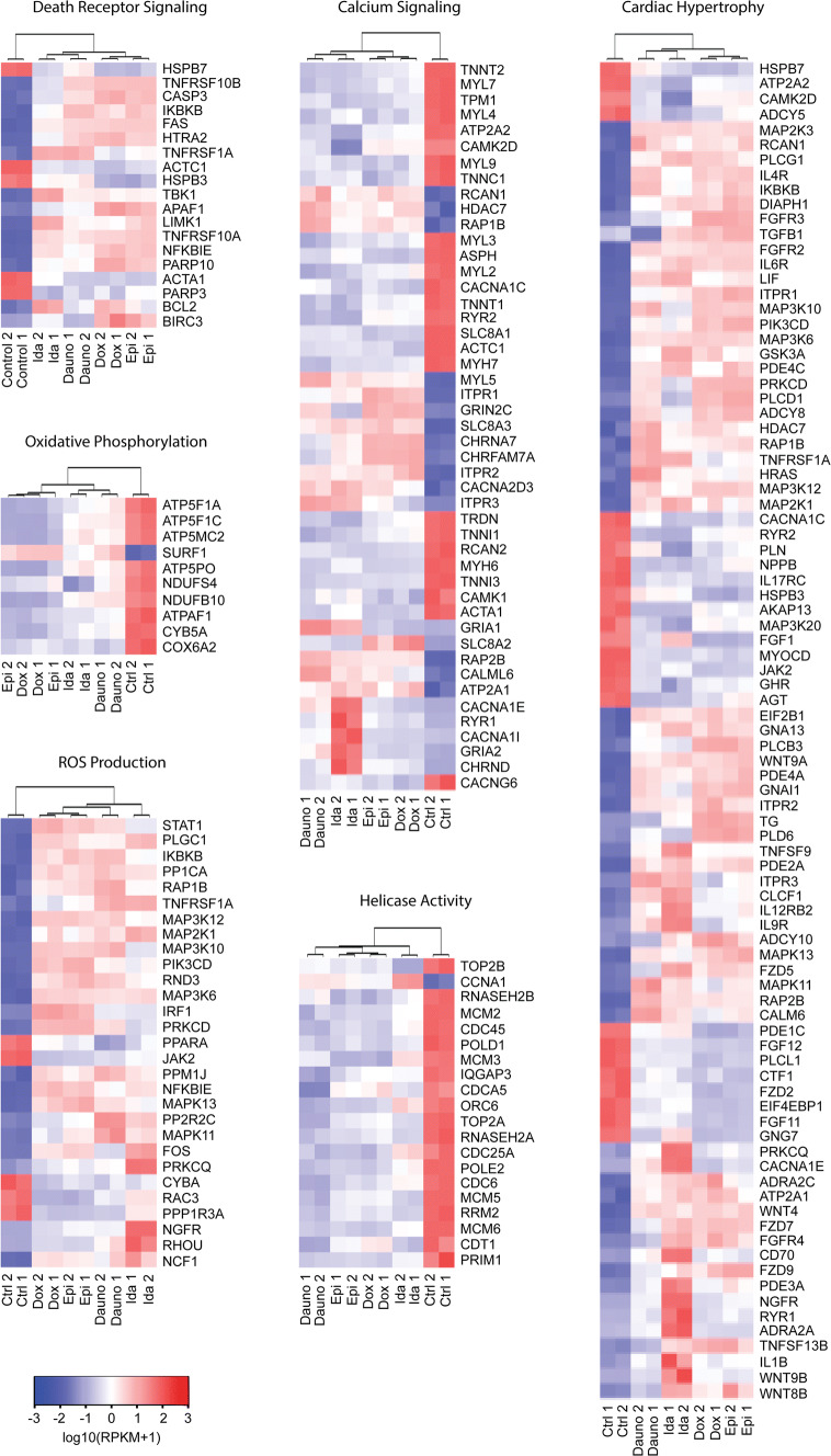
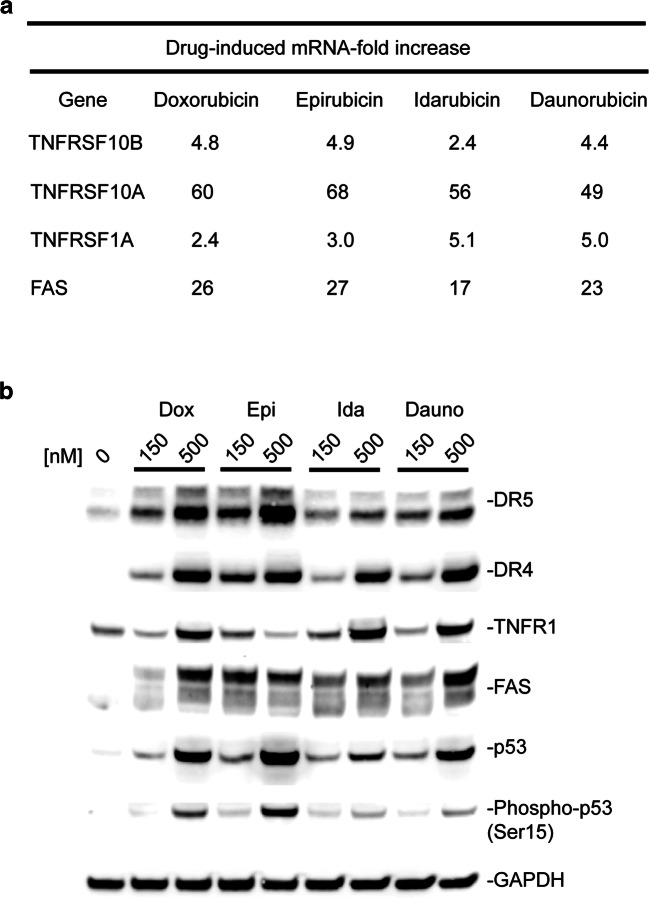
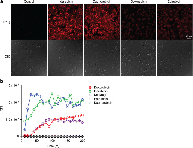
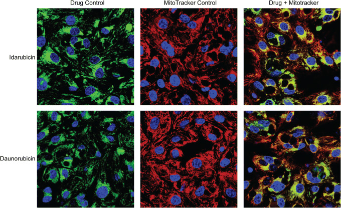
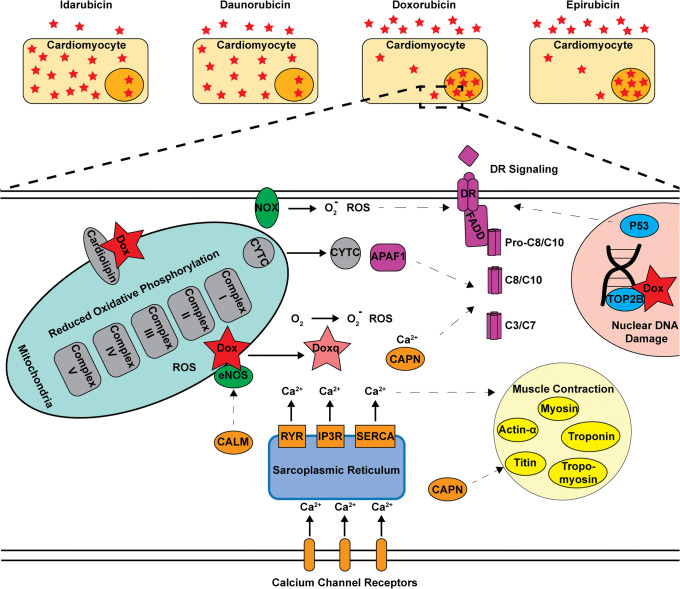
Anthracyclines are a class of chemotherapy drugs that are highly effective for the treatment of human cancers, but their clinical use is limited by associated dose-dependent cardiotoxicity. The precise mechanisms by which individual anthracycline induces cardiotoxicity are not fully understood. Human-induced pluripotent stem cell-derived cardiomyocytes (hiPSC-CMs) are emerging as a physiologically relevant model to assess drugs cardiotoxicity. Here, we describe an assay platform by coupling hiPSC-CMs and impedance measurement, which allows real-time monitoring of cardiomyocyte cellular index, beating amplitude, and beating rate. Using this approach, we have performed comparative studies on a panel of four anthracycline drugs (doxorubicin, epirubicin, idarubicin, and daunorubicin) which share a high degree of structural similarity but are associated with distinct cardiotoxicity profiles and maximum cumulative dose limits. Notably, results from our hiPSC-CMs impedance model (dose-dependent responses and EC50 values) agree well with the recommended clinical dose limits for these drugs. Using time-lapse imaging and RNAseq, we found that the differences in anthracycline cardiotoxicity are closely linked to extent of cardiomyocyte uptake and magnitude of activation/inhibition of several cellular pathways such as death receptor signaling, ROS production, and dysregulation of calcium signaling. The results provide molecular insights into anthracycline cardiac interactions and offer a novel assay system to more robustly assess potential cardiotoxicity during drug development.

Keywords: anthracycline; cardiotoxicity; cellular model; hiPSC-CMs.


Figures

Fig. 1
Fig. 1
Anthracycline effect on cardiomyocyte beating. hiPSC-CMs were treated with anthracycline (0–5 μM) and impedance-based contractility measurements were recorded in real-time every 90 min for 48 h. a After 48 h, cardiomyocyte beating profiles displayed visible decreases in beating amplitudes and increases in beating rates. Normalized cardiomyocyte beating amplitude (b) and rate (c) were quantified and plotted versus time for each anthracycline treatment condition
Fig. 2
Fig. 2
Anthracycline effect on cardiomyocyte cellular index. a Impedance-based cellular index of hiPSC-CMs was recorded in real time every 90 min for 48 h (vehicle = black, 10 nM = green, 20 nM = red, 100 nM = blue, 500 nM = purple, 1.5 μM = gray, 2.5 μM = orange, and 5.0 μM = cyan). b Normalized 48-h time point cellular index data were plotted versus log anthracycline concentration to generate dose response curves. c Dose response curves were fitted using nonlinear regression analysis to yield EC50 values. Notably, EC50 values agreed well with recommended maximum cumulative dose limits for those drugs
Fig. 3
Fig. 3
Transcriptomic analysis of anthracycline-induced cardiotoxicity. hiPSC-CMs were treated with 500 nM anthracycline for 48 h before RNA extraction and sequencing. a Regularized linear discriminant analysis (RLDA) algorithm was used to identify significant differential mRNA gene expressions for each anthracycline treatment compared to untreated control samples. Data sets were filtered using a p value significance < 0.05, fold-changes > 2, and RPKM ≠ 0. Only well-established gene transcripts were included (no LOC and LINC genes). b Ingenuity pathway analysis (IPA) algorithms were used to score canonical pathways and upstream regulator networks reported in literature based on inputted gene expression data. Activation Z-scores were shown using heat maps to compare each anthracycline
Fig. 4
Fig. 4
Hierarchical gene expression clustering of dysregulated cardiotoxicity molecular mechanisms. Heat mapping and hierarchical clustering were performed using RPKM values of dysregulated hiPSC-CMs genes identified in IPA cardiotoxicity pathways
Fig. 5
Fig. 5
Upregulation of death receptor signaling as a critical component of anthracycline-induced cardiotoxicity. hiPSC-CMs were treated with 150 or 500 nM anthracycline for 48 h. a RNAseq results showing anthracyclines increased mRNA expression of death receptors in hiPSC-CMs. b Immunoblotting results confirmed anthracyclines induced cardiomyocyte overexpression of death receptors and p53 at the protein level
Fig. 6
Fig. 6
Dynamics of anthracycline cellular uptake. a Confocal microscopy images of hiPSC-CMs were taken after 24-h treatment with 5 μM anthracycline. Images were acquired using a 40× objective lens and laser excitation at 488 nm. Anthracycline fluorescence is shown in red. b Time-lapse imaging was performed every 10 min for 200 min. The RFI of representative cells was quantified and plotted versus time to compare anthracycline uptake rates. Shown are representatives of two replicates
Fig. 7
Fig. 7
Mitochondrial localization of idarubicin and daunorubicin. hiPSC-CMs mitochondria were stained with Mitotracker Deep Red before treating with 5 μM idarubicin or daunorubicin for 2 h. Confocal microscopy images were acquired using a 40× objective lens and laser excitation at 405 nm for nucleus detection (shown in blue), 488 nm for anthracycline detection (shown in green), and 633 nm for mitochondria detection (shown in red). All images were acquired using the same red, blue, and green laser settings
Fig. 8
Fig. 8
A mechanistic view of anthracycline-induced cardiotoxicity. Anthracyclines can be taken up by hiPSC-CMs at significantly different rates and amounts due to their differences in lipophilicity. Once inside the cell, they induce alterations in several inter-linked canonical pathways (dashed arrows) such as DR signaling (purple), DNA damage/helicase inhibition (blue), dysregulation of calcium signaling (orange), reduced oxidative phosphorylation (gray), ROS production (green), and cardiac structural damage (yellow). However, the extent to which each pathway is activated or inhibited is specific for each anthracycline drug and appears to be associated with the drug’s cellular uptake

Similar articles

Cited by

References

    1. Minotti G, Menna P, Salvatorelli E, Cairo G, Gianni L. Anthracyclines: molecular advances and pharmacologic developments in antitumor activity and cardiotoxicity. Pharmacol Rev. 2004;56(2):185–229. doi: 10.1124/pr.56.2.6. - DOI - PubMed
    1. Venkatesh P, Kasi A. Anthracyclines. Treasure Island (FL): StatPearls; 2020.
    1. Ryberg M, Nielsen D, Skovsgaard T, Hansen J, Jensen BV, Dombernowsky P. Epirubicin cardiotoxicity: an analysis of 469 patients with metastatic breast cancer. J Clin Oncol. 1998;16(11):3502–3508. doi: 10.1200/JCO.1998.16.11.3502. - DOI - PubMed
    1. Anderlini P, Benjamin RS, Wong FC, Kantarjian HM, Andreeff M, Kornblau SM, O'Brien S, Mackay B, Ewer MS, Pierce SA. Idarubicin cardiotoxicity: a retrospective study in acute myeloid leukemia and myelodysplasia. J Clin Oncol. 1995;13(11):2827–2834. doi: 10.1200/JCO.1995.13.11.2827. - DOI - PubMed
    1. Octavia Y, Tocchetti CG, Gabrielson KL, Janssens S, Crijns HJ, Moens AL. Doxorubicin-induced cardiomyopathy: from molecular mechanisms to therapeutic strategies. J Mol Cell Cardiol. 2012;52(6):1213–1225. doi: 10.1016/j.yjmcc.2012.03.006. - DOI - PubMed

Publication types

MeSH terms

Grants and funding