

Indirect White Matter Pathways Are Associated With Treated Naming Improvement in Aphasia
- PMID: 33719732
- PMCID: PMC8068606
- DOI: 10.1177/1545968321999052
Indirect White Matter Pathways Are Associated With Treated Naming Improvement in Aphasia
Abstract
Background: White matter disconnection of language-specific brain regions associates with worse aphasia recovery. Despite a loss of direct connections, many stroke survivors may maintain indirect connections between brain regions.
Objective: To determine (1) whether preserved direct connections between language-specific brain regions relate to better poststroke naming treatment outcomes compared to no direct connections and (2) whether for individuals with a loss of direct connections, preserved indirect connections are associated with better treatment outcomes compared to individuals with no connections.
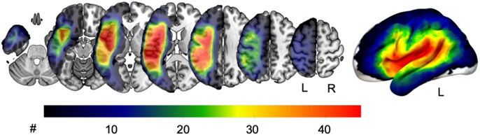
Methods: We computed structural whole-brain connectomes from 69 individuals with chronic left-hemisphere stroke and aphasia who completed a 3-week-long language treatment that was supplemented by either anodal transcranial direct current stimulation (A-tDCS) or sham stimulation (S-tDCS). We determined differences in naming improvement between individuals with direct, indirect, and no connections using 1-way analyses of covariance and multivariable linear regressions.
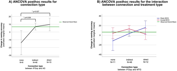
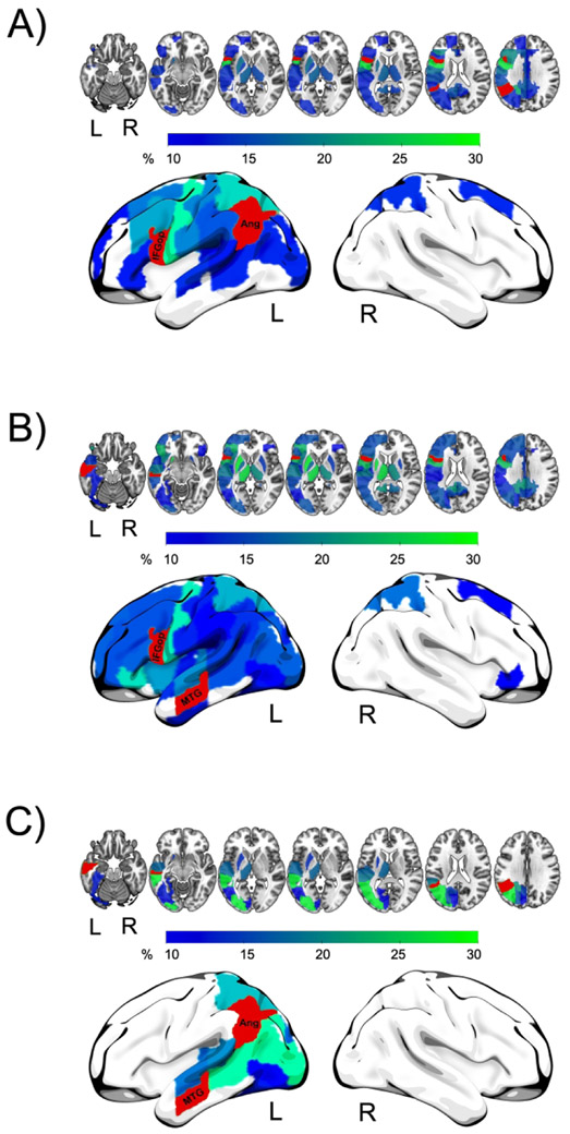
Results: Independently of tDCS modality, direct or indirect connections between the inferior frontal gyrus pars opercularis and angular gyrus were both associated with a greater increase in correct naming compared to no connections (P = .027 and P = .039, respectively). Participants with direct connections between the inferior frontal gyrus pars opercularis and middle temporal gyrus who received S-tDCS and participants with indirect connections who received A-tDCS significantly improved in naming accuracy.
Conclusions: Poststroke preservation of indirect white matter connections is associated with better treated naming improvement in aphasia even when direct connections are damaged. This mechanistic information can be used to stratify and predict treated naming recovery in individuals with aphasia.
Keywords: aphasia; brain connectomics; magnetic resonance imaging; rehabilitation; stroke; white matter.
Figures
References
-
- Dragoy O, Akinina Y, Dronkers N. Toward a functional neuroanatomy of semantic aphasia: A history and ten new cases. Cortex. 2017;97:164–182. - PubMed
Publication types
MeSH terms
Grants and funding
LinkOut - more resources
Full Text Sources
Other Literature Sources
Medical
Miscellaneous
