

Radiological evaluation of postoperative osteomyelitis in long bones: Which is the best tool?
- PMID: 33719739
- PMCID: PMC8750142
- DOI: 10.1177/1750458920961347
Radiological evaluation of postoperative osteomyelitis in long bones: Which is the best tool?
Abstract
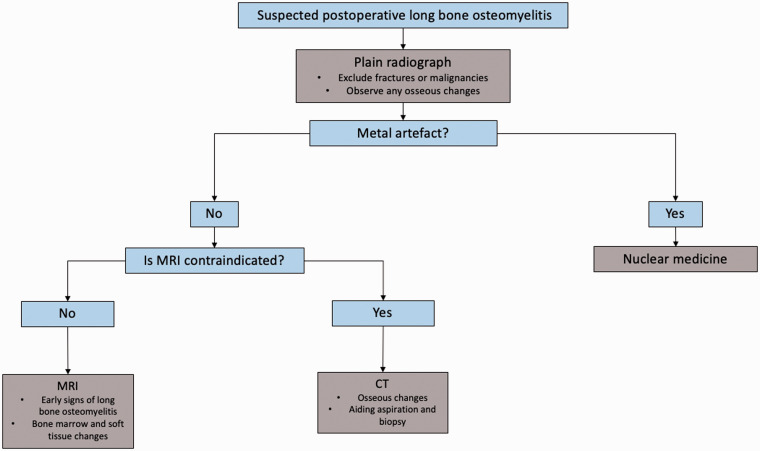
Currently, definitive diagnosis of osteomyelitis involves a combination of clinical signs, symptoms, laboratory tests, imaging modalities and cultures from blood, joint or body fluid. Imaging plays a critical role in the osteomyelitis diagnosis. Each of these tests incurs an additional cost to the patient or healthcare system and their use varies according to the preference of the healthcare professional and the healthcare setup. Imaging plays a critical role in the diagnosis and management of postoperative long bone osteomyelitis, with the aim of reducing long-term complications such as non-union, amputation and pathological fractures. In this review, we discuss the key findings on different radiological modalities and correlate them with disease pathophysiology. Currently, magnetic resonance imaging is the best available imaging modality due to its sensitivity in detecting early signs of long bone osteomyelitis and high soft tissue resolution. Other modalities such as radio-nuclear medicine, computed tomography and ultrasound have been proved to be useful in different clinical scenarios as described in this narrative review.
Keywords: Diagnostic tools; Health economics; Long bone infection; Postoperative osteomyelitis; Radiological evaluation.
Figures
Similar articles
-
Osteomyelitis: a review of currently used imaging techniques.Eur Radiol. 1999;9(5):894-900. doi: 10.1007/s003300050763. Eur Radiol. 1999. PMID: 10369987 Review.
-
Imaging of osteomyelitis and musculoskeletal soft tissue infections: current concepts.Rheum Dis Clin North Am. 2003 Feb;29(1):89-109. doi: 10.1016/s0889-857x(02)00078-9. Rheum Dis Clin North Am. 2003. PMID: 12635502 Review.
-
The accuracy of different imaging techniques in diagnosis of acute hematogenous osteomyelitis.Medicina (Kaunas). 2009;45(8):624-31. Medicina (Kaunas). 2009. PMID: 19773621
-
Diagnostic imaging of pediatric hematogenous osteomyelitis: lessons learned from a multi-modality approach.Eur Radiol. 2006 Sep;16(9):2109-19. doi: 10.1007/s00330-006-0187-4. Epub 2006 Mar 16. Eur Radiol. 2006. PMID: 16541223 Review.
-
[Hematogenous osteomyelitis of the calcaneus in children: 26 cases].Rev Chir Orthop Reparatrice Appar Mot. 2008 Sep;94(5):434-42. doi: 10.1016/j.rco.2008.02.004. Epub 2008 May 2. Rev Chir Orthop Reparatrice Appar Mot. 2008. PMID: 18774017 French.
Cited by
-
The usefulness of different imaging modalities in mandibular osteonecrosis and osteomyelitis diagnosis.Sci Rep. 2025 Apr 10;15(1):12272. doi: 10.1038/s41598-025-96910-x. Sci Rep. 2025. PMID: 40210732 Free PMC article.
-
Positron Emission Tomography-Computed Tomography and Magnetic Resonance Imaging Assessments in a Mouse Model of Implant-Related Bone and Joint Staphylococcus aureus Infection.Microbiol Spectr. 2023 Jun 15;11(3):e0454022. doi: 10.1128/spectrum.04540-22. Epub 2023 Apr 3. Microbiol Spectr. 2023. PMID: 37010409 Free PMC article.
References
-
- Christian S, Kraas J, Conway WF. 2007. Musculoskeletal infections. Seminars in Roentgenology 42 92–101. - PubMed
-
- Collins MS, Schaar MM, Wenger DE, Mandrekar JN. 2005. T1-weighted MRI characteristics of pedal osteomyelitis. American Journal of Roentgenology 185 386–393. - PubMed
-
- Donovan A, Schweitzer ME. 2010. Use of MR imaging in diagnosing diabetes-related pedal osteomyelitis. Radiographics 30 723–736. - PubMed
Publication types
MeSH terms
LinkOut - more resources
Full Text Sources
Other Literature Sources
