. 2021 Dec;10(1):651-663.
doi: 10.1080/22221751.2021.1903343.

High dose of vesicular stomatitis virus-vectored Ebola virus vaccine causes vesicular disease in swine without horizontal transmission

Affiliations

High dose of vesicular stomatitis virus-vectored Ebola virus vaccine causes vesicular disease in swine without horizontal transmission

Igor Morozov et al. Emerg Microbes Infect. 2021 Dec.

Abstract

ABSTRACTThe recent impact of Ebola virus disease (EVD) on public health in Africa clearly demonstrates the need for a safe and efficacious vaccine to control outbreaks and mitigate its threat to global health. ERVEBO® is an effective recombinant Vesicular Stomatitis Virus (VSV)-vectored Ebola virus vaccine (VSV-EBOV) that was approved by the FDA and EMA in late 2019 for use in prevention of EVD. Since the parental virus VSV, which was used to construct VSV-EBOV, is pathogenic for livestock and the vaccine virus may be shed at low levels by vaccinated humans, widespread deployment of the vaccine requires investigation into its infectivity and transmissibility in VSV-susceptible livestock species. We therefore performed a comprehensive clinical analysis of the VSV-EBOV vaccine virus in swine to determine its infectivity and potential for transmission. A high dose of VSV-EBOV resulted in VSV-like clinical signs in swine, with a proportion of pigs developing ulcerative vesicular lesions at the nasal injection site and feet. Uninoculated contact control pigs co-mingled with VSV-EBOV-inoculated pigs did not become infected or display any clinical signs of disease, indicating the vaccine is not readily transmissible to naïve pigs during prolonged close contact. In contrast, virulent wild-type VSV Indiana had a shorter incubation period and was transmitted to contact control pigs. These results indicate that the VSV-EBOV vaccine causes vesicular illness in swine when administered at a high dose. Moreover, the study demonstrates the VSV-EBOV vaccine is not readily transmitted to uninfected pigs, encouraging its safe use as an effective human vaccine.

Keywords: Ebola virus disease; VSV; safety study; swine; virus-vectored vaccine.


Conflict of interest statement

Sheri Dubey, Sean P. Troth, and Beth-Ann Coller are employees of Merck & Co., Inc., Kenilworth, NJ USA and may have shares in the company and/or patents relating to the technology. Richard Nichols reports personal fees from NewLink Genetics and Crozet BioPharma. Thomas P. Monath reports personal fees from NewLink Genetics Corp. and Merck Inc. Thomas P. Monath, Richard Nichols, and Brian K. Martin were associated with Bioprotection Systems, Inc., during the course of the study. Bioprotection Systems, Inc., licensed the VSV-EBOV vaccine from Public Health Canada and sub-licensed the vaccine to Merck & Co., Inc.

Figures

Figure 1.
Figure 1.
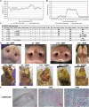
Clinical analysis of wtVSV-inoculated pigs. (A) Average daily temperatures of wtVSV-inoculated pigs compared to uninoculated pigs. (B) Average lesion scores of wtVSV-inoculated pigs compared to uninoculated environmental controls on each day of the study. (C) Summary of RT-qPCR analysis performed to detect viral RNA in clinical samples collected throughout the study, indicating the presence (+) or absence (−) of viral RNA. P-LN – parotid lymph node; RP-LN – retropharyngeal lymph node; SC-LN – superficial cervical lymph node; M-LN – mandibular lymph node; NP – nasal planum; NA – Not Applicable because not present or collected; DPI - days post inoculation in which positive samples were detected. (D) Representative pictures showing vesicular lesions in pigs. (E) Immunohistochemistry analysis performed on nasal planum lesion tissue collected on 2 DPI necropsy from wtVSV-infected pig #3 showing positive (red) immunostaining using anti-VSV-G rabbit polyclonal antibody localized to the stratum spinosum and granulosum of the epidermis.
Figure 2.
Figure 2.
Clinical analysis of VSV-EBOV-inoculated pigs. (A) Average daily temperatures of VSV-EBOV-inoculated pigs compared to uninoculated controls. (B) Average lesion scores of VSV-EBOV-inoculated pigs compared to uninoculated controls on each day of the study. (C) Summary of RT-qPCR analysis performed to detect viral RNA in clinical samples collected throughout the study, indicating the presence (+) or absence (−) of viral RNA. P-LN – parotid lymph node; RP-LN – retropharyngeal lymph node; SC-LN – superficial cervical lymph node; M-LN – mandibular lymph node; NP – nasal planum; NA – Not Applicable because not present or collected; DPI - days post inoculation in which positive samples were detected. (D) Representative pictures showing vesicular lesions in VSV-EBOV-inoculated pigs. (E) Immunohistochemistry analysis performed on foot lesion tissue (hoof and skin) collected on 10 DPI necropsy from VSV-EBOV-inoculated pig #15 showing positive (red) immunostaining using anti-EBOV-GP rabbit polyclonal antibody localized to the stratum spinosum and granulosum of the epidermis.
Figure 3.
Figure 3.
Clinical analysis of wtVSV-contact control pigs. (A) Average daily temperatures of wtVSV- contact control pigs compared to uninoculated controls. (B) Average lesion scores of wtVSV-contact control pigs compared to uninoculated controls on each day of the study. (C) Summary of RT-qPCR analysis performed to detect viral RNA in clinical samples collected throughout the study, indicating the presence (+) or absence (−) of viral RNA. P-LN – parotid lymph node; RP-LN – retropharyngeal lymph node; SC-LN – superficial cervical lymph node; M-LN – mandibular lymph node; NP – nasal planum; NA – Not Applicable because not present or collected; DPI - days post inoculation of principal infected pigs in which positive samples were detected in this group. (D) Representative pictures showing vesicular lesions in wtVSV-contact control pigs.
Figure 4.
Figure 4.
Clinical analysis of VSV-EBOV-contact control pigs. (A) Average daily temperatures of VSV-EBOV contact control pigs compared to uninoculated controls. (B) Average lesion scores of VSV-EBOV contact control pigs compared to uninoculated controls on each day of the study. * – one pig (#10) showed a focal <2 cm reddening on a hind limb on 14 DPI that was transient and did not show vesicular or ulcerative features typical of VSV infection and was ultimately determined to be a mechanical injury. (C) Summary of RT-qPCR analysis performed to detect viral RNA in clinical samples collected throughout the study, indicating the presence (+) or absence (−) of viral RNA. P-LN – parotid lymph node; RP-LN – retropharyngeal lymph node; SC-LN – superficial cervical lymph node; M-LN – mandibular lymph node; NP – nasal planum; NA – Not Applicable because lesions not present or collected; DPI - days post inoculation of principal infected pigs. * – suspected foot lesion swabs and suspected lesion tissues were collected on any tissue that was discolored even if the cause was likely a mechanical injury or benign discoloration and tested for viral RNA. All such samples were negative for VSV-specific RNA. (D) Representative pictures showing a lack of vesicular lesions in VSV-EBOV-contact control pigs.

Similar articles

Cited by

References

    1. Rojas M, Monsalve DM, Pacheco Y, et al. . Ebola virus disease: An emerging and re-emerging viral threat. J Autoimmun. 2020 Jan;106:102375. - PubMed
    1. Emanuel J, Marzi A, Feldmann H.. Filoviruses: ecology, molecular biology, and evolution. Adv Virus Res. 2018;100:189–221. - PMC - PubMed
    1. Bausch DG. West Africa 2013 Ebola: from virus outbreak to humanitarian crisis. Curr Top Microbiol Immunol. 2017;411:63–92. - PubMed
    1. Higgs ES, Dubey SA, Coller BAG, et al. . Accelerating vaccine development during the 2013–2016 West African Ebola virus disease outbreak. Curr Top Microbiol Immunol. 2017;411:229–261. - PubMed
    1. Regules JA, Beigel JH, Paolino KM, et al. . A recombinant vesicular stomatitis virus Ebola vaccine. N Engl J Med. 2017 Jan 26;376(4):330–341. - PMC - PubMed

MeSH terms