. 2021 Feb 24:(168):10.3791/61937.
doi: 10.3791/61937.

Simple, Affordable, and Modular Patterning of Cells using DNA

Affiliations

Simple, Affordable, and Modular Patterning of Cells using DNA

Katelyn A Cabral et al. J Vis Exp. .

Abstract

The relative positioning of cells is a key feature of the microenvironment that organizes cell-cell interactions. To study the interactions between cells of the same or different type, micropatterning techniques have proved useful. DNA Programmed Assembly of Cells (DPAC) is a micropatterning technique that targets the adhesion of cells to a substrate or other cells using DNA hybridization. The most basic operations in DPAC begin with decorating cell membranes with lipid-modified oligonucleotides, then flowing them over a substrate that has been patterned with complementary DNA sequences. Cells adhere selectively to the substrate only where they find a complementary DNA sequence. Non-adherent cells are washed away, revealing a pattern of adherent cells. Additional operations include further rounds of cell-substrate or cell-cell adhesion, as well as transferring the patterns formed by DPAC to an embedding hydrogel for long-term culture. Previously, methods for patterning oligonucleotides on surfaces and decorating cells with DNA sequences required specialized equipment and custom DNA synthesis, respectively. We report an updated version of the protocol, utilizing an inexpensive benchtop photolithography setup and commercially available cholesterol modified oligonucleotides (CMOs) deployed using a modular format. CMO-labeled cells adhere with high efficiency to DNA-patterned substrates. This approach can be used to pattern multiple cell types at once with high precision and to create arrays of microtissues embedded within an extracellular matrix. Advantages of this method include its high resolution, ability to embed cells into a three-dimensional microenvironment without disrupting the micropattern, and flexibility in patterning any cell type.


Figures

Figure 1:
Figure 1:. Overview of CMO-DPAC protocol.
First, a DNA-patterned slide is created by coating an aldehyde-functionalized glass slide with a positive photoresist, covering it with a transparency mask in the desired pattern, and exposing it to UV light. The UV-exposed photoresist is washed away with developer, leaving exposed regions of the aldehyde slide and allowing the binding of amine-functionalized DNA to the surface. Cells are then labeled with CMOs and flowed over the surface. The DNA on the cell membrane hybridizes to the DNA on the surface, resulting in adhesion.
Figure 2:
Figure 2:. Cells are labeled with CMOs in a stepwise process.
First, the cholesterol-modified Universal Anchor Strand is pre-hybridized with the Adapter Strand. Next, the Universal Anchor + Adapter solution is mixed with the cell suspension. The cholesterol on the Universal Anchor + Adapter complex inserts into the cell membrane. After incubation, the cholesterol-modified Universal Co-Anchor Strand is added to the cell suspension, where it hybridizes with the Universal Anchor Strand and inserts into the cell membrane. The addition of the second cholesterol molecule increases the net hydrophobicity of the DNA complex and stabilizes it within the membrane. After washing out the excess DNA, the cells are concentrated and added to a PDMS flow cell on top of the patterned surface. The 3’ end of the Adapter Strand hybridizes with the Surface DNA Strand on the glass slide, resulting in adhesion to the slide specifically in regions functionalized with complementary DNA.
Figure 3:
Figure 3:. Photolithography is used to create the DNA-patterned slides that will ultimately dictate the placement of cells.
(A) Overview of photolithography process. An aldehyde-functionalized slide is spin-coated with a positive photoresist. UV light shines onto the slide through a transparency photomask that is transparent where cell adhesion is desired. After the slide is developed, the regions that were previously exposed to UV light now have exposed aldehyde groups. A 20 µM solution of an amine-functionalized DNA oligo is then dropped onto the slide and spread over the patterned regions. The slide is then baked to induce the formation of Schiff bonds (C=N) between the amine and aldehyde groups, a reversible covalent bond. Subsequent reductive amination with 0.25% sodium borohydride in PBS converts the Schiff base to a secondary amine by reductive amination, resulting in an irreversible bond between the DNA and the slide. The remaining photoresist can then be removed by rinsing with acetone. (B) This process can be repeated to create multi-component DNA patterns and therefore perform experiments with multiple cell populations. (i) After the first oligo is patterned, the slide is again coated in photoresist and the protocol proceeds as before. Alignment of the photomasks using fiduciary markers is necessary for patterning multiple DNA strands. (ii) Each cell type being patterned differs in the 20-base modular domain of the Adapter Strand. By using orthogonal sets of complementary oligos, multiple cell types can be patterned without cross-adhesion.
Figure 4:
Figure 4:. Adhesion of CMO-labeled cells to DNA patterns increases as a function of CMO concentration during labeling.
In this experiment, the Universal Anchor + Adapter Strand (pre-hybridized) and the Universal Co-Anchor were used at equal concentrations. Concentration refers to the concentration of CMO in the cell suspension during CMO labeling of cells. (A) Quantification of the percentage of 15 µm diameter DNA spots that were occupied by CMO-labeled MCF10A cells as a function of CMO concentration during cell labeling. Data represented as the mean ± standard deviation from three experiments. (B) Representative images of the DNA patterns (magenta) and adhered MCF10As (cyan) at different concentrations of CMO. Scale bar = 100 µm.
Figure 5:
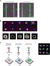
Figure 5:. CMO-DPAC can be used to create two-dimensional cell patterns that can subsequently be embedded into a three-dimensional hydrogel for culture and/or layered to create multilayered structures.
(A) Direct comparison between CMO-labeled human umbilical vein endothelial cells (HUVECs) and LMO-labeled HUVECs adhered to a linear DNA pattern. Both methods of cell labeling result in nearly 100% occupancy of the DNA pattern. (B) Single Madin-Darby Canine Kidney cells (MDCKs) expressing H2B-RFP were patterned onto 15 µm diameter spots spaced 200 µm apart and subsequently embedded in Matrigel. After 120 h of culture, the resulting epithelial cysts were fixed and stained for E-cadherin, actin, and collagen IV. Spheroid in white box is shown in detail. Scale bar = 50 µm. (C) Multilayered cellular structures can be created by labeling separate cell populations with complementary Adapter Strands and patterning sequentially so that each new addition of cells adheres to the cell layer before it. (i) A schematic of the sequential patterning of cell populations to create multilayered structures. (ii)Three-layered cell aggregates of MCF10As (visualized using dyes) were created using this process. Scale bar = 50 µm.
Figure 6:
Figure 6:. Multiple cell types can be patterned without cross-contamination or loss of adhesion.
Multiple amine-modified DNA oligos were patterned sequentially onto an aldehyde slide and aligned through use of metal fiduciary markers. Three populations of MCF10As (cyan, magenta, yellow) were stained with unique dyes labeled with complementary CMOs, and patterned onto the slide, resulting in an image of the UC Berkeley and UCSF logos. Scale bar 1 mm.

Similar articles

Cited by

References

    1. Kreeger PK, Strong LE, Masters KS Engineering approaches to study cellular decision-making. Annual Review of Biomedical Engineering (December 2017), 49–72 (2018). - PMC - PubMed
    1. Goubko C. a., Cao X Patterning multiple cell types in co-cultures: A review. Materials Science and Engineering C 29 (6), 1855–1868 (2009).
    1. Sun W et al. The bioprinting roadmap. Biofabrication 12 (2), 022002 (2020). - PubMed
    1. Liu WF, Chen CS Cellular and multicellular form and function. Advanced Drug Delivery Reviews 59 (13), 1319–1328 (2007). - PMC - PubMed
    1. Duffy RM, Sun Y, Feinberg AW Understanding the role of ECM protein composition and geometric micropatterning for engineering human skeletal muscle. Annals of Biomedical Engineerin.g 44 (6), 2076–2089 (2016). - PMC - PubMed

Publication types