. 2021 Mar 1;4(3):e211489.
doi: 10.1001/jamanetworkopen.2021.1489.

Association of Clinical, Biological, and Brain Magnetic Resonance Imaging Findings With Electroencephalographic Findings for Patients With COVID-19

Collaborators, Affiliations

Association of Clinical, Biological, and Brain Magnetic Resonance Imaging Findings With Electroencephalographic Findings for Patients With COVID-19

Virginie Lambrecq et al. JAMA Netw Open. .

Erratum in

  • Group Members Added to Supplement.
    [No authors listed] [No authors listed] JAMA Netw Open. 2022 Jun 1;5(6):e2219899. doi: 10.1001/jamanetworkopen.2022.19899. JAMA Netw Open. 2022. PMID: 35708695 Free PMC article. No abstract available.

Abstract

Importance: There is evidence of central nervous system impairments associated with coronavirus disease 2019 (COVID-19) infection, including encephalopathy. Multimodal monitoring of patients with COVID-19 may delineate the specific features of COVID-19-related encephalopathy and guide clinical management.

Objectives: To investigate clinical, biological, and brain magnetic resonance imaging (MRI) findings in association with electroencephalographic (EEG) features for patients with COVID-19, and to better refine the features of COVID-19-related encephalopathy.

Design, setting, and participants: This retrospective cohort study conducted in Pitié-Salpêtrière Hospital, Paris, France, enrolled 78 hospitalized adults who received a diagnosis of severe acute respiratory syndrome coronavirus 2 (SARS-Cov2) and underwent EEG between March 30 and June 11, 2020.

Exposures: Detection of SARS-CoV-2 from a nasopharyngeal specimen using a reverse transcription-polymerase chain reaction assay or, in the case of associated pneumonia, on a computed tomography scan of the chest.

Main outcomes and measures: Data on the clinical and paraclinical features of the 78 patients with COVID-19 were retrieved from electronic patient records.

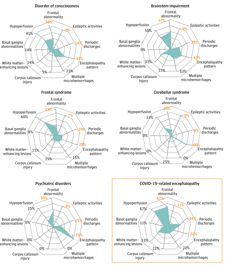
Results: Of 644 patients who were hospitalized for COVID-19, 78 (57 men [73%]; mean [SD] age, 61 [12] years) underwent EEG. The main indications for EEG were delirium, seizure-like events, and delayed awakening in the intensive care unit after stopping treatment with sedatives. Sixty-nine patients showed pathologic EEG findings, including metabolic-toxic encephalopathy features, frontal abnormalities, periodic discharges, and epileptic activities. Of 57 patients who underwent brain MRI, 41 showed abnormalities, including perfusion abnormalities, acute ischemic lesions, multiple microhemorrhages, and white matter-enhancing lesions. Fifty-five patients showed biological abnormalities, including dysnatremia, kidney failure, and liver dysfunction, the same day as the EEG. The results of cerebrospinal fluid analysis were negative for SARS-Cov-2 for all tested patients. Nine patients who had no identifiable cause of brain injury outside COVID-19 were further isolated; their brain injury was defined as COVID-19-related encephalopathy. They represented 1% (9 of 644) of patients with COVID-19 requiring hospitalization. Six of these 9 patients had movement disorders, 7 had frontal syndrome, 4 had brainstem impairment, 4 had periodic EEG discharges, and 3 had MRI white matter-enhancing lesions.

Conclusions and relevance: The results from this cohort of patients hospitalized with COVID-19 suggest there are clinical, EEG, and MRI patterns that could delineate specific COVID-19-related encephalopathy and guide treatment strategy.


Conflict of interest statement

Conflict of Interest Disclosures: Dr Huberfeld reported receiving personal fees from Advicenne, GW Pharma, and EISAI outside the submitted work. Dr Morélot-Panzini reported receiving personal fees from Astra-Zeneca, GSK, SOS Oxygène, ADEP, ISIS, Resmed, Chiesi, Menarini, Vivisol, Air Liquide, and Lowenstein outside the submitted work. Dr Raux reported receiving personal fees from Chiesi outside the submitted work. Dr Luyt reported receiving personal fees from Bayer Healthcare, Merck, ThermoFischer Brahms, Carmat, and Biomérieux outside the submitted work. Dr Corvol reported receiving grants from Fédération Internationale pour l’Automobile and Investissements d’avenir program during the conduct of the study; personal fees from Biogen, UCB, Prevail Therapeutic, Idorsia, Ever Pharma, Denali, Air Liquide, and Theranexus; and grants from Sanofi outside the submitted work. Dr Lubetzki reported receiving personal fees from Biogen, Merck-Serono, Roche, Rewind, and Ipsen outside the submitted work. Dr Pyatigorskaya reported receiving personal fees from GE Healthcare and Biogen; and grants from ANR outside the submitted work. Dr Navarro reported receiving personal fees from UCB Pharma, Liva Nova, and EISAI outside the submitted work. No other disclosures were reported.

Figures

Figure 1.
Figure 1.. Examples of Electroencephalogram Recordings and Magnetic Resonance Imaging Findings
Eight electrodes, longitudinal bipolar montage, 20-second epoch, low frequency filter 0.53 Hz, high frequency filter 70 Hz. A, Intermittent slow biphasic delta waves in bifrontal areas, with low-voltage continuous background activity. B, Diphasic and triphasic anterior slow waves, with slow continuous background activity, C, Periodic discharges in the bifrontal areas, with low voltage background activity, D, Bilateral lesions in the supratentorial white matter (arrowhead), hyperintense on axial fluid-attenuated inversion recovery images, E, Multiple microhemorrhages (arrowheads) involving the corpus callosum on T2 star images, F, Left frontotemporal hypoperfusion (arrowhead). ECG indicates electrocardiogram.
Figure 2.
Figure 2.. Representation of Electroencephalogram (EEG) and Brain Magnetic Resonance Imaging (MRI) Findings According to Neurologic Syndromes
Each radar chart is a graphical representation of the proportion of patients with the selected neurologic syndrome who had EEG findings (frontal abnormality, epileptic activities, periodic discharges, and encephalopathy pattern; percentage in orange) or MRI findings (multiple microhemorrhages, corpus callosum injury, white matter–enhancing lesions, basal ganglia abnormalities, and hypoperfusion; percentage in black). Each concentric circle represents a proportion of 20%. A blue line is drawn connecting the values (percentages) for each finding (EEG or MRI), giving the blue polygon appearance. The polygon represents the prevalence of EEG and MRI findings: the larger the area of the polygon is, the more abnormal EEG or MRI findings the patients had. Data are represented as proportion to take into account missing data. COVID-19 indicates coronavirus disease 2019.
Figure 3.
Figure 3.. Clinical, Electroencephalogram (EEG), and Magnetic Resonance Imaging (MRI) Disturbances in Patients With Coronavirus Disease 2019 (COVID-19)–Related Encephalopathy
Receiver operating characteristic curve for the model, evaluating the performance of movement disorders, brainstem impairment, frontal syndrome, EEG periodic discharges, and white matter–enhancing MRI lesions to identify patients with COVID-19–related encephalopathy. AUC indicates area under the curve.

References

    1. Ellul MA, Benjamin L, Singh B, et al. . Neurological associations of COVID-19. Lancet Neurol. 2020;19(9):767-783. doi:10.1016/S1474-4422(20)30221-0 - DOI - PMC - PubMed
    1. Nepal G, Rehrig JH, Shrestha GS, et al. . Neurological manifestations of COVID-19: a systematic review. Crit Care. 2020;24(1):421. doi:10.1186/s13054-020-03121-z - DOI - PMC - PubMed
    1. Chougar L, Shor N, Weiss N, et al. ; CoCo Neurosciences Study Group . Retrospective observational study of brain MRI findings in patients with acute SARS-CoV-2 infection and neurologic manifestations. Radiology. 2020;297(3):E313-E323. doi:10.1148/radiol.2020202422 - DOI - PMC - PubMed
    1. Paterson RW, Brown RL, Benjamin L, et al. . The emerging spectrum of COVID-19 neurology: clinical, radiological and laboratory findings. Brain. 2020;143(10):3104-3120. doi:10.1093/brain/awaa240 - DOI - PMC - PubMed
    1. Flamand M, Perron A, Buron Y, Szurhaj W. Pay more attention to EEG in COVID-19 pandemic. Clin Neurophysiol. 2020;131(8):2062-2064. doi:10.1016/j.clinph.2020.05.011 - DOI - PMC - PubMed

Publication types