. 2021 Apr;41(5):631-641.
doi: 10.1002/pd.5931. Epub 2021 Mar 22.

Chromosomal mosaicism: Origins and clinical implications in preimplantation and prenatal diagnosis

Affiliations

Chromosomal mosaicism: Origins and clinical implications in preimplantation and prenatal diagnosis

Brynn Levy et al. Prenat Diagn. 2021 Apr.

Abstract

The diagnosis of chromosomal mosaicism in the preimplantation and prenatal stage is fraught with uncertainty and multiple factors need to be considered in order to gauge the likely impact. The clinical effects of chromosomal mosaicism are directly linked to the type of the imbalance (size, gene content, and copy number), the timing of the initial event leading to mosaicism during embryogenesis/fetal development, the distribution of the abnormal cells throughout the various tissues within the body as well as the ratio of normal/abnormal cells within each of those tissues. Additional factors such as assay noise and culture artifacts also have an impact on the significance and management of mosaic cases. Genetic counseling is an important part of educating patients about the likelihood of having a liveborn with a chromosome abnormality and these risks differ according to the time of ascertainment and the tissue where the mosaic cells were initially discovered. Each situation needs to be assessed on a case-by-case basis and counseled accordingly. This review will discuss the clinical impact of finding mosaicism through: embryo biopsy, chorionic villus sampling, amniocentesis, and noninvasive prenatal testing using cell-free DNA.


Conflict of interest statement

COI statement: F.R.G., is full-time employee of TomaLab without ownership shares; she is an expert panel member for Roche and consultant for Menarini Silicon. B.L. is a consultant for Igenomix and Natera.

Figures

Figure 1.
Figure 1.
Origins of mosaic preimplantation embryos in human. a, Incidence of aneuploid sperm and eggs (single cells) and mosaic embryos at the cleavage and blastocyst stages (% of cases). b-e, examples of mechanisms resulting in chromosomally mosaic embryos. b, anaphase lag. c, failed chromosome capture. d, tripolar spindle formation. e, endoduplication or failed cytokinesis result in tetraploid cells.
Figure 2.
Figure 2.
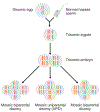
Trisomy rescue. This example illustrates a maternal meiosis I segregation error that has produced a disomic egg. Fertilization by the normal haploid sperm leads to a trisomic embryo. Depending on which chromosome is lost during trisomic rescue, there are three possible outcomes for the rescued disomic homologues. When either of the maternal chromosomes is lost during trisomic rescue, the resulting disomic cell line will contain both a maternal and paternal homologue (biparental inheritance). When the paternal chromosome is lost during the rescue event, the resulting disomic homologue will only derive from one parent, the mother (uniparental disomy, UPD). In all cases, mosaicism for a trisomic and disomic cell line will be present.
Figure 3:
Figure 3:
Schematic representation showing the final classification of mosaicism detected at the time of chorionic villus sampling after follow-up confirmatory amniocentesis. CPM, Confined Placental Mosaicism; TFM, True Fetal Mosaicism. If the chromosome abnormality is only detected in the trophoblast and is not confirmed at the time of amniocentesis, it is classified as CPM I. If it is confirmed, it is referred to as TFM IV. Similarly, if the abnormality is only detected in the mesenchyme and is not confirmed at the time of amniocentesis, it is classified as CPM II. If it is confirmed, it is referred to as TFM V. If the abnormality is detected in both layers and not confirmed at the time of amniocentesis, it is classified as CPM III. If confirmed, it is referred to as TFM VI. Data derived from Malvestiti et al. and Benn et al.
Figure 4:
Figure 4:
Frequency of the type of mosaicism observed at the time of CVS (Pie Chart) and likelihood of each type being confirmed at the time of amniocentesis (Bar Charts). CPM: Confined Placental Mosaicism. TFM: True Fetal Mosaicism. Data derived from Malvestiti et al. and Benn et al.
Figure 5:
Figure 5:
Overall likelihood of confirming true fetal mosaicism at amniocentesis for select chromosomes. Data derived from Malvestiti et al. and Benn et al.

Similar articles

Cited by

References

    1. Benn P, Grati FR. Aneuploidy in first trimester chorionic villi and spontaneous abortions: Windows into the origin and fate of aneuploidy through embryonic and fetal development. Prenat Diagn. 2020. - PubMed
    1. Chatzimeletiou K, Morrison EE, Prapas N, Prapas Y, Handyside AH. Spindle abnormalities in normally developing and arrested human preimplantation embryos in vitro identified by confocal laser scanning microscopy. Human reproduction. 2005;20(3):672–682. - PubMed
    1. Hardy K, Winston RM, Handyside AH. Binucleate blastomeres in preimplantation human embryos in vitro: failure of cytokinesis during early cleavage. J Reprod Fertil. 1993;98(2):549–558. - PubMed
    1. Holubcova Z, Blayney M, Elder K, Schuh M. Human oocytes. Error-prone chromosome-mediated spindle assembly favors chromosome segregation defects in human oocytes. Science. 2015;348(6239):1143–1147. - PMC - PubMed
    1. Levy B, Sigurjonsson S, Pettersen B, et al. Genomic imbalance in products of conception: single-nucleotide polymorphism chromosomal microarray analysis. Obstet Gynecol. 2014;124(2 Pt 1):202–209. - PubMed

Supplementary concepts

LinkOut - more resources