. 2021 Mar 15;17(3):e1009434.
doi: 10.1371/journal.pgen.1009434. eCollection 2021 Mar.

The velvet protein Vel1 controls initial plant root colonization and conidia formation for xylem distribution in Verticillium wilt

Affiliations

The velvet protein Vel1 controls initial plant root colonization and conidia formation for xylem distribution in Verticillium wilt

Annalena M Höfer et al. PLoS Genet. .

Abstract

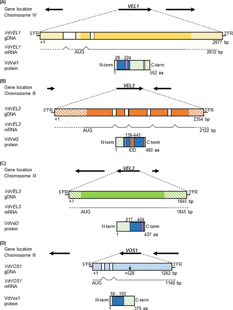
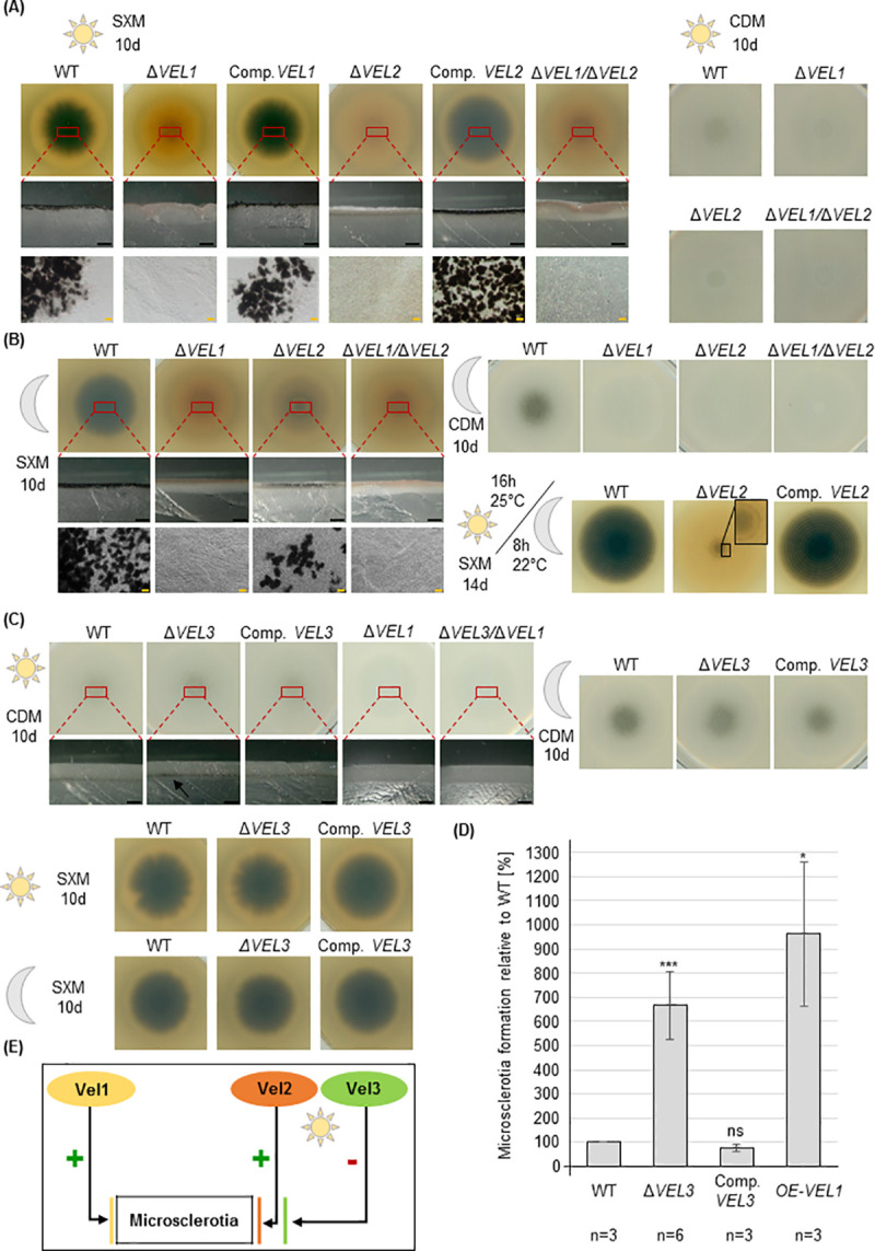
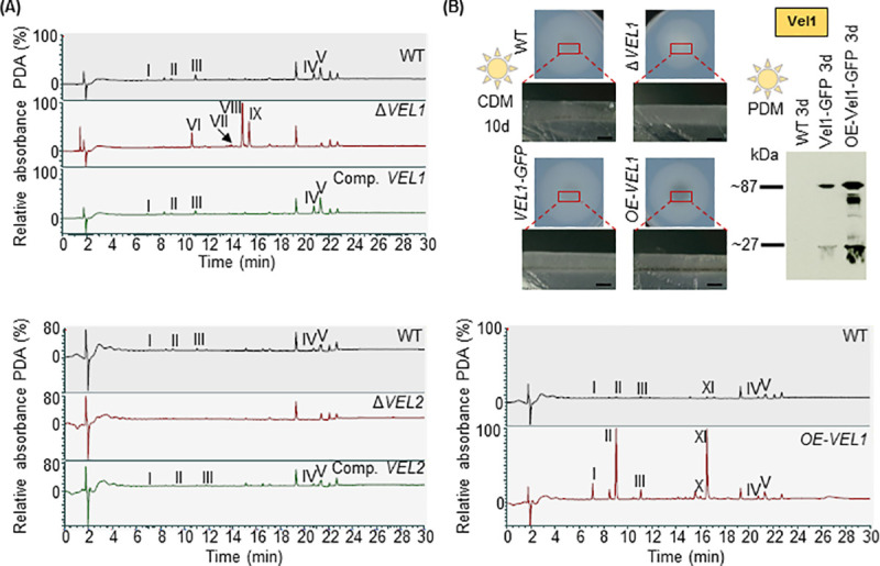
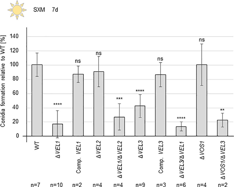
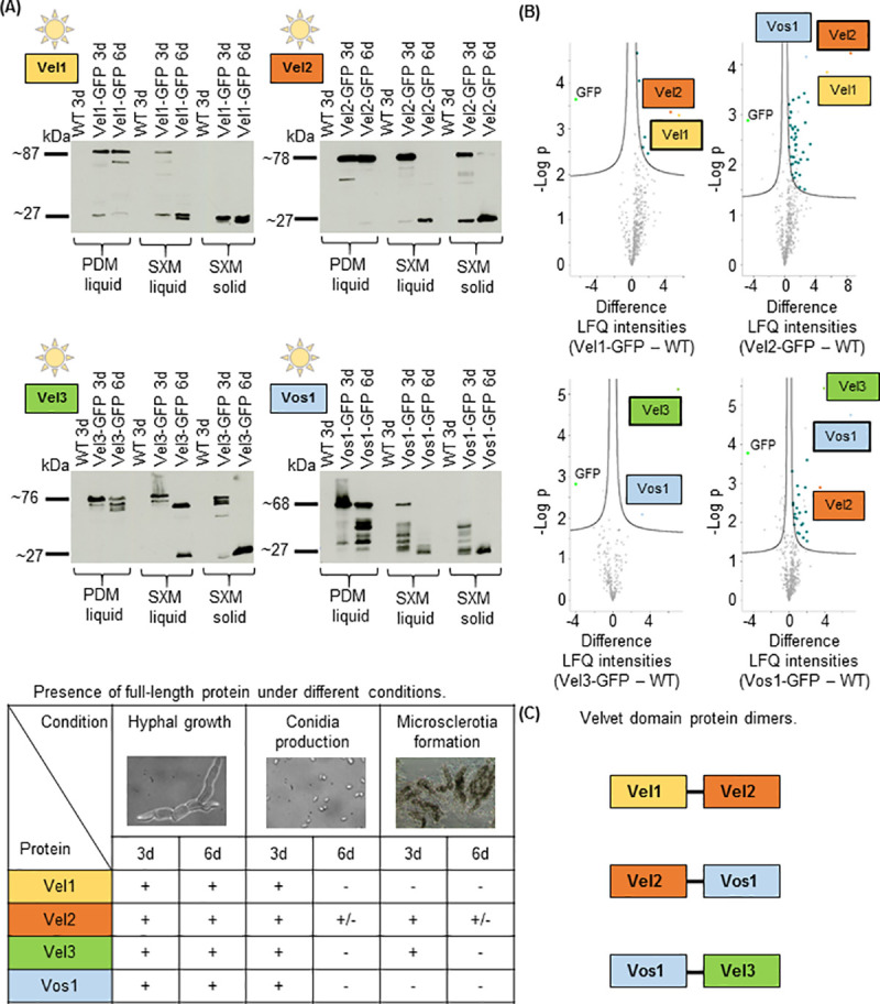
The conserved fungal velvet family regulatory proteins link development and secondary metabolite production. The velvet domain for DNA binding and dimerization is similar to the structure of the Rel homology domain of the mammalian NF-κB transcription factor. A comprehensive study addressed the functions of all four homologs of velvet domain encoding genes in the fungal life cycle of the soil-borne plant pathogenic fungus Verticillium dahliae. Genetic, cell biological, proteomic and metabolomic analyses of Vel1, Vel2, Vel3 and Vos1 were combined with plant pathogenicity experiments. Different phases of fungal growth, development and pathogenicity require V. dahliae velvet proteins, including Vel1-Vel2, Vel2-Vos1 and Vel3-Vos1 heterodimers, which are already present during vegetative hyphal growth. The major novel finding of this study is that Vel1 is necessary for initial plant root colonization and together with Vel3 for propagation in planta by conidiation. Vel1 is needed for disease symptom induction in tomato. Vel1, Vel2, and Vel3 control the formation of microsclerotia in senescent plants. Vel1 is the most important among all four V. dahliae velvet proteins with a wide variety of functions during all phases of the fungal life cycle in as well as ex planta.


Conflict of interest statement

The authors have declared that no competing interests exist.

Figures

Fig 1
Fig 1. V. dahliae genes encoding velvet domain proteins.
Genomic loci (gDNA) including transcription directions of neighboring genes and assignments to fungal chromosomes are indicated. Introns are depicted in white, exons are depicted in color; color hatched areas show the predicted untranslated region (UTR). Velvet domains (dark blue) were predicted by InterProScan according to entry IPR037525. Nuclear localization sequences (NLS, in grey) were predicted by using cNLS mapper. Potential PEST motifs (purple) were identified using epestfind. Details are given in S1–S4 Figs. (A) VEL1 (VDAG_JR2_Chr7g04890a) consists of three exons and two introns. The deduced Vel1 protein holds a velvet domain and a PEST motif at the N-terminus and an NLS at the C-terminus. (B) VEL2 (VDAG_JR2_Chr3g06150a) consists of five exons and four introns. The Vel2 velvet domain includes an NLS and is interrupted by an intrinsically disordered domain (IDD). (C) VEL3 (VDAG_JR2_Chr6g00630a) consists of a single exon. The Vel3 protein has one velvet domain, a PEST motif and an NLS at the C-terminus. (D) VOS1 (VDAG_JR2_Chr3g12090a) original annotation (starting point indicated by black arrow) was corrected by sequencing and verified by mass spectrometry. The corrected VOS1 annotation results in three exons and two introns and a Vos1 protein with N-terminal velvet domain.
Fig 2
Fig 2. V. dahliae microsclerotia formation depends on Vel1 with Vel2 as additional positive and Vel3 as negative control regulator primarily in light.
5x10⁴ spores of the indicated strains were spotted on indicated plates (SXM: simulated xylem medium; CDM: minimal Czapek-Dox-Medium) and incubated for 10 days at 25°C. Single colonies on SXM are shown from the back and colonies on CDM are shown from the top of the plate. Cross sections were made through the middle of the colony. Microscopy images show colony material scraped from the surface. (A) Vel1 and Vel2 are required for microsclerotia development in the presence of light. Microsclerotia formation of the ΔVEL1 or ΔVEL2 strains was compared to wild type (WT) and respective complementation strains (Comp.) on single colony pictures (first row), cross sections (second row) and in micrographs (third row). (B) Vel1 is primarily required for microsclerotia formation in darkness with a smaller contribution of Vel2. Incubation as in (A) but in darkness instead of light. Wild type as well as the VEL2 deletion strain produce microsclerotia in darkness, but absence of VEL2 leads to reduced amounts of microsclerotia. Lower right part: Vel2 function is independent of the fungal light/dark control. Phenotypical analysis during long day/night cycling conditions (16 h, 25°C: 8 h, 22°C, light: dark). Wild type displays a ring-like structure around the inoculation point with melanized microsclerotia and reduced color of the microsclerotia towards the margins of the colony. The VEL2 deletion strain is also capable to form the ring-like structure but with hardly any melanization (black box). (C) Vel3 inhibits microsclerotia formation in light without strong impact in dark on minimal medium. Colony pictures (first row) and cross sections (second row) are shown. Black scale bar = 1 mm, yellow scale bar = 20 μm. (D) Microsclerotia quantification of the wild type, the VEL3 deletion and complementation strain as well as the VEL1 overexpression strain. The VEL3 deletion and the VEL1 overexpression strains produce significantly more microsclerotia than the wild type and complementation strain. The experiment was repeated three times with three technical replicates each. Significant differences to wild type were calculated by t-test and indicate*:p<0.05; ***:p<0.001; ns: not significant. (E) Interplay between the velvet domain proteins Vel1, Vel2 and Vel3 in V. dahliae microsclerotia formation in the presence or absence of illumination.
Fig 3
Fig 3. V. dahliae Vel1 and Vel2 control functions in secondary metabolite production.
Displayed are chromatograms of metabolites extracted from ΔVEL1 and ΔVEL2 (A) and from the VEL1 overexpression strain (OE-VEL1) (B). LC/MS combined with photodiode array detection (PDA) analysis is shown. Secondary metabolites were extracted from two-week-old fungal mycelium grown on Czapek-Dox-Medium (CDM) supplemented with glucose in constant light. (A) Upper part: Deletion of VEL1VEL1) leads to reduced formation of five substances (I-V) and production of four substances (VI-IX) which were not abundant in metabolite extracts of wild type (WT) or complementation (Comp. VEL1) control strains. Lower part: The same five substances (I-V), which were not detected in the metabolite extracts of the VEL1 deletion strain, are also absent in the VEL2 deletion strain extracts. (B) VEL1 overexpression increases melanization and alters the expression of secondary metabolites. Upper part, left: Phenotypical analysis of wild type, VEL1 deletion strain, Vel1 tagged with GFP and overexpression of Vel1 tagged with GFP. 5x10⁴ spores of the indicated strains were spotted on Czapek-Dox-Medium (CDM) and incubated for 10 days at 25°C in light. An overview of a single colony and a cross section are displayed. The VEL1 deletion strain (ΔVEL1) is hindered in formation of melanized microsclerotia; in contrast the overexpression of VEL1 tagged with GFP (OE-VEL1) melanizes more than wild type (WT) or a strain with endogenously integrated Vel1-GFP as controls. Black scale bar = 1 mm. Upper part, right: Western hybridization of wild type, Vel1 tagged with GFP and overexpression of Vel1 fused to GFP. Liquid potato dextrose medium (PDM) cultures inoculated with 1x10⁶ freshly harvested spores were grown for three days at 25°C in light. Free GFP (27 kDa) or increased Vel1-GFP (87 kDa) in the overexpression strain were detected by Western hybridization with a GFP antibody in crude extracts. Lower part: Chromatogram of secondary metabolites of wild type and VEL1 overexpression strain extracts from two-week-old fungal mycelium grown on Czapek-Dox-Medium (CDM) supplemented with glucose. Depicted is LC/MS analysis with photodiode array detection (PDA) analysis. Seven substances (I-V, X and XI) are more abundant in OE-VEL1 than in wild type.
Fig 4
Fig 4. V. dahliae conidiospore formation requires the VEL1 and VEL3 genes.
2x10⁵ freshly harvested spores were inoculated in liquid simulated xylem medium (SXM) and incubated at 25°C for seven days under constant agitation and light. The conidia formation was quantified relative to the wild type (WT). Each experiment was performed with three technical replicates (n = 1). Two individual transformants were used for the following strains: ΔVEL1, ΔVEL2, ΔVEL1VEL2, ΔVEL3, ΔVEL3VEL1 and ΔVOS1. Bars represent the mean values of all experiments and error bars correspond to standard deviations. VEL1 and VEL3 deletion and double deletion strains are impaired in conidia formation. VEL2 and VOS1 are dispensable for conidiation. Observed defects were restored in the corresponding complementation strains. Significant differences were calculated by t-test and indicate: **:p<0.01; ***:p<0.001; ****:p = 0.0001; ns: not significant.
Fig 5
Fig 5. Velvet domain proteins during vegetative growth and development of V. dahliae.
(A) Western hybridization with a GFP antibody for velvet domain proteins fused to GFP in extracts from V. dahliae strains of VEL1-GFP, VEL2-GFP, VEL3-GFP and VOS1-GFP grown during conditions stimulating filamentous growth (PDM: liquid potato dextrose medium), conidia (SXM: pectin-rich simulated xylem medium) or microsclerotia (SXM plates covered by a nylon membrane) formation. 1x10⁶ freshly harvested spores were inoculated and extracts were prepared after growth for three and six days at 25°C in light (free GFP: 27 kDa; Vel1-GFP: 87 kDa; Vel2-GFP: 78 kDa; Vel3-GFP 76 kDa; Vos1-GFP: 68 kDa). Presence (+) or absence (-) of full-length proteins during different conditions is summarized in the table below. (B) Vel1-3-GFP and Vos1-GFP interacting proteins during filamentous vegetative V. dahliae growth. Spores from the strains mentioned in (A) were grown in liquid PDM during constant light at 25°C for 72 h, subjected to GFP-trap-pull-down, trypsin digested and resulting peptides were analyzed by LC/MS. Volcano plots show differences of examined velvet-GFP fusion proteins in comparison to wild type (WT) on x-axis and -Log p values on y-axis. Displayed is the mean of three independent experiments. Significant hits of the pull-down are visible in the upper right part. Missing values were replaced four times with imputed values to obtain reliable interaction candidates. Proteins that were significant in all four repetitions are colored (Vel1: yellow, Vel2: orange, Vel3: green, Vos1: light blue, other proteins: dark blue). GFP is indicated in lime and the bait is framed by a black line. Vel1 interacts with six other proteins including Vel2; Vel2 interacts with Vel1 and Vos1 and 40 other proteins; Vel3 interacts with Vos1; Vos1 interacts with Vel2 and Vel3 as well as 21 other proteins. (C) Summary of the velvet domain dimer complexes identified during V. dahliae vegetative growth.
Fig 6
Fig 6. V. dahliae requires Vel1 to induce disease symptoms in tomato plants.
Tomato plants were grown for 10 days and inoculated by root dipping with the same amount of fungal spores of V. dahliae wild type and indicated mutant strains defective in genes for velvet domain proteins or a VEL1 complementation strain. The mock control was treated with demineralized water. Single experiments included at least 14 plants and two different transformants for each deletion and double deletion strain were used. Each strain was analyzed in at least two independent experiments. (A) Disease symptoms were scored after 21 days (16 h, 25°C: 8 h, 22°C, light: dark). Measurements included heights of plants until the vegetation point, length of the biggest leaf and fresh weight of aerial plant parts. Parameters were calculated into a disease score leading to the categories healthy plant, weak symptoms, strong symptoms and heavy symptoms. The diagram shows the relative number of plants of each category and a representative plant for each strain. Mock treated plants were mostly healthy with some plants with weak symptoms. More than 60% of plants inoculated with wild type developed heavy or strong disease symptoms. (B) Images of hypocotyl cross sections with arrows indicating discolorations, which are not found in uninfected plants and in approximately 40% of plants treated with the ΔVEL1 strain. Scale bar = 500 μm. (C) Quantification of plants without disease symptoms. Means and error bars represent the standard deviation and mean of two independent experiments including at least 14 plants and at least two deletion strains. Significant differences calculated by t-test are indicated: ***:p<0.001; ****:p = 0.0001; ns: not significant. (D) Fungal growth from stem sections of tomato plants. Stems of tomato plants treated with indicated V. dahliae strains were surface sterilized and incubated for seven days at 25°C on PDM plates supplemented with chloramphenicol (34 μg/ml). Wild type as well as VEL1 complementation strain could be re-isolated from the stem. The VEL1 deletion strain could only be re-isolated from plants displaying disease symptoms. (E) Verification of isolated fungal strains by PCR. Fungal material isolated from plate was inoculated in PDM for mycelia production. Spores of wild type were inoculated in PDM as positive control (PC). Genomic DNA was extracted and PCR was conducted to verify the wild type (WT: 2130 bp) or VEL1 deletion (ΔVEL1: 4342 bp) genotype.
Fig 7
Fig 7. Vel1 is required for colonization and penetration of Arabidopsis thaliana roots.
Fluorescence microscopy images of wild type ectopically overexpressing green fluorescent protein (GFP, WT/OE-GFP) and the ΔVEL1 strain ectopically overexpressing GFP (ΔVEL1/OE-GFP). Surface sterilized A. thaliana seeds were grown for three weeks during long-day conditions (16 h, 25°C: 8 h, 22°C, light: dark) on Murashige and Skoog Medium. After inoculation with fungal spores by root dipping the plants were further incubated for five days on 1% agarose. A. thaliana roots were stained with propidium iodide solution for microscopy (0.0025% propidium iodide, 0.005% silwet). Differential interference contrast (DIC), green fluorescent filter view (GFP), red fluorescent filter view (RFP) and a merge of GFP and RFP channels are presented. White arrows indicate potential fungal penetration points on the root surface, which are absent in the ΔVEL1 strain. (A) Overview projections of stacks of single images of wild type and ΔVEL1 strains, both overexpressing GFP; scale bar = 50 μm. (B) Close-up view of the same strains as in (A); scale bar = 20 μm. (C) 3D volume view of single picture stacks displaying the same position as in (B).
Fig 8
Fig 8. V. dahliae velvet protein functions in development, plant interaction and disease.
Vel1 (yellow) forms a heterodimer with Vel2 (orange) during vegetative growth and activates V. dahliae conidiation, microsclerotia formation and a specific secondary metabolism (SM). Vel1 is required for root colonization, penetration and propagation within the host, which requires conidia formation. Som1 (grey) might activate Vel1. Vel2 contributes to a specific SM and is also present during microsclerotia formation when Vel1 is already absent and supports resting structure formation in light. Vel3 (green) reduces Vel1 activated SM as well as microsclerotia formation in light, but activates conidiation (+ indicates supportive functions,—reducing functions).

Similar articles

Cited by

References

    1. Fradin EF, Thomma BPHJ. Physiology and molecular aspects of Verticillium wilt diseases caused by V. dahliae and V. albo-atrum. Mol Plant Pathol. 2006;7:71–86. 10.1111/j.1364-3703.2006.00323.x - DOI - PubMed
    1. Klosterman SJ, Atallah ZK, Vallad GE, Subbarao KV. Diversity, pathogenicity, and management of Verticillium species. Annu Rev Phytopathol. 2009;47:39–62. 10.1146/annurev-phyto-080508-081748 - DOI - PubMed
    1. Inderbitzin P, Bostock RM, Davis RM, Usami T, Platt HW, Subbarao KV. Phylogenetics and taxonomy of the fungal vascular wilt pathogen Verticillium, with the descriptions of five new species. PLoS One. 2011;6:e28341. 10.1371/journal.pone.0028341 - DOI - PMC - PubMed
    1. Wilhelm S. Longevity of the Verticillium wilt fungus in the laboratory and field. Phytopathology. 1955;45:180–181.
    1. Depotter JRL, Deketelaere S, Inderbitzin P, von Tiedemann A, Höfte M, Subbarao KV, et al.. Verticillium longisporum, the invisible threat to oilseed rape and other brassicaceous plant hosts. Mol Plant Pathol. 2016;17:1004–1016. 10.1111/mpp.12350 - DOI - PMC - PubMed

Publication types