Review
. 2021 Apr;19(2):206-221.
doi: 10.1007/s11914-020-00647-7. Epub 2021 Mar 15.

Skeletal Functions of Voltage Sensitive Calcium Channels

Affiliations
Review

Skeletal Functions of Voltage Sensitive Calcium Channels

Christian S Wright et al. Curr Osteoporos Rep. 2021 Apr.

Abstract

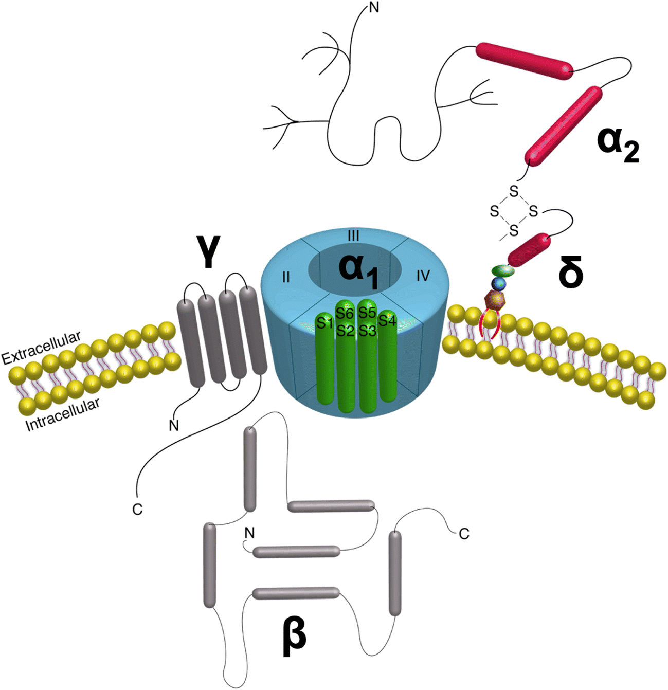
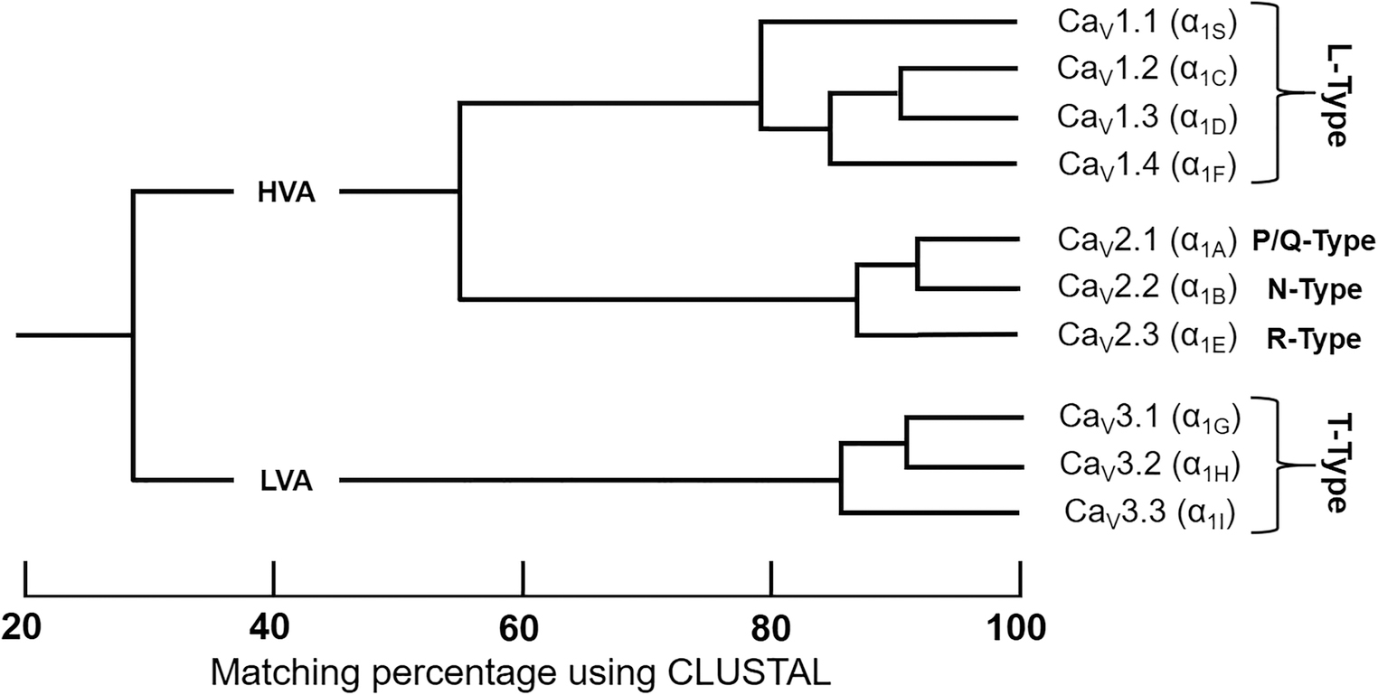
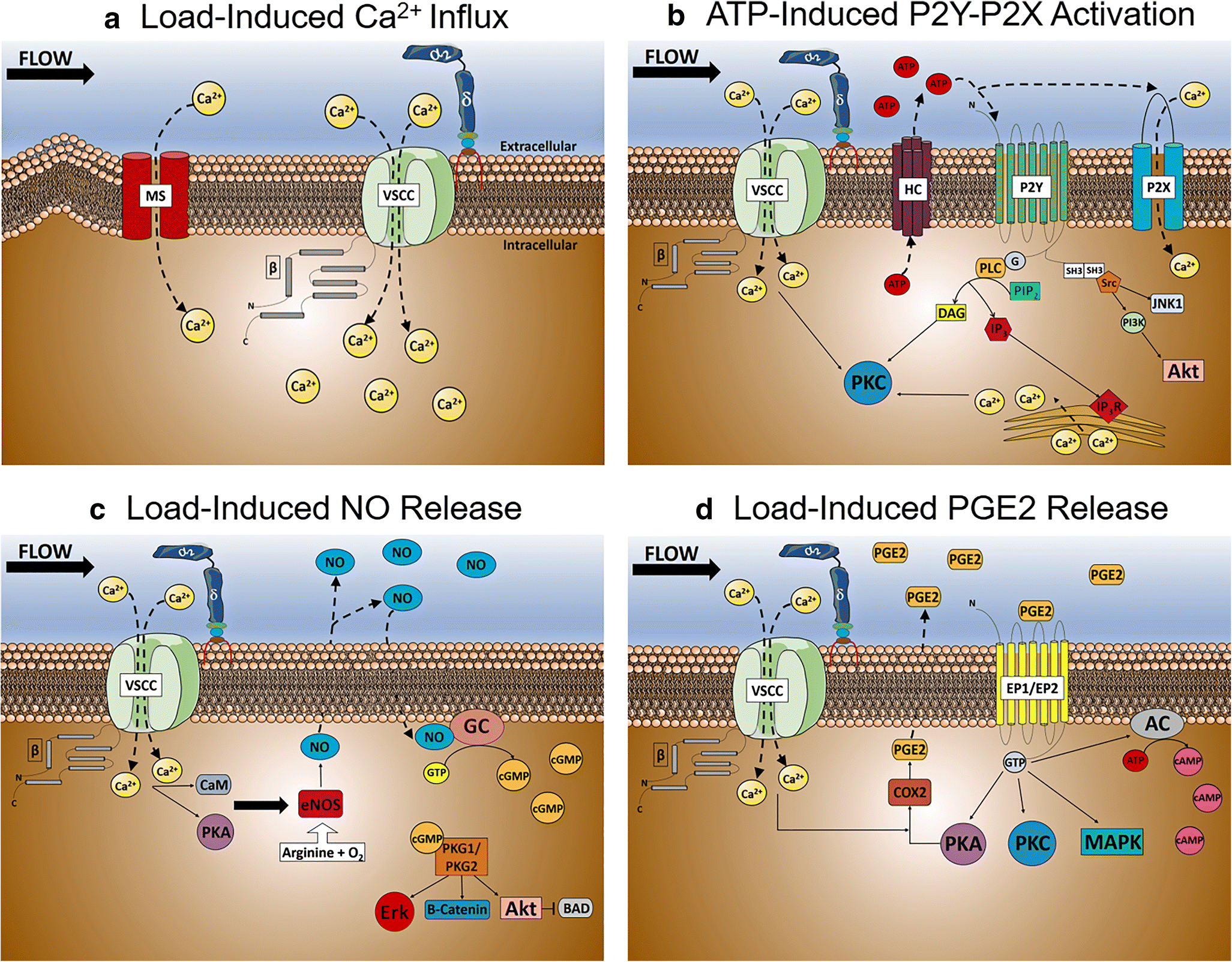
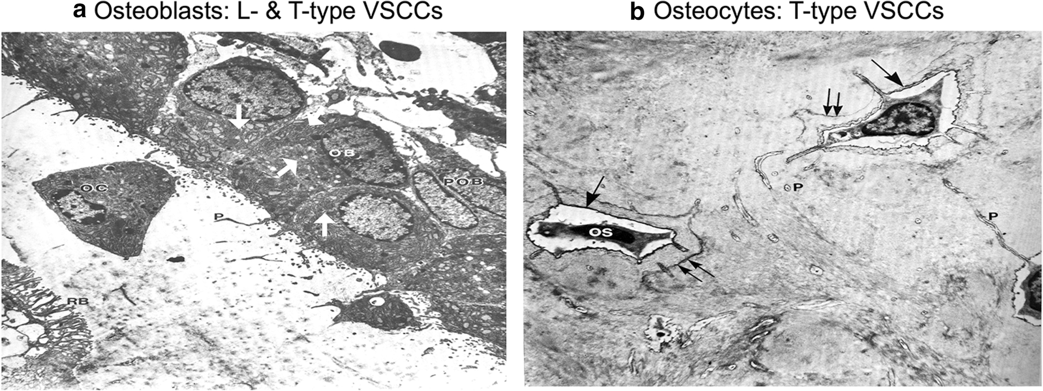
Voltage-sensitive calcium channels (VSCCs) are ubiquitous multimeric protein complexes that are necessary for the regulation of numerous physiological processes. VSCCs regulate calcium influx and various intracellular processes including muscle contraction, neurotransmission, hormone secretion, and gene transcription, with function specificity defined by the channel's subunits and tissue location. The functions of VSCCs in bone are often overlooked since bone is not considered an electrically excitable tissue. However, skeletal homeostasis and adaptation relies heavily on VSCCs. Inhibition or deletion of VSCCs decreases osteogenesis, impairs skeletal structure, and impedes anabolic responses to mechanical loading. RECENT FINDINGS: While the functions of VSCCs in osteoclasts are less clear, VSCCs have distinct but complementary functions in osteoblasts and osteocytes. PURPOSE OF REVIEW: This review details the structure, function, and nomenclature of VSCCs, followed by a comprehensive description of the known functions of VSCCs in bone cells and their regulation of bone development, bone formation, and mechanotransduction.

Keywords: Bone; Calcium channels; Mechanical loading; Osteoblast; Osteoclast; Osteocyte.


Conflict of interest statement

Conflicts of Interest: None

Figures

Fig 1.
Fig 1.. VSCC Structure.
The VSCC complex is composed of the α1 pore-forming subunit with auxiliary β, γ, and α2δ subunits bound to the pore, positioned to alter gating kinetics of the channel. The entirely extracellular α2δ subunit is anchored to the membrane via a GPI-anchor.
Figure 2.
Figure 2.. Phylogeny of Voltage-Sensitive Calcium Channel α1 Subunit.
Only the membrane-spanning segments and the pore loops (~350 amino acids) were compared. By only comparing sequencing pairs, three distinct VSCCs families can be identified with intrafamily sequence identities above 80% (CaV1.x, CaV2.x, CaV3.x). High voltage-activated (HVA); low voltage-activated (LVA). Adapted from Ertel EA et al [44] & Dolphin AC [43].
Figure 3.
Figure 3.. Role of L-type VSCCs in Osteoblast Mechanotransduction.
A) Calcium influx following load-induced shear stress in osteoblasts. Following a relatively small, but rapid influx of calcium via mechanosensitive channels, the plasma membrane is depolarized triggering VSCCs to further increasing intracellular calcium influx. B) Load-induced ATP release following calcium influx by osteoblast VSCCs. Increases in intracellular calcium concentrations by VSCCs increases ATP efflux through hemichannels, activating the ATP-gated purinergic P2X and P2Y receptors to increase P2X-dependent calcium influx and release of endoplasmic reticulum calcium stores. C) Load-induced Nitric Oxide (NO) release by osteoblast VSCCs. Endothelial nitric oxide synthase (eNOS) increases NO expression following VSCC calcium influx and activation by calmodulin (CaM) and protein kinase A (PKA), causing NO-induced guanylate cyclase activation and increases in cyclic guanosine monophosphate (cGMP) intracellular concentrations. D) Load-induced Prostaglandin E2 (PGE2) release by VSCCs in osteoblasts. Following VSCC calcium influx and PKA activation, PGE2 is synthesized by cyclooxygenase-2 (COX-2) and prostanoid synthases, causing an increase in PGE2 release and the activation of Prostaglandin E1/E2 Receptors (EP1/EP2). ATP, Adenosine triphosphate; AC; Adenylate cyclase; BAD, Bcl-2-associated death promoter; cAMP, Cyclic adenosine monophosphate; JNK1, c-Jun N-terminal kinase; DAG, diacylglycerol; eNOS, endothelial nitric oxide synthase; Erk, Extracellular-signal-regulated kinase; IP3, Inositol triphosphate; IP3R, Inositol triphosphate receptor; MAPK, Mitogen-activated protein kinase; PI3K, Phosphoinositide 3-kinases; PLC, Phospholipase C; PIP2, Phosphoinositol-4,5-bisphosphate; PKA, Protein kinase A; Akt, Protein kinase B; PKC, Protein kinase C; PKG1/PKG2, Protein kinase G; Src, Src kinase; SH3, Src homology 3 domain
Fig. 4:
Fig. 4:. Predominate VSCC composition coincides with the morphology and function of osteoblasts and osteocytes.
A). The enlarged ER and golgi secretory organelles (white arrows) of osteoblasts (OB) gives its characteristic “plump”, cuboidal morphology and high output of extracellular matrix. The predominate CaV1.2 L-type VSCCs in osteoblasts have a large single channel conductance and slow voltage-dependent inactivation, allowing for „long-lasting‟ calcium influx, the full release of ER calcium stores, and the stimulus necessary for bone formation and remodeling. B) With its flat, stellate shape, mature osteocytes (OS) have much smaller secretory organelles in comparison to osteoblasts. By shifting from expression of L-type to T-type VSCCs during differentiation, the presence of CaV3.2 VSCCs in osteocytes enables faster and more complete channel inactivation than L-type VSCCs, allowing osteocytes to retain its sensitivity to calcium influx without generating cytotoxic Ca2+ levels. Figures adapted from Butler TW [142].

References

    1. Catterall WA, Perez-Reyes E, Snutch TP, Striessnig J. International Union of Pharmacology. XLVIII. Nomenclature and structure-function relationships of voltage-gated calcium channels. Pharmacol Rev. 2005;57(4):411–25. doi:57/4/411 [pii] 10.1124/pr.57.4.5. - DOI - PubMed
    1. Yu FH, Catterall WA. The VGL-chanome: a protein superfamily specialized for electrical signaling and ionic homeostasis. Sci STKE. 2004;2004(253):re15. doi:stke.2532004re15 [pii] 10.1126/stke.2532004re15. - DOI - PubMed
    1. Arikkath J, Campbell KP. Auxiliary subunits: essential components of the voltage-gated calcium channel complex. Curr Opin Neurobiol. 2003;13(3):298–307. doi:S0959438803000667 [pii]. - PubMed
    1. Garcia K, Nabhani T, Garcia J. The calcium channel alpha2/delta1 subunit is involved in extracellular signalling. J Physiol. 2008;586(3):727–38. doi:jphysiol.2007.147959 [pii] 10.1113/jphysiol.2007.147959. - DOI - PMC - PubMed
    1. Takahashi M, Seagar MJ, Jones JF, Reber BF, Catterall WA. Subunit structure of dihydropyridine-sensitive calcium channels from skeletal muscle. Proc Natl Acad Sci U S A. 1987;84(15):5478–82. doi:10.1073/pnas.84.15.5478. - DOI - PMC - PubMed

Publication types

Substances

LinkOut - more resources