. 2021 Oct;36(5):958-967.
doi: 10.1007/s12250-021-00359-x. Epub 2021 Mar 15.

Establishment of a Reverse Genetic System of Severe Fever with Thrombocytopenia Syndrome Virus Based on a C4 Strain

Affiliations

Establishment of a Reverse Genetic System of Severe Fever with Thrombocytopenia Syndrome Virus Based on a C4 Strain

Mingyue Xu et al. Virol Sin. 2021 Oct.

Erratum in

Abstract

Severe fever with thrombocytopenia syndrome virus (SFTSV) is an emerging tick-borne bunyavirus that causes hemorrhagic fever-like disease (SFTS) in humans with a case fatality rate up to 30%. To date, the molecular biology involved in SFTSV infection remains obscure. There are seven major genotypes of SFTSV (C1-C4 and J1-J3) and previously a reverse genetic system was established on a C3 strain of SFTSV. Here, we reported successfully establishment of a reverse genetics system based on a SFTSV C4 strain. First, we obtained the 5'- and 3'-terminal untranslated region (UTR) sequences of the Large (L), Medium (M) and Small (S) segments of a laboratory-adapted SFTSV C4 strain through rapid amplification of cDNA ends analysis, and developed functional T7 polymerase-based L-, M- and S-segment minigenome assays. Then, full-length cDNA clones were constructed and infectious SFTSV were recovered from co-transfected cells. Viral infectivity, growth kinetics, and viral protein expression profile of the rescued virus were compared with the laboratory-adapted virus. Focus formation assay showed that the size and morphology of the foci formed by the rescued SFTSV were indistinguishable with the laboratory-adapted virus. However, one-step growth curve and nucleoprotein expression analyses revealed the rescued virus replicated less efficiently than the laboratory-adapted virus. Sequence analysis indicated that the difference may be due to the mutations in the laboratory-adapted strain which are more prone to cell culture. The results help us to understand the molecular biology of SFTSV, and provide a useful tool for developing vaccines and antivirals against SFTS.

Keywords: Bunyavirus; C4 strain; Minigenome; Reverse genetic system; Severe fever with thrombocytopenia syndrome virus (SFTSV); T7 polymerase.


Conflict of interest statement

The authors declare that they have no conflict of interest.

Figures

Fig. 1
Fig. 1
Schematic of the 3′- and 5′ UTRs of complementary viral RNA. The sequences of 5′ UTR were present in capital letters, and the sequence of 3′ UTR were present in lowercase letters. The corrected nucleotide of the adapted-SFTSV-WCH by RACE analysis was shown in red compared to these published SFTSV-WCH sequences (blue bases in the brackets). Potential to form Watson–Crick base pairs (dash in red) or noncanonical U-G pairings (dash in green) is indicated. The first 9 nt of both 3′ and 5′ termini are relatively conserved between all segments and comprise the conserved complementary region (red boxed sequences), except noncanonical U-G pairings. The variable complementary region of each segment is shown as blue boxed sequences.
Fig. 2
Fig. 2
Creation of the L/M/S-based minigenome constructs. A Schematic diagram of the generation of L-, M-, and S-based reporter minigenomes. B Effect of increasing amounts of pCAGGS-RdRp (upper panel) or pCAGGS-NP (lower panel) on M-segment minigenome assay. BSR-T7 cells were co-transfected with pT7-M-UTR-eGFP (0.5 μg), and the indicated amounts of pCAGGS-RdRp and pCAGGS-NP. C Generation of L/M/S-based minigenomes. BSR-T7 cells were transfected with the minigenome plasmid of pT7-L- UTR-eGFP, pT7-M-UTR-eGFP or pT7-delNSs: eGFP (UTR-eGFP); or co-transfected with the minigenome plasmid and pCAGGS-NP (NP + UTR-eGFP); the minigenome plasmid and pCAGGS-RdRp (RdRP + UTR-eGFP); or the minigenome plasmid with pCAGGS-NP and pCAGGS-RdRp (RdRp + NP + UTR-eGFP). At 36 h post transfection, fluorescence of eGFP was observed. Scale bar: 400 μm.
Fig. 3
Fig. 3
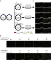
Rescue of infectious virus from cDNA. A General outline of the procedure to generate the rescued virus from cDNA. BSR-T7 cells were transfected pCAGGS-NP, pCAGGS-RdRp, pT7-S, pT7-M, and pT7-L. B Detection of the infectivity of the collected supernatants from passage 0 (P0) to P3 by immune fluorescence using the anti-NP antibody. The infectivity of the rescued virus was increasing with passaging from P0–P3, and the positive rate of fluorescent cells had reached 100% in P3. Scale bar: 400 μm.
Fig. 4
Fig. 4
Characterization of rSFTSV-WCH in comparison to the adapted-SFTSV-WCH. A Comparison of immune-stained foci of rSFTSV-WCH and adapted-SFTSV-WCH. B The titration of rSFTSV-WCH collected at different passages. The titer of rSFTSV-WCH increased from P3 to P8 but was lower than that of the adapted-SFTSV-WCH. C One-step growth curves of rSFTSV-WCH and adapted-SFTSV-WCH conducted in Vero cells at an MOI of 1. P values of < 0.01 indicated extremely statistical differences between two groups at any time point. D Time-course analysis of nucleoprotein (NP) expression in infected cells. Vero cells were infected with rSFTSV-WCH or adapted-SFTSV-WCH at an MOI of 1, and cellular samples were harvested at the indicated time points for Western blot analysis using anti-NP antibody.

References

    1. Amroun A, Priet S, de Lamballerie X, Querat G. Bunyaviridae RdRps: structure, motifs, and RNA synthesis machinery. Crit Rev Microbiol. 2017;43:753–778. doi: 10.1080/1040841X.2017.1307805. - DOI - PubMed
    1. Barr JN, Wertz GW. Bunyamwera bunyavirus RNA synthesis requires cooperation of 3′- and 5′-terminal sequences. J Virol. 2004;78:1129–1138. doi: 10.1128/JVI.78.3.1129-1138.2004. - DOI - PMC - PubMed
    1. Barr JN, Wertz GW. Role of the conserved nucleotide mismatch within 3′- and 5′-terminal regions of Bunyamwera virus in signaling transcription. J Virol. 2005;79:3586–3594. doi: 10.1128/JVI.79.6.3586-3594.2005. - DOI - PMC - PubMed
    1. Bergeron E, Albarino CG, Khristova ML, Nichol ST. Crimean-Congo hemorrhagic fever virus-encoded ovarian tumor protease activity is dispensable for virus RNA polymerase function. J Virol. 2010;84:216–226. doi: 10.1128/JVI.01859-09. - DOI - PMC - PubMed
    1. Bergeron E, Zivcec M, Chakrabarti AK, Nichol ST, Albarino CG, Spiropoulou CF. Recovery of recombinant Crimean Congo hemorrhagic fever virus reveals a function for non-structural glycoproteins cleavage by Furin. Plos Pathog. 2015;11:e1004879. doi: 10.1371/journal.ppat.1004879. - DOI - PMC - PubMed

LinkOut - more resources