Review
. 2021 Jun;5(6):447-458.
doi: 10.1016/S2352-4642(20)30313-8. Epub 2021 Mar 12.

Fetal body MRI and its application to fetal and neonatal treatment: an illustrative review

Affiliations
Review

Fetal body MRI and its application to fetal and neonatal treatment: an illustrative review

Joseph R Davidson et al. Lancet Child Adolesc Health. 2021 Jun.

Abstract

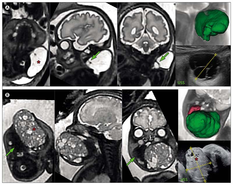
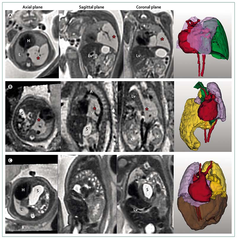
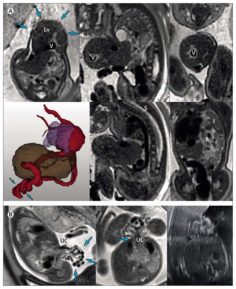
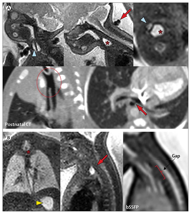
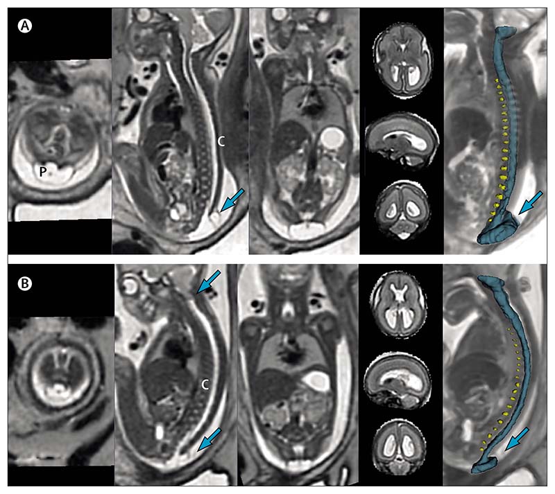
This Review depicts the evolving role of MRI in the diagnosis and prognostication of anomalies of the fetal body, here including head and neck, thorax, abdomen and spine. A review of the current literature on the latest developments in antenatal imaging for diagnosis and prognostication of congenital anomalies is coupled with illustrative cases in true radiological planes with viewable three-dimensional video models that show the potential of post-acquisition reconstruction protocols. We discuss the benefits and limitations of fetal MRI, from anomaly detection, to classification and prognostication, and defines the role of imaging in the decision to proceed to fetal intervention, across the breadth of included conditions. We also consider the current capabilities of ultrasound and explore how MRI and ultrasound can complement each other in the future of fetal imaging.


Conflict of interest statement

Declaration of interests

JRD and AD declare grants from the Wellcome Trust and Engineering and Physical Sciences Research Council (EPSRC; GIFT-Surg Project). PDC is supported by a UK National Institute for Health Research (NIHR) professorship. MD declares grants from the Wellcome Trust, EPSRC, and NIHR. Other authors declare no competing interests.

Figures

Figure 1
Figure 1. T2-weighted MRIs of two fetuses with neck masses
(A) Fetus at 31 weeks and 6 days’ gestation with a lymphatic malformation (marked by an asterisk) of the posterolateral left neck, with arrows pointing to internal septations. The 3D model (top right) shows the lesion (green) lying separate to the fetal airway, major vessels, and CNS structures. The corresponding fetal ultrasound is shown bottom right. (B) Fetus at 33 weeks and 4 days’ gestation with an oropharyngeal teratoma (marked by an asterisk) in relation to the tongue (green arrow). The sagittal and coronal views in the two central images show the relationship to the fetal airway. The corresponding fetal ultrasound is shown bottom right. The 3D model (top right) shows the 85 cm3 tumour mass (green) and tongue (pink). 3D=three-dimensional. USS=ultrasound scan.
Figure 2
Figure 2. T2-weighted MRI and three-dimensional models of three fetuses with lesions of the thorax
(A) Fetus of 24 weeks’ gestation with a lesion of left lower lung showing a microcystic congenital pulmonary airway malformation of the left lower lobe (marked by an asterisk), also shown in 3D (green), with heart (red) and lungs (lilac). (B) Fetus of 24 weeks’ gestation with bronchopulmonary sequestration (marked by an asterisk) and right congenital diaphragmatic hernia. Reorientation to true sagittal shows a feeding vessel arising from the aorta. Note the right-sided diaphragmatic hernia containing the bowel (yellow), gallbladder, and stomach. Kidneys are shown in the coronal plane. 3D models show bronchopulmonary sequestration (green), lungs (lilac), and heart (red). (C) Fetus of 33 weeks’ gestation with a left-sided congenital diaphragmatic hernia, containing stomach and intestine (yellow), and liver (brown) within the hernia, and showing the heart (red) and lung (lilac) in relation to hernial content. 3D=three-dimensional. A=aorta. B=bowel. G=gallbladder. H=heart. K=kidney. L=lung. Lv=Liver. S=stomach. V=vessel.
Figure 3
Figure 3. MRIs of two fetuses with abdominal wall defects
(A) Fetus at 34 weeks and 6 days of gestation with exomphalos (arrows on axial image, top left). The 3D image shows that the exomphalos contains the liver (brown) and the prominent umbilical vein (red) follows a twisting and irregular course (arrows; lung shown in lilac). The lower middle panel shows marked angulation of the thoracic spine. Bowel in the associated diaphragmatic hernia is shown in the lower right image with the compressed left lung. (B) Gastroschisis, with no sign of bowel dilatation on the MRI. The exteriorised bowel (arrows, left image) and insertion (arrow, central image) of umbilical cord are shown. Image on the right shows the referring ultrasound scan. B=bowel. I=lung. Lv=liver. S=spine. UC=umbilical cord. V=vein.
Figure 4
Figure 4. MRIs of two fetuses with oesophageal atresia
(A) Fetus at 32 weeks and 5 days of gestation has a dilated upper pouch (marked by an asterisk), displacing the trachea (blue arrowhead) in the axial view (upper right image). A two-vessel umbilical cord (red arrow) can be seen in the sagittal plane (top middle image). The postnatal CT (lower two images) shows a long-segment common tracheoesophageal channel (encircled, left) and distal fistula arising from the left main bronchus (red arrow, right). (B) Fetus at 30 weeks and 5 days of gestation has a dilated upper pouch (marked by an asterisk in the left image and an arrow in the central image) and normal stomach (arrowhead), suggesting a distal fistula. Gap measurement is possible in the sagittal image (right). bSSFP=balanced steady-state free precession MRI.
Figure 5
Figure 5. MRIs of two fetuses with spina bifida
(A) Open spina bifida (non-eligible for fetal surgery). First and second sacral vertebral defect with cord, dorsal sac (arrow), and placode. The three-dimensional model shows intervertebral discs (yellow) and spinal canal (blue). (B) Open spina bifida (eligible for fetal surgery). Low sacral defect with hindbrain herniation (upper arrow on sagittal view). C=spinal cord. P=placode.

References

    1. Kyriakopoulou V, Vatansever D, Davidson A, et al. Normative biometry of the fetal brain using magnetic resonance imaging. Brain Struct Funct. 2017;222:2295–307. - PMC - PubMed
    1. Lloyd DFA, Pushparajah K, Simpson JM, et al. Three-dimensional visualisation of the fetal heart using prenatal MRI with motion-corrected slice-volume registration: a prospective, single-centre cohort study. Lancet. 2019;393:1619–27. - PMC - PubMed
    1. Aughwane R, Ingram E, Johnstone ED, Salomon LJ, David AL, Melbourne A. Placental MRI and its application to fetal intervention. Prenat Diagn. 2020;40:38–48. - PMC - PubMed
    1. Melbourne A, Aughwane R, Sokolska M, et al. Separating fetal and maternal placenta circulations using multiparametric MRI. Magn Reson Med. 2019;81:350–61. - PMC - PubMed
    1. Uus A, Zhang T, Jackson LH, et al. Deformable slice-to-volume registration for motion correction of fetal body and placenta MRI. IEEE Trans Med Imaging. 2020;39:1. - PMC - PubMed

MeSH terms