. 2021 Mar-Apr;18(2):1479164121999033.
doi: 10.1177/1479164121999033.

Endothelial deletion of PKCδ prevents VEGF inhibition and restores blood flow reperfusion in diabetic ischemic limb

Affiliations

Endothelial deletion of PKCδ prevents VEGF inhibition and restores blood flow reperfusion in diabetic ischemic limb

Laura Croteau et al. Diab Vasc Dis Res. 2021 Mar-Apr.

Abstract

Aims: Peripheral artery disease is a complication of diabetes leading to critical hindlimb ischemia. Diabetes-induced inhibition of VEGF actions is associated with the activation of protein kinase Cδ (PKCδ). We aim to specifically investigate the role of PKCδ in endothelial cell (EC) function and VEGF signaling.

Methods: Nondiabetic and diabetic mice, with (ec-Prkcd-/-) or without (ec-Prkcdf/f) endothelial deletion of PKCδ, underwent femoral artery ligation. Blood flow reperfusion was assessed up to 4 weeks post-surgery. Capillary density, EC apoptosis and VEGF signaling were evaluated in the ischemic muscle. Src homology region 2 domain-containing phosphatase-1 (SHP-1) phosphatase activity was assessed in vitro using primary ECs.

Results: Ischemic muscle of diabetic ec-Prkcdf/f mice exhibited reduced blood flow reperfusion and capillary density while apoptosis increased as compared to nondiabetic ec-Prkcdf/f mice. In contrast, blood flow reperfusion and capillary density were significantly improved in diabetic ec-Prkcd-/- mice. VEGF signaling pathway was restored in diabetic ec-Prkcd-/- mice. The deletion of PKCδ in ECs prevented diabetes-induced VEGF unresponsiveness through a reduction of SHP-1 phosphatase activity.

Conclusions: Our data provide new highlights in mechanisms by which PKCδ activation in EC contributed to poor collateral vessel formation, thus, offering novel therapeutic targets to improve angiogenesis in the diabetic limb.

Keywords: Diabetes; PKCδ; SH2 domain-containing phosphatase 1; peripheral arterial disease; vascular endothelial growth factor.


Conflict of interest statement

Declaration of conflicting interests: The author(s) declared no potential conflicts of interest with respect to the research, authorship, and/or publication of this article.

Figures

Figure 1.
Figure 1.
Blood flow reperfusion and vascular density in the ischemic limb of diabetic and nondiabetic mice: (a) laser Doppler imaging of nondiabetic (NDM) and diabetic (DM) ec-Prkcdf/f and ec-Prkcd−/− mice, pre, post, and 4 weeks following femoral artery ligation, (b) quantification of the percentage of blood flow reperfusion in the ischemic limb, (c) immunofluorescence of α-smooth muscle actin (green) and endothelial cells (CD31-red), and (d) quantification of vascular density in the ischemic muscle (number of vessel smaller than 30 μm) of three cross-sections per animal. Results are shown as mean ± SD of 12 (ec-Prkcd−/−) and 15 (ec-Prkcdf/f) mice per group (a and b) and 6–8 mice per group (c and d). *p = 0.0009 versus NDM ec-Prkcdf/f. †p = 0.0089 versus DM ec-Prkcd−/−.
Figure 2.
Figure 2.
Histological analysis of muscle integrity and endothelial cells apoptosis: (a) histological cross-sections of diabetic and nondiabetic ischemic muscle stained with hematoxylin-eosin, (b) quantification of the percentage of the inter-fiber area per muscle fiber, (c) immunofluorescence of α-smooth muscle actin (green) and TUNEL endothelial positive cells (CD31-red) in the ischemic muscle of nondiabetic and diabetic ec-Prkcdf/f and ec-Prkcd−/− mice, and (d) quantification of the percentage of endothelial apoptotic cells per mm² in the cross-sections of ischemic muscle in each group. Results are shown as mean ± SD of 12–15 mice per group (a and b) and 10–12 mice per group (c and d).
Figure 3.
Figure 3.
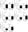
Decreased mRNA expression of angiogenic factors and eNOS in the muscle of diabetic ec-Prkcdf/f mice. Quantitative real-time PCR of: (a) VEGF-A, (b) PDGF-B, (c) PDGF-β, (d) FLK-1, and (e) eNOS in ischemic muscle of nondiabetic (NDM) and diabetic (DM) ec-Prkcdf/f and ec-Prkcd−/− mice. Results are shown as mean ± SD of 10–12 mice per group.
Figure 4.
Figure 4.
Decreased activation of the VEGF signaling pathway and increased expression of SHP-1 in the muscle of diabetic ec-Prkcdf/f mice: (a) expression of phospho-VEGFR (Y1175), VEGFR, phospho-eNOS (S1177), eNOS, phospho-Akt (S473), (b) phospho-PLC-γ (Y783), PLC-γ, phospho-Src (Y416), Src, and (c) SHP-1 and GAPDH in ischemic muscle of nondiabetic (NDM) and diabetic (DM) ec-Prkcdf/f and ec-Prkcd−/− mice. Protein expression was detected by immunoblot blot, and densitometric quantitation was measured. Results are shown as mean ± SD of 8–10 mice per group.
Figure 5.
Figure 5.
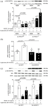
Akt expression and activation in lung EC and SHP-1 phosphatase activity in BAEC: (a) expression of phospho-Akt (S473) and total Akt in EC isolated from the lungs of diabetic (DM) and nondiabetic (NDM) ec-Prkcdf/f and ec-Prkcd−/− mice exposed to NG (clear bars) or HG (black bars) concentrations followed by VEGF stimulation. Protein expression was detected by immunoblot blot, and densitometric quantitation was measured, (b) phosphatase activity of SHP-1 in BAEC infected or not with the adenoviral vector of dominant negative form of PKCδ, and (c) expression of phospho-Akt (S473), total Akt in BAECs infected with either Ad-GFP or Ad-dnSHP-1 and exposed to NG (clear bars) or HG (black bars) concentrations followed by VEGF stimulation. Protein expression was detected by immunoblot blot, and densitometric quantitation was measured. Results are shown as mean ± SD test of 4 (a) and 5 (b, c) independent experiments.

References

    1. Dieter RS, Chu WW, Pacanowski JP, et al.. The significance of lower extremity peripheral arterial disease. Clin Cardiol 2002; 25(1): 3–10. - PMC - PubMed
    1. American Diabetes Association. Peripheral arterial disease in people with diabetes. Clin Diabetes 2004; 22(4): 181–189.
    1. Carmeliet P.Angiogenesis in health and disease. Nat Med 2003; 9(6): 8. - PubMed
    1. Lamalice L, Le Boeuf F, Huot J.Endothelial Cell Migration During Angiogenesis. Circ Res 2007; 100(6): 782–794. - PubMed
    1. Takeshita S, Rossow ST, Kearney M, et al.. Time course of increased cellular proliferation in collateral arteries after administration of vascular endothelial growth factor in a rabbit model of lower limb vascular insufficiency. Am J Pathol 1995; 147(6): 1649–1660. - PMC - PubMed

Publication types

MeSH terms

Substances