. 2021 Mar 16;22(1):83.
doi: 10.1186/s13059-021-02304-3.

Prime editing in mice reveals the essentiality of a single base in driving tissue-specific gene expression

Affiliations

Prime editing in mice reveals the essentiality of a single base in driving tissue-specific gene expression

Pan Gao et al. Genome Biol. .

Abstract

Background: Most single nucleotide variants (SNVs) occur in noncoding sequence where millions of transcription factor binding sites (TFBS) reside. Here, a comparative analysis of CRISPR-mediated homology-directed repair (HDR) versus the recently reported prime editing 2 (PE2) system was carried out in mice over a TFBS called a CArG box in the Tspan2 promoter.

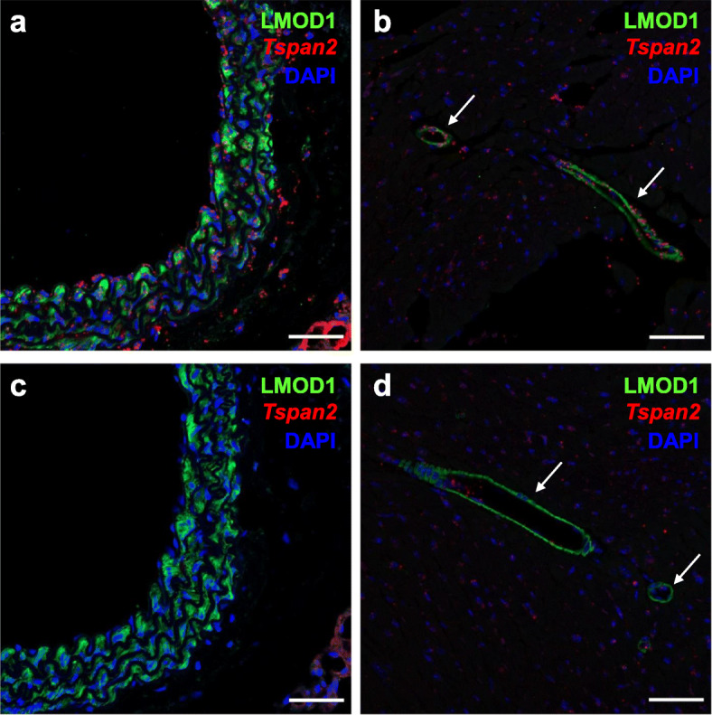
Results: Quantitative RT-PCR showed loss of Tspan2 mRNA in aorta and bladder, but not heart or brain, of mice homozygous for an HDR-mediated three base pair substitution in the Tspan2 CArG box. Using the same protospacer, mice homozygous for a PE2-mediated single-base substitution in the Tspan2 CArG box displayed similar cell-specific loss of Tspan2 mRNA; expression of an overlapping long noncoding RNA was also nearly abolished in aorta and bladder. Immuno-RNA fluorescence in situ hybridization validated loss of Tspan2 in vascular smooth muscle cells of HDR and PE2 CArG box mutant mice. Targeted sequencing demonstrated variable frequencies of on-target editing in all PE2 and HDR founders. However, whereas no on-target indels were detected in any of the PE2 founders, all HDR founders showed varying levels of on-target indels. Off-target analysis by targeted sequencing revealed mutations in many HDR founders, but none in PE2 founders.

Conclusions: PE2 directs high-fidelity editing of a single base in a TFBS leading to cell-specific loss in expression of an mRNA/long noncoding RNA gene pair. The PE2 platform expands the genome editing toolbox for modeling and correcting relevant noncoding SNVs in the mouse.

Keywords: CRISPR; Gene expression; Genome editing; Mouse; Prime editing; Transcription.


Conflict of interest statement

DRL is a consultant and co-founder of Editas Medicine, Pairwise Plants, Beam Therapeutics, and Prime Medicine, companies that use genome-editing technologies. KH, JAW, and APK are employees of Synthego Corporation. CRL and SQT have filed a patent application on CHANGE-seq. SQT is a member of the scientific advisory board of Kromatid.

Figures

Fig. 1
Fig. 1
HDR-mediated editing of Tspan2 CArG box. a Targeting strategy with CArG box sequence (red), protospacer sequence (green), PAM sequence (blue shade), and 3 bp substitution (blue) in ssODN repair template. b Allele-specific PCR of some founder mice with evidence of wild type (top) or mutant (bottom) edit and corresponding Sanger sequencing of CArG box (shaded). c qRT-PCR of Tspan2 in indicated tissues and genotypes (n = 5–7 mice/genotype). Black, blue, and red bars here and below represent relative (wild type set to value of 1) mean Tspan2 mRNA (± STD) in wild type, heterozygous, and homozygous genotypes, respectively. d Average Ct values for indicated tissues (n = 6–7 mice/tissue). Asterisks indicate p < 0.05. sg, sgRNA edited Tspan2 CArG box
Fig. 2
Fig. 2
Spatial localization of Tspan2 mRNA in HDR-edited mouse tissues. Immunofluorescence (LMOD1) and RNA FISH (Tspan2) of wild type aorta (a) and heart (b) versus Tspan2sg/sg CArG box mutant (HDR) aorta (c) and heart (d). Arrows point to coronary vessels of the heart. Note decrease in Tspan2 mRNA (red dots) in vascular smooth muscle cells (labeled green with LMOD1 antibody) of aorta and coronary vessels in CArG box mutants (c, d). Scale bars are 50 μm. Representative images from n = 3 mice
Fig. 3
Fig. 3
PE2-mediated editing of Tspan2 CArG box. a Targeting strategy with CArG box in red sequence, protospacer in green sequence, PAM in blue shaded sequence, and 1 bp substitution within RT template in blue. b Representative genotyping of several founders with correct installation of 1 bp transversion (left) and Sanger sequencing of CArG box showing correct edit in a mutant founder (right, shaded). c qRT-PCR of Tspan2 mRNA in indicated tissues and genotypes (n = 4 mice per genotype). peg, pegRNA edited mouse; RT, reverse transcriptase; PBS, primer binding site
Fig. 4
Fig. 4
Spatial localization of Tspan2 mRNA in PE2-edited mouse tissues. Immunofluorescence (LMOD1) and RNA FISH (Tspan2) of blood vessels in wild type heart (a) and brain (b) versus Tspan2peg/peg CArG box mutant heart (c) and brain (d). The Tspan2 mRNA is indicated by the red dots in vascular smooth muscle cells (labeled green with LMOD1 antibody) of wild type blood vessels (a, b), but is nearly absent in CArG mutant vessels (c, d). Scale bars are 50 μm. Representative images from n = 2 mice
Fig. 5
Fig. 5
Mouse Tspan2 and Tspan2os loci. a UCSC Genome Browser screenshot of the 5′ mouse Tspan2 locus and overlapping, divergently transcribed lncRNA, Tspan2os. The sequence of the CArG box shown here is the complement of that shown in Supplementary Fig. 1 due to direction of transcription in mouse versus human Tspan2. Note the CArG box falls within a high degree of mammalian conservation (red arrow). b qRT-PCR of Tspan2os RNA in aorta and bladder of indicated PE2-mediated genotypes. n = 4 aortae for each genotype
Fig. 6
Fig. 6
On-target sequence fidelity at the Tspan2 CArG box. Percent editing across HDR (a) and PE2 (b) founder mice. CRISPResso sequence output for individual founders from sgRNA (c) and pegRNA (d) study. Protospacer (blue line) and PAM (red box) are indicated as are numbers indicating frequency of correct edits. Black boxes in panel c indicate deletions. Please note, a 150 bp deletion could not be aligned in CRISPResso for HDR founders 7, 10, and 33, but is included in the quantitative data of panel a. PE2 founders 32 and 38 represent littermate controls that did not exhibit on-target editing
Fig. 7
Fig. 7
Bulk RNA-seq of aortae from HDR and PE2-edited mice. Scatter plots between a HDR (sgRNA) and b PE2 (pegRNA) mice. The position of differential Tspan2 normalized reads is indicated in red. There was no overlap in genes up- or downregulated between the HDR and PE2 scatter plots. Many of the upregulated transcripts, particularly in the pegRNA experiment, are due to large deviations in reads among single replicates. For a listing of the significantly regulated genes, please see Supplementary Table S2. n = 4 aortae for each genotype
Fig. 8
Fig. 8
Genome-wide off-target analysis of HDR and PE2 edited founder mice. a Bar plot of number of CHANGE-seq sites detected using Cas9 WT and synthetic sgRNA or pegRNA targeting Tspan2, on WT genomic DNA from same strain of mice used in HDR and PE2 editing experiments. b Venn diagram depicting common predicted off-target sites for sgRNA (orange) and pegRNA (blue) groups. c Manhattan plots of CHANGE-seq detected on- and off-target sites organized by chromosomal position, for sgRNA and pegRNA, with bar heights representing CHANGE-seq read count. Arrow indicates the on-target site. d, e Indel frequencies evaluated by rhAmpSeq at on- and off-target sites detected by CHANGE-seq for sgRNA founders (d) and for pegRNA founders (e)

References

    1. Kadonaga JT. Regulation of RNA polymerase II transcription by sequence-specific DNA binding factors. Cell. 2004;116:247–257. doi: 10.1016/S0092-8674(03)01078-X. - DOI - PubMed
    1. Maurano MT, Humbert R, Rynes E, Thurman RE, Haugen E, Wang H, Reynolds AP, Sandstrom R, Qu H, Brody J, et al. Systematic localization of common disease-associated variation in regulatory DNA. Science. 2012;337:1190–1195. doi: 10.1126/science.1222794. - DOI - PMC - PubMed
    1. Vierstra J, Lazar J, Sandstrom R, Halow J, Lee K, Bates D, Diegel M, Dunn D, Neri F, Haugen E, et al. Global reference mapping of human transcription factor footprints. Nature. 2020;583:729–736. doi: 10.1038/s41586-020-2528-x. - DOI - PMC - PubMed
    1. Haraksingh RR, Snyder MP. Impacts of variation in the human genome on gene regulation. J Mol Biol. 2013;425:3970–3977. doi: 10.1016/j.jmb.2013.07.015. - DOI - PubMed
    1. Studer M, Gavalas A, Marshall H, Ariza-McNaughton L, Rijli FM, Chambon P, Krumlauf R. Genetic interactions between Hoxa1 and Hoxb1 reveal new roles in regulation of early hindbrain patterning. Development. 1998;125:1025–1036. - PubMed

Publication types