. 2021 May:167:105548.
doi: 10.1016/j.phrs.2021.105548. Epub 2021 Mar 15.

Resveratrol-mediated attenuation of superantigen-driven acute respiratory distress syndrome is mediated by microbiota in the lungs and gut

Affiliations

Resveratrol-mediated attenuation of superantigen-driven acute respiratory distress syndrome is mediated by microbiota in the lungs and gut

Hasan Alghetaa et al. Pharmacol Res. 2021 May.

Abstract

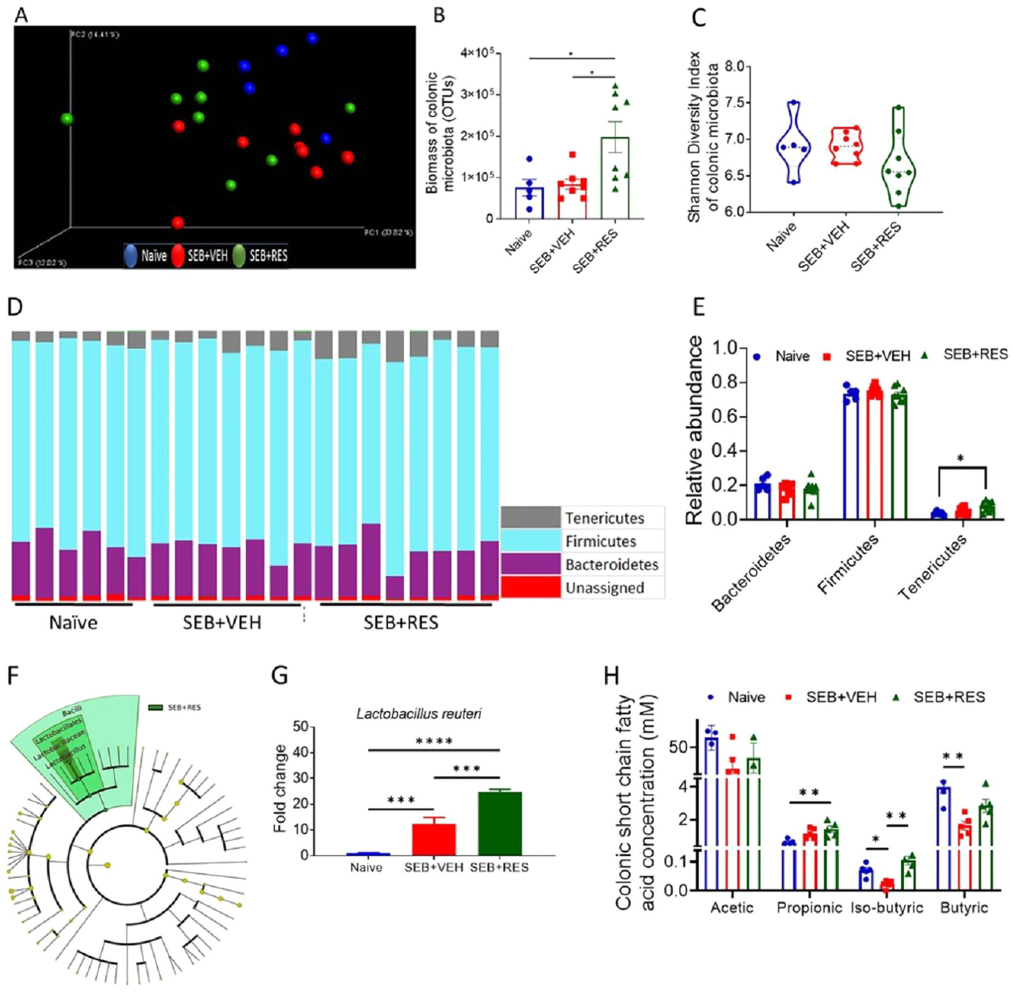
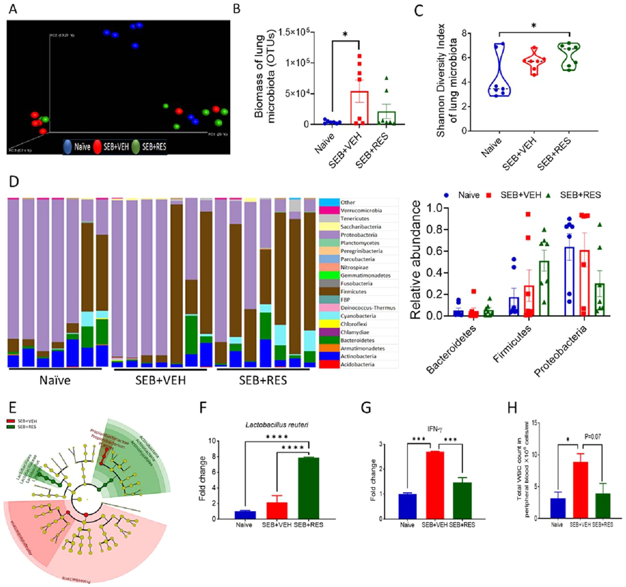
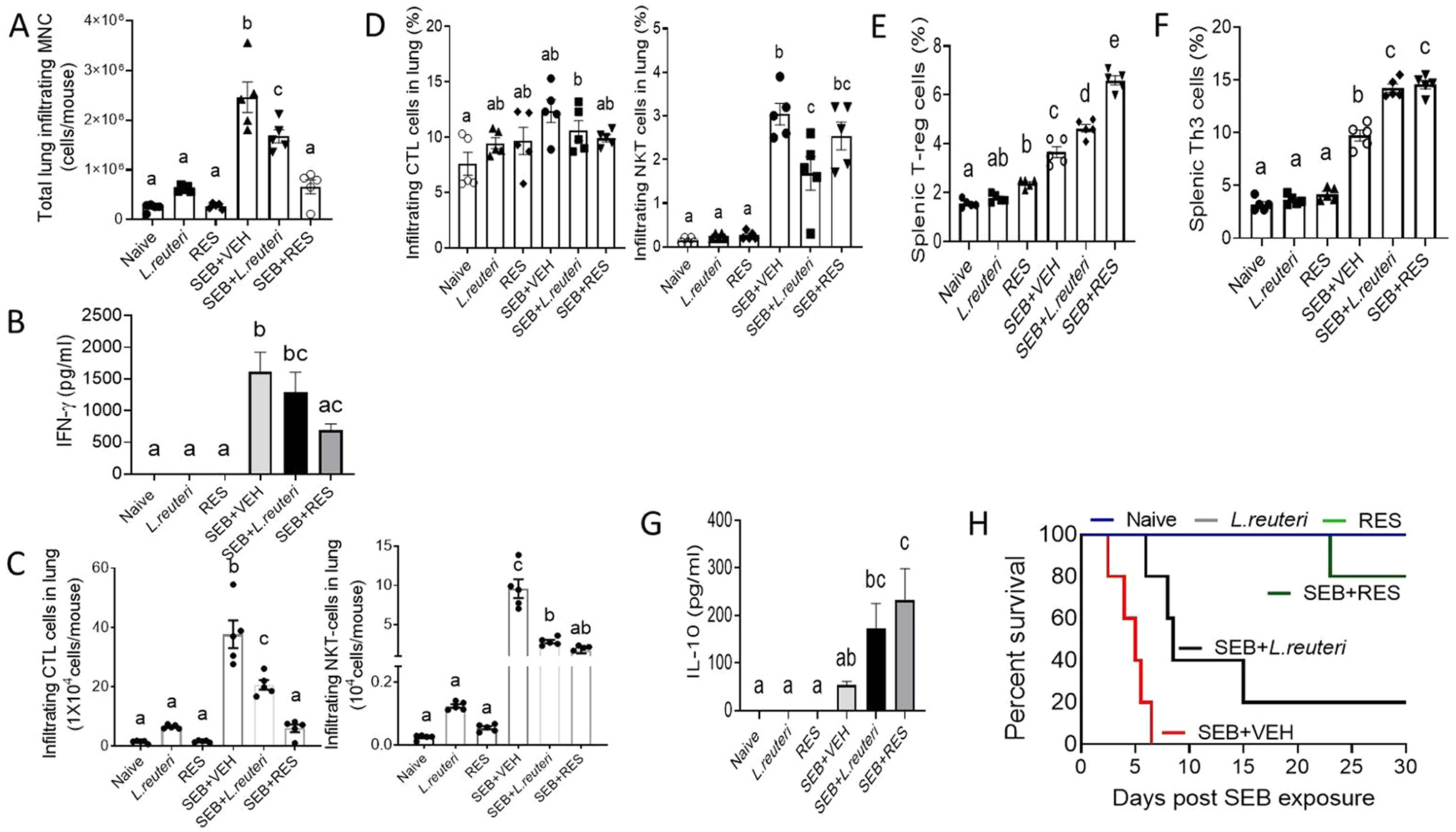
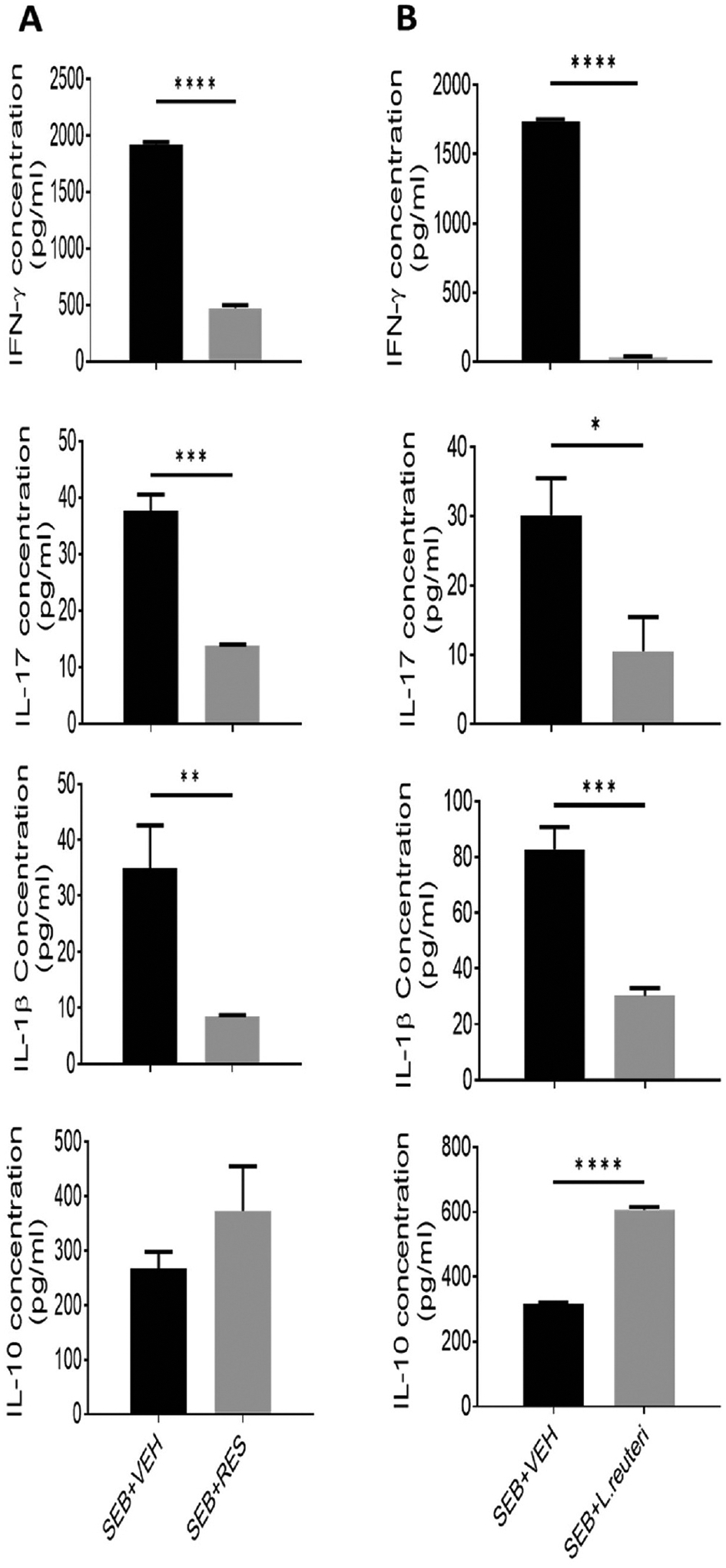
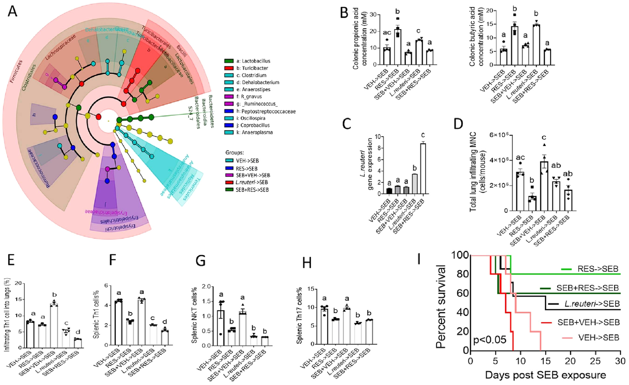
Acute Respiratory Distress Syndrome (ARDS) is triggered by a variety of agents, including Staphylococcal Enterotoxin B (SEB). Interestingly, a significant proportion of patients with COVID-19, also develop ARDS. In the absence of effective treatments, ARDS results in almost 40% mortality. Previous studies from our laboratory demonstrated that resveratrol (RES), a stilbenoid, with potent anti-inflammatory properties can attenuate SEB-induced ARDS. In the current study, we investigated the role of RES-induced alterations in the gut and lung microbiota in the regulation of ARDS. Our studies revealed that SEB administration induced inflammatory cytokines, ARDS, and 100% mortality in C3H/HeJ mice. Additionally, SEB caused a significant increase in pathogenic Proteobacteria phylum and Propionibacterium acnes species in the lungs. In contrast, RES treatment attenuated SEB-mediated ARDS and mortality in mice, and significantly increased probiotic Actinobacteria phylum, Tenericutes phylum, and Lactobacillus reuteri species in both the colon and lungs. Colonic Microbiota Transplantation (CMT) from SEB-injected mice that were treated with RES as well as the transfer of L. reuteri into recipient mice inhibited the production of SEB-mediated induction of pro-inflammatory cytokines such as IFN-γ and IL-17 but increased that of anti-inflammatory IL-10. Additionally, such CMT and L. reuteri recipient mice exposed to SEB, showed a decrease in lung-infiltrating mononuclear cells, cytotoxic CD8+ T cells, NKT cells, Th1 cells, and Th17 cells, but an increase in the population of regulatory T cells (Tregs) and Th3 cells, and increase in the survival of mice from SEB-mediated ARDS. Together, the current study demonstrates that ARDS induced by SEB triggers dysbiosis in the lungs and gut and that attenuation of ARDS by RES may be mediated, at least in part, by alterations in microbiota in the lungs and the gut, especially through the induction of beneficial bacteria such as L. reuteri.

Keywords: ARDS; Cytokine storm; DiOC(6)(3) (3,3′-Dihexyloxacarbocyanine iodide, PubChem CID: 9894321); Microbiota; Resveratrol; SEB; Superantigen; butyric acid (PubChem CID: 264); carboxymethylcellulose (PubCID: 24748); interleukin 1beta (PubChem CID: 159483); iso butyric acid (PubChem CID: 6590) and acetic acid (PubChem CID: 176); lipopolysaccharide (LPS, PubChem CID: 451715); metronidazole (Bioxtra, PubChem CID: 24896667); propionic acid (PubChem CID: 1032); resveratrol (PubChem CID: 445154).


Conflict of interest statement

Conflict of Interest

The authors declare no conflict of interest in this study.

Figures

Fig. 1.
Fig. 1.
Gating strategy of multi-parametric flow-cytometry analysis of T-lymphocyte subsets. A) Discrimination of lymphocytes and singlets. B) Major T cell components, from top; CD3+NK1.1+ (NKT), CD3+CD8+ (CTL), CD3+CD4+ cells and histogram of CD3+ cells population. C) T-helper subsets gated on CD3+ cells; from top CD4+IFN-γ+ (Th1), CD4+IL4+ (Th2) and CD4+IL17+ (Th17). D) T regulatory cells and FoxP3 T-reg (upper panel) and FoxP3 Treg subsets, IL10+ cells (Tr1) or TGF-β+ cells (Th3). NKT: natural killer T cells; CTL: cytotoxic T lymphocytes; Th: T helper lymphocytes; Tr1: T regulatory cells type-1.
Fig. 2.
Fig. 2.
Resveratrol treatment alters gut microbial composition and SCFA production in SEB-induced ARDS. Naïve mice or those treated with SEB + VEH or SEB + RES were studied for alterations in gut microbiota 48 h after SEB injection as described in Methods. A) Principal coordinate analysis plot reveals the clustering of individual commensal bacteria in colon based on their 16S rRNA content similarity. B) Comparison of bacterial biomass in the lung and colon represented by number of OTUs. C) Shannon index shows the diversity of microbiome in various groups in colonic lumen. D) Stack bar plot shows the relative abundance of major colonic bacterial phyla among experimental groups with statistical analysis. E) Comparison of abundance of bacteroidetes, firmicutes and tenericutes in colon. F) Cladogram generated from LEfSe analysis of taxonomic levels of colon microbiota shows biomarker bacteria between SEB + RES group when compared to SEB + VEH group. G) qPCR validation of Lactobacillus reuteri gene expression in isolated bacterial DNA from colons of experimental groups. H) Detection of SCFAs in the colon flushes measured by GC-FID technology. One-way ANOVA test was used and p-value < 0.05 is considered a significant difference. *p < 0.05, **p < 0.01, ***p < 0.001 and ****p < 0.0001.
Fig. 3.
Fig. 3.
Resveratrol treatment alters lung microbiota and increased enrichment of beneficial bacteria and suppression of SEB-mediated inflammation. Mice were treated with SEB + VEH or SEB + RES as described in Fig. 1 legend. A) Principal coordinate analysis plot reveals the clustering of individual commensal bacteria in the lungs based on their 16S rRNA content similarity. B) Comparison of lung bacterial biomass represented by abundance of OTUs. C) Shannon index shows the diversity of microbiome in lung tissue. D) Stack bar plot shows the fluctuations of major bacterial phyla among groups in lung tissue with statistical analysis of major enriched phyla (right panel). E) Cladogram depicting biomarkers between SEB-induced mouse group and both SEB-induced and RES-treated group. F) qPCR validation of Lactobacillus reuteri gene expression in isolated bacterial DNA from experimental groups. G) qPCR validation of pro-inflammatory gene, IFN-γ, in infiltrating mononuclear cells (MNCs) in lungs H) VetScan results show the systemic response in SEB exposure depicted by total number of white blood cells in peripheral blood of experimental mice. One-way ANOVA test was used and p-value < 0.05 is considered a significant difference. *p < 0.05, **p < 0.01, ***p < 0.001 and ****p < 0.0001.
Fig. 4.
Fig. 4.
Administration of RES or Lactobacillus attenuates SEB-induced ARDS. Naïve mice were untreated or treated with RES or Lactobacillus reuteri. Also, SEB injected mice were treated with VEH, L. reuteri or RES as described in Materials and methods. A) Infiltrating mononuclear cells counts in the lungs of SEB-injected mice. ELISA results show the pro-inflammatory (B) and anti-inflammatory cytokines (G) in the sera of experimental mice. C) Absolute number of infiltrating CD3+CD8+ and NKT cell populations in the lungs. D) Percentage of infiltrating CD3+CD8+ and NKT cell populations in the lungs. E) Treg populations in spleen gated on CD3+CD4+. F) T-helper3 population in spleen gated on CD3+CD4+Foxp3. H) Survival rate test for 30 days post-SEB exposure. One-way ANOVA test was used and p-value < 0.05 is considered a significant difference. Log-rank (Mantel-Cox) test was used to compare the survival curves. Bars denote Mean±SEM and any significant differences (p < 0.05) are indicated by lowercase letters among groups.
Fig. 5.
Fig. 5.
Effect of RES or L. reuteri on the induction of inflammatory cytokines in vitro. Naïve splenocytes were harvested and pre-treated for 1 h with either 50uM RES or L. reuteri at a ratios of 1:5 (splenocytes: L. reuteri) prior to activation of these cells with 1 μg/ml SEB for 48 h. ELISA was performed to measure IFN-γ, IL-β, IL-17 and IL-10 cytokine levels on supernatants collected 48 h post-SEB activation from, A) RES-pretreated cells. B) L. reuteri co-cultured cells. Student’s t-test was used and p-value < 0.05 is considered significant difference. *p < 0.05, **p < 0.01, ***p < 0.001 and ****p < 0.0001.
Fig. 6.
Fig. 6.
RES administration increases the apoptosis of effector immune cells and protects the mucosal barrier. Lung-infiltrating mononuclear cells were isolated from the SEB-injected mice treated with RES or VEH and stained with DiOC6 [3] dye to study apoptosis. (A) Shows a representative experiment and B) shows statistical analysis of multiple samples. (C) Alveolar epithelial cells type II (MLE15) was cultured to form monolayer cells before adding SEB-activated splenocytes pre-treated with RES or VEH to evaluate the influence of the inflammatory process on the integrity of the mucosal barrier. Student’s t-test was used for B and Mann-Whitney test was used for statistical differences in the curve of barrier function (C). p-value < 0.05 is considered significant difference. ****p < 0.0001.
Fig. 7.
Fig. 7.
Effect of CMT from mice treated with RES or L. reuteri on SEB-mediated ARDS. Recipient mice were pre-treated with extensive regimen of 8 antibiotics (ABX) for four weeks followed by CMT from RES, VEH, SEB + RES, SEB + VEH donors or L. reuteri, and then exposed to SEB, as detailed in Methods. The various groups are represented as follows: CMT from Vehicle treated mice into ABX mice followed by SEB (VEH→SEB), CMT from RES treated mice into ABX mice followed by SEB (RES→SEB), CMT from SEB + VEH treated mice into ABX mice followed by SEB (SEB + VEH→SEB), L. reuteri transfer into ABX mice followed by SEB (L. Reuteri →SEB), and CMT from SEB + RES treated mice into ABX mice followed by SEB (SEB + RES→SEB). A) Cladogram shows the least significant discriminative changes in gut microbiota amongst various groups. B) Gut content assayed for SCFA including propionic and butyric acids. C) qPCR quantification of L. reuteri gene expression in bacterial DNA isolated from the gut. D) Infiltrating mononuclear cells (MNCs) counts in lung tissue. E-H) Flow cytometry results showing percentages of: E) Th1 in lungs. F) Th1 in the spleen G) NKT in the spleen and H) Th17 in the spleen. I) Survival rate observed for 30 days post-SEB exposure. One-way ANOVA test was used. Log-rank (Mantel-Cox) test was used to compare the survival curves. Bars denote Mean ± SEM and any significant differences (p < 0.05) are indicated by lowercase letters among groups.

Similar articles

Cited by

References

    1. Fan E, Brodie D, Slutsky AS, Acute respiratory distress syndrome: advances in diagnosis and treatment, JAMA 319 (7) (2018) 698–710, 10.1001/jama.2017.21907. - DOI - PubMed
    1. Nadeem A, Al-Harbi NO, Ahmad SF, Al-Harbi MM, Alhamed AS, Alfardan AS, Assiri MA, Ibrahim KE, Albassam H, Blockade of interleukin-2-inducible T-cell kinase signaling attenuates acute lung injury in mice through adjustment of pulmonary Th17/Treg immune responses and reduction of oxidative stress, Int. Immunopharmacol 83 (2020), 106369, 10.1016/j.intimp.2020.106369. - DOI - PubMed
    1. Kong F, Sun Y, Song W, Zhou Y, Zhu S, MiR-216a alleviates LPS-induced acute lung injury via regulating JAK2/STAT3 and NF-kappaB signaling, Hum. Cell 33 (1) (2020) 67–78, 10.1007/s13577-019-00289-7. - DOI - PubMed
    1. Qiu N, Xu X, He Y, LncRNA TUG1 alleviates sepsis-induced acute lung injury by targeting miR-34b-5p/GAB1, BMC Pulm. Med 20 (1) (2020) 49, 10.1186/s12890-020-1084-3. - DOI - PMC - PubMed
    1. Rieder SA, Nagarkatti P, Nagarkatti M, CD1d-independent activation of invariant natural killer T cells by staphylococcal enterotoxin B through major histocompatibility complex class II/T cell receptor interaction results in acute lung injury, Infect. Immun 79 (8) (2011) 3141–3148, 10.1128/IAI.00177-11. - DOI - PMC - PubMed

Publication types

MeSH terms