. 2021 Apr 1;148(7):dev199133.
doi: 10.1242/dev.199133. Epub 2021 Apr 15.

Igf signaling couples retina growth with body growth by modulating progenitor cell division

Affiliations

Igf signaling couples retina growth with body growth by modulating progenitor cell division

Clara Becker et al. Development. .

Abstract

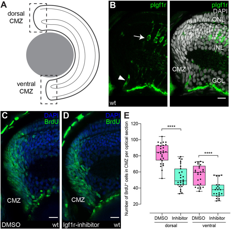
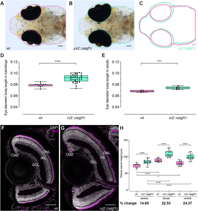
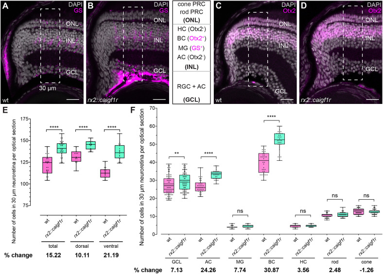
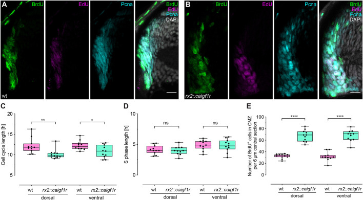
How the body and organs balance their relative growth is of key importance for coordinating size and function. This is of particular relevance in organisms, which continue to grow over their entire life span. We addressed this issue in the neuroretina of medaka fish (Oryzias latipes), a well-studied system with which to address vertebrate organ growth. We reveal that a central growth regulator, Igf1 receptor (Igf1r), is necessary and sufficient for proliferation control in the postembryonic retinal stem cell niche: the ciliary marginal zone (CMZ). Targeted activation of Igf1r signaling in the CMZ uncouples neuroretina growth from body size control, and we demonstrate that Igf1r operates on progenitor cells, stimulating their proliferation. Activation of Igf1r signaling increases retinal size while preserving its structural integrity, revealing a modular organization in which progenitor differentiation and neurogenesis are self-organized and highly regulated. Our findings position Igf signaling as a key module for controlling retinal size and composition, with important evolutionary implications.

Keywords: Medaka; Organ size; Scaling; Self-organization.


Conflict of interest statement

Competing interests The authors declare no competing or financial interests.

Figures

Fig. 1.
Fig. 1.
Igf1r signaling regulates proliferation in the ciliary marginal zone. (A) Schematic representation of a transverse retinal section. Dashed squares represent dorsal (d) and ventral (v) ciliary marginal zone (CMZ). All CMZ sections in this article depict the dorsal CMZ, with separate quantifications for dorsal and ventral CMZ, unless stated otherwise. (B) Cryosection of wild-type hatchling with anti-pIgf1r (green) staining shows Igf1r activity in single CMZ cells (arrowhead, n=35 cells in 15 sections from five fish) and in Müller glia (MG) (arrow) in the inner nuclear layer (INL). Scale bar: 10 μm. GCL, ganglion cell layer; ONL, outer nuclear layer. (C,D) Wild-type hatchlings were incubated for 24 h in BrdU and 10 μM Igf1r inhibitor NVP-AEW541 (D) or DMSO (C). Cryosections of DMSO- (C) and Igf1r inhibitor- (D) treated retinae with BrdU staining (green). Scale bars: 10 μm. (E) Quantification of BrdU-positive cell number in one optical section per central section shows a decrease in Igf1r-inhibitor-treated retinae [n=26 (dorsal)/23 (ventral) sections from 10 retinae in six fish] compared with DMSO [n=28 (dorsal)/27 (ventral) sections from 10 retinae in six fish] (data obtained from two independent experiments; t-test, ****Pd<0.0001; Mann–Whitney test, ****Pv<0.0001). Box plots show median, and 25th and 75th percentiles, with whiskers from minimum to maximum data points.
Fig. 2.
Fig. 2.
Constitutive activation of Igf1r in retinal stem and progenitor cells results in increased neuroretina size. (A-C) Eye size of rx2::caigf1r hatchlings (B, green in C) is increased compared with wild-type siblings (A, pink in C). Scale bars: 100 μm. (D,E) Quantification of relative eye size (eye diameter normalized to body length) of wild-type (D: n=12; E: n=11) and rx2::caigf1r (D: n=38; E: n=15) hatchlings (D), and 3-month-old adults (E) (t-test, ****Phatchling<0.0001, ***Padult=0.0009). (F,G) Cryosections of wild-type (F) and rx2::caigf1r (G) hatchling retinae with staining for Rx2 (magenta) display neuroretinal expansion. Wild-type section corresponds to fish 5 and rx2::caigf1r section to fish 16 in Table S1. Scale bars: 50 μm. (H) Neuroretinal thickness was measured perpendicular to the inner plexiform layer (IPL) in the fully laminated, CMZ-proximal region in wild-type and rx2::caigf1r retinae. Quantification of retinal column height in the central (wild type, n=11 sections from eight retinae in five fish; rx2::caigf1r, n=23 sections from ten retinae in eight fish), dorsal and ventral (wild type, n=18 sections from 12 retinae in six fish; rx2::caigf1r, n=24 sections from 14 retinae in eight fish) retina shows an increase in rx2::caigf1r compared with wild-type fish and in CMZ-derived compared with embryonic retina (t-test, ****P<0.0001, *Pwt c-v=0.0130). Box plots show median, and 25th and 75th percentiles, with whiskers from minimum to maximum data points.
Fig. 3.
Fig. 3.
Cell type composition of rx2::caigf1r retinae is shifted towards INL neurons. (A-D) Cell type numbers were quantified in a 30 μm wide region (dashed rectangles) in the differentiated peripheral neuroretina in cryosections of wild-type (A,C) and rx2::caigf1r (B,D) hatchlings: cone and rod PRCs, and cells in the retinal ganglion layer (GCL) are identified by location; Müller glia (MG) are GS positive (A,B, magenta), bipolar cells (BCs) are Otx2 positive (C,D, magenta), and horizontal (HCs) and amacrine cells (ACs) are Otx2 negative (C,D). Scale bars: 20 μm. (E) Quantification of total cell number shows an increase in rx2::caigf1r [n=36 (total)/18 (dorsal/ventral) sections from six retinae in three fish] compared with wild-type [n=36 (total)/18 (dorsal/ventral) sections from six retinae in three fish] retinae (t-test, ****P<0.0001). The percentage change of mean cell number from wild type to rx2::caigf1r shows notable differences. (F) Quantification of the number of each cell type shows an increase in the number of GCL, ACs and BCs in rx2::caigf1r (n≥35 sections from six retinae in three fish) compared with wild-type (n≥36 sections from six retinae in three fish) retinae (t-test, ****PAC<0.0001; Mann–Whitney test, **PGCL=0.0076, nsPMG=0.0724, ****PBC<0.0001, nsPHC=0.3444, nsProd=0.1771 and nsPcone=0.3114). Percentage change of mean cell number of wild type and rx2::caigf1r shows the largest changes in AC and BC number. ns, not significant. Box plots show median, and 25th and 75th percentiles, with whiskers from minimum to maximum data points.
Fig. 4.
Fig. 4.
Constitutive activation of Igf1r signaling decreases cell cycle length in the CMZ. (A,B) Cryosections of wild-type (A) and rx2::caigf1r (B) hatchling retinae incubated for 2 h with BrdU and for 30 min with EdU to determine cell cycle length. BrdU (green), EdU (magenta) and Pcna (cyan) staining partially overlap in the CMZ. Scale bars: 10 μm. (C) Quantification of cell cycle length shows a reduction of 1-2 h in rx2::caigf1r (n=11 sections from four retinae in four fish) compared with wild-type (n=11 sections from four retinae in four fish) retinae (data obtained from two independent experiments; Mann–Whitney test, **Pd=0.0018; t-test, *Pv=0.0188). (D) Quantification of S-phase length in rx2::caigf1r (n=11 sections from four retinae in four fish) compared with wild-type (n=11 sections from four retinae in four fish) retinae (data obtained from two independent experiments; t-test, nsPd=0.6764, nsPv=0.8223). S-phase length is not altered in rx2::caigf1r retinae. (E) Quantification of BrdU-positive cell number in the CMZ per 6 μm central section shows that numbers have more than doubled in rx2::caigf1r (n=11 sections from four retinae in four fish) compared with wild-type (n=11 sections from four retinae in four fish) retinae (data obtained from two independent experiments; t-test, ****Pd/v<0.0001). ns, not significant. Box plots show median, and 25th and 75th percentiles, with whiskers from minimum to maximum data points.
Fig. 5.
Fig. 5.
Cndp is expressed in multipotent neuroretinal stem cells. (A) Cryosection of a cndp::eGFP-caax hatchling retina with GFP staining (green) in a peripheral subset of the Rx2 (magenta) domain in the CMZ. Scale bar: 10 μm. (B) Schematic representation of the constructs used for lineage tracing. Upon tamoxifen induction, mCherry is floxed out and H2B-eGFP is expressed in GaudíRSG fish. (C) Experimental outline. cndp::CreERT2 is injected in one-cell stage GaudíRSG embryos. At hatching, fish are incubated in tamoxifen overnight and grown for 2-3 weeks before analysis. (D) Schematic representation of a whole-mount neuroretina containing stem cell clones (arrowheads) and progenitor clones (asterisks). Progenitor clones are not connected to the CMZ. (E,F) Whole-mount immunostaining of cndp::CreERT2, GaudíRSG retinae using GFP (green) with neuroretinal clones (E, arrowheads) labeling the whole retinal column (F), originating from multipotent neuroretinal stem cells (n=9 clones in four retinae from four fish, data obtained from two independent experiments). Scale bars: 100 μm in E; 20 μm in F.
Fig. 6.
Fig. 6.
Constitutive activation of Igf1r signaling expands retinal progenitor cell numbers. (A) Schematic representation of the dorsal CMZ of a retinal section with cndp (magenta) and rx2 (green) expression in stem and progenitor cells. (B,C) Cryosections of wild-type (B) and rx2::caigf1r (C) cndp::H2A-mCherry reporter hatchling retinae. mCherry (magenta) is visible in peripheral-most cells in the CMZ (arrowheads). (D) Quantification of H2A-mCherry-positive cell numbers in the CMZ per 16 µm central section does not indicate a major expansion of cndp-positive stem cells in rx2::caigf1r (n=18 sections from six retinae in three fish) compared with wild-type (n=18 sections from six retinae in three fish) retinae (Mann–Whitney test, *Pd=0.0402; t-test, nsPv=0.2177). (E,F) Cryosections of wild-type (E) and rx2::caigf1r (F) hatchling retinae. Rx2 staining (green) marks peripheral cells in the CMZ. (G) Quantification of Rx2-positive cell numbers in the CMZ per 16 µm central section demonstrates that Rx2-positive stem and progenitor cell numbers are more than doubled in rx2::caigf1r (n=9 sections from six retinae in three fish) compared with wild-type (n=9 sections from six retinae in three fish) retinae (t-test, ****Pd/v<0.0001). (H) Igf1r inhibition decreases proliferating cells (green) in the CMZ by 30%, while Igf1r activation increases proliferation by at least 100%. (I) Igf1r activation in the CMZ expands progenitor numbers (rx2-positive, green) in rx2::caigf1r fish, but does not enlarge the stem cell population (cndp-positive, magenta). Cell cycle duration in the CMZ is shortened from 12 h in wild-type fish to 10 h upon Igf1r activation in rx2::caigf1r fish. ns, not significant. Box plots show median, and 25th and 75th percentiles, with whiskers from minimum to maximum data points. Scale bars: 10 μm.

Similar articles

Cited by

References

    1. Aghaallaei, N., Gruhl, F., Schaefer, C. Q., Wernet, T., Weinhardt, V., Centanin, L., Loosli, F., Baumbach, T. and Wittbrodt, J. (2016). Identification, visualization and clonal analysis of intestinal stem cells in fish. Development 143, 3470-3480. 10.1242/dev.134098 - DOI - PMC - PubMed
    1. Alonso-Barba, J. I., Rahman, R.-U., Wittbrodt, J. and Mateo, J. L. (2016). MEPD: medaka expression pattern database, genes and more. Nucleic Acids Res. 44, D819-D821. 10.1093/nar/gkv1029 - DOI - PMC - PubMed
    1. Andersen, D. S., Colombani, J. and Léopold, P. (2013). Coordination of organ growth: principles and outstanding questions from the world of insects. Trends Cell Biol. 23, 336-344. 10.1016/j.tcb.2013.03.005 - DOI - PubMed
    1. Attias-Geva, Z., Bentov, I., Fishman, A., Werner, H. and Bruchim, I. (2011). Insulin-like growth factor-I receptor inhibition by specific tyrosine kinase inhibitor NVP-AEW541 in endometrioid and serous papillary endometrial cancer cell lines. Gynecol. Oncol. 121, 383-389. 10.1016/j.ygyno.2011.01.008 - DOI - PubMed
    1. Ayaso, E., Nolan, C. M. and Byrnes, L. (2002). Zebrafish insulin-like growth factor-I receptor: molecular cloning and developmental expression. Mol. Cell. Endocrinol. 191, 137-148. 10.1016/S0303-7207(02)00083-7 - DOI - PubMed

Publication types

MeSH terms

Substances