Review
. 2021 Mar 15;148(6):dev189217.
doi: 10.1242/dev.189217.

Mammalian primordial germ cell specification

Affiliations
Review

Mammalian primordial germ cell specification

Grace V Hancock et al. Development. .

Abstract

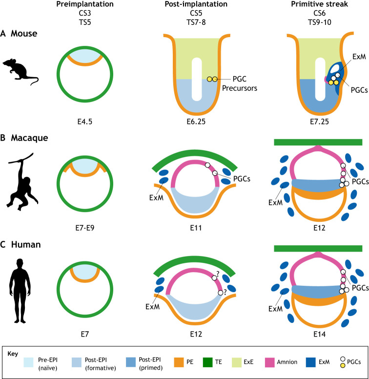
The peri-implantation window of mammalian development is the crucial window for primordial germ cell (PGC) specification. Whereas pre-implantation dynamics are relatively conserved between species, the implantation window marks a stage of developmental divergence between key model organisms, and thus potential variance in the cell and molecular mechanisms for PGC specification. In humans, PGC specification is very difficult to study in vivo To address this, the combined use of human and nonhuman primate embryos, and stem cell-based embryo models are essential for determining the origin of PGCs, as are comparative analyses to the equivalent stages of mouse development. Understanding the origin of PGCs in the peri-implantation embryo is crucial not only for accurate modeling of this essential process using stem cells, but also in determining the role of global epigenetic reprogramming upon which sex-specific differentiation into gametes relies.

Keywords: Embryo; Germ cells; Implantation; Peri-implantation; Pluripotency; Primordial germ cells.


Conflict of interest statement

Competing interestsThe authors declare no competing or financial interests.

Figures

Fig. 1.
Fig. 1.
Representation of peri-implantation embryo development in mammals. Mouse (A), monkey (B) and human (C) development. Formative pluripotency is described only in mouse and is therefore inferred in the other species. (A) In mouse embryos, the egg cylinder stages begin at Theiler stage (TS) 7. A pre-streak embryo is shown at embryonic day E6.25, which can be assigned to late TS8 with the specification of Blimp1+ primordial germ cell (PGC) precursors (yellow). Primitive streak formation begins at TS9, with mPGC specification from the precursors occurring late in TS9 and continuing in TS10. (B) In the monkey (cyno), cyPGCs are first identified at E11 [Carnegie stage (CS) 5c] in the amnion, with morphological evidence of primitive streak formation occurring in CS6. (C) In humans, ex vivo culture indicates that hPGCs can be identified before primitive streak formation at E12. ‘?’ indicates that the cell layer of origin is not yet known for humans. At CS6, cyPGCs are found in the posterior primitive streak. In the mouse embryo, bone morphogenetic protein 4 (BMP4) and BMP8b are produced from extra-embryonic endoderm (ExE) to induce mPGC specification. In the nonhuman primate embryo, BMP4 is reported as being produced by amnion cells and possibly by extra-embryonic mesoderm (ExM). In the human embryo, the cell of origin is not yet known, and the source of BMP for inducing PGC specification is also unclear. Supporting embryonic and extra-embryonic tissue types are depicted as: preimplantation epiblast (pre-EPI), represented in culture by the naïve state; post-implantation epiblast (post-EPI), represented by the formative and primed states; primitive endoderm (PE); and trophectoderm (TE).
Fig. 2.
Fig. 2.
Stem cell models of primordial germ cell induction. Modeling primordial germ cell (PGC) development with embryonic stem cells (ESCs) or induced pluripotent stem cells (iPSCs) uses either aggregate models or embryo models. (A) Aggregate models involve the generation of disorganized three-dimensional aggregates with primordial germ cell (PGC)-like cells (PGCLCs) induced from either a progenitor intermediate or by direct induction in response to BMP4 and other cytosines. Aggregate models have been created in the mouse (Hayashi et al., 2011, 2012), rhesus (Sosa et al., 2018), cyno (Sakai et al., 2020) and human (Irie et al., 2015; Sasaki et al., 2015; Chen et al., 2017, 2019). Growth factors, inhibitors and media types used for each PGCLC induction protocol are depicted. Proteins used to identify PGCLCs in each species are shown. (B) Embryo models involve modeling the spatial organization of some lineages in the peri-implantation embryo; these models have identified PGCLCs in the embryonic stem cell (ESC) and trophoblast stem cell (TSC) embryo model generated with mouse cells (Harrison et al., 2017), as well as in the posteriorized embryonic-like sac model generated with human cells (Zheng et al., 2019). AITS, 50% advanced RPMI, 50% neurobasal medium, and 1× insulin, transferrin and selenoprotein; BMP, bone morphogenetic protein; cyno, cynomolgus; DK20, DMEM/F-12 and 20% KSR; EGF, epidermal growth factor; EpiLC, epiblast-like cell; ETS, ES- and TS-derived stem cell embryo; FGF, fibroblast growth factor; GK15, GMEM and 15% KSR; iMELCs, incipient mesoderm-like cell; iPSC, induced pluripotent stem cell; KSR, knockout serum replacer; mTeSR, commercial media; RDNF, 50% RPMI, 25% DMEM, 25% neurobasal medium and 10% fetal bovine serum (FBS); RF, RPM1 and 20% FBS; SCF, stem cell factor; TeSR-E8, TeSR-Essential 8 commercial media. Figure created with BioRender.com.

References

    1. Aramaki, S., Hayashi, K., Kurimoto, K., Ohta, H., Yabuta, Y., Iwanari, H., Mochizuki, Y., Hamakubo, T., Kato, Y., Shirahige, K.et al. (2013). A mesodermal factor, T, specifies mouse germ cell fate by directly activating germline determinants. Dev. Cell 27, 516-529. 10.1016/j.devcel.2013.11.001 - DOI - PubMed
    1. Armant, D. R. (2005). Blastocysts don't go it alone. Extrinsic signals fine-tune the intrinsic developmental program of trophoblast cells. Dev. Biol. 280, 260-280. 10.1016/j.ydbio.2005.02.009 - DOI - PMC - PubMed
    1. Bao, S., Tang, F., Li, X., Hayashi, K., Gillich, A., Lao, K. and Surani, M. A. (2009). Epigenetic reversion of post-implantation epiblast to pluripotent embryonic stem cells. Nature 461, 1292-1295. 10.1038/nature08534 - DOI - PMC - PubMed
    1. Bardot, E. S. and Hadjantonakis, A.-K. (2020). Mouse gastrulation: coordination of tissue patterning, specification and diversification of cell fate. Mech. Dev. 163, 103617. 10.1016/j.mod.2020.103617 - DOI - PMC - PubMed
    1. Bedzhov, I., Leung, C. Y., Bialecka, M. and Zernicka-Goetz, M. (2014). In vitro culture of mouse blastocysts beyond the implantation stages. Nat. Protoc. 9, 2732-2739. 10.1038/nprot.2014.186 - DOI - PubMed

Publication types

LinkOut - more resources