. 2021 Mar 23;118(12):e2012529118.
doi: 10.1073/pnas.2012529118.

Immunoediting role for major vault protein in apoptotic signaling induced by bacterial N-acyl homoserine lactones

Affiliations

Immunoediting role for major vault protein in apoptotic signaling induced by bacterial N-acyl homoserine lactones

Josep Rayo et al. Proc Natl Acad Sci U S A. .

Abstract

The major vault protein (MVP) mediates diverse cellular responses, including cancer cell resistance to chemotherapy and protection against inflammatory responses to Pseudomonas aeruginosa Here, we report the use of photoactive probes to identify MVP as a target of the N-(3-oxo-dodecanoyl) homoserine lactone (C12), a quorum sensing signal of certain proteobacteria including P. aeruginosa. A treatment of normal and cancer cells with C12 or other N-acyl homoserine lactones (AHLs) results in rapid translocation of MVP into lipid raft (LR) membrane fractions. Like AHLs, inflammatory stimuli also induce LR-localization of MVP, but the C12 stimulation reprograms (functionalizes) bioactivity of the plasma membrane by recruiting death receptors, their apoptotic adaptors, and caspase-8 into LR. These functionalized membranes control AHL-induced signaling processes, in that MVP adjusts the protein kinase p38 pathway to attenuate programmed cell death. Since MVP is the structural core of large particles termed vaults, our findings suggest a mechanism in which MVP vaults act as sentinels that fine-tune inflammation-activated processes such as apoptotic signaling mediated by immunosurveillance cytokines including tumor necrosis factor-related apoptosis inducing ligand (TRAIL).

Keywords: bacterial signaling; cross-kingdom communication; immunoediting; immunosurveillance.


Conflict of interest statement

The authors declare no competing interest.

Figures

Fig. 1.
Fig. 1.
Chemical proteomic analysis identifies MVP as a potential target of C12. (A) General scheme of the chemical proteomics strategy, utilizing the probe P6 (Left, Bottom). (B) Representative LC–MS profiles of identified MVP peptides. SILAC ratios and peptide sequences are shown on the top. (C) Representative example of a competition effect of C12 on P6-mediated MVP labeling. Labeling by the probe in the presence of the native molecule C12 (red) is reduced when compared to labeling with the probe alone (blue). (D) The left panel shows barrel-like structure of vault, with MVP unit marked in green; top and bottom arrows mark vault cap and shoulder regions, respectively. The right panel shows the results of in silico docking simulation of MVP-C12 interaction, suggesting C12 (white) localization in a close proximity to shoulder region of vault (yellow). (E) Representative example using the hydrazine (N2H4) cleavable levulinic linker for the identification of P6 peptide adducts from a vault labeling experiment. The m/z values for matched pair of light (14N) and heavy (15N) adducts identifying MVP peptide are shown, resulting in a characteristic doublet signal. Images and molecules shown in A, D, and E are not drawn to scale. For additional information relevant to experiments described above, see SI Appendix, Figs. S2–S4.
Fig. 2.
Fig. 2.
C12-producing P. aeruginosa and C12 alone induce MVP translocation to LR fractions. (A) Comparison of NHBE cell responsiveness to P. aeruginosa wild type (wt) or lasI mutant (ΔlasI) by using WB analysis of cellular fractions containing cytosol (C) or LRs for expression of MVP, vimentin (a marker of LR fractions), and HSP70 (a marker of cytosol fraction). (B) NHBE cells were stimulated with C12 or its unnatural R-stereoisomer (C12R), and cellular samples were analyzed as in A as well as for expression of PARP4. (C, D) A549 lung cancer cell line (C) and normal human CBM (D) were stimulated with C12 and analyzed as indicated in B. For additional information relevant to experiments described above, see SI Appendix, Figs. S5 and S6.
Fig. 3.
Fig. 3.
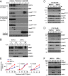
MVP attenuates C12-induced apoptotic signaling. (A) A549 cells were stimulated with C12, and the subcellular fractions were prepared and analyzed by WB for expression of vault components, apoptotic caspases, and loading control markers as indicated. (B) BMDM from wild-type (MVP+/+) and MVP-deficient (MVP−/−) mice were treated with C12 as indicated, and the subcellular fractions (cytosolic [C] and LR) were analyzed by WB for expression of casp8, vault components (MVP and PARP4), and vimentin. (C, D) WB analysis monitoring PARP and casp3 cleavage as well as the phosphorylation of eIF2α (p-eIF2α) in extracts from wild-type and MVP-deficient MEF (C) or BMDM (D) stimulated with C12 as indicated. (E) Profiles of casp8, casp9, or casp3 activity in BMDM treated with C12. (F) WB analysis monitoring activation of initiator caspases (casp8 and casp9) in extracts from MVP+/+ or MVP−/− BMDM treated with C12 as indicated. For additional information relevant to experiments described above, see SI Appendix, Figs. S7 and S8.
Fig. 4.
Fig. 4.
MVP deficiency selectively affects C12-induced prosurvival activity of p38 signaling. (A) WB analysis monitoring expression of TNF and interleukin 1 beta (IL-1β) in extracts from wild-type (WT) and MVP-deficient BMDM stimulated with LPS in the presence of indicated doses of C12. (B) TNF secretion from WT or MVP-deficient (MVP ko) BMDM stimulated with LPS in the presence of indicated doses of C12. (C) WB analysis for p38, eIF2α, JNK, ERK1/2, and their phosphorylated form as well as MVP in extracts from WT and MVP-deficient BMDM stimulated with C12 as indicated. (D) Three independent cytosol and LR fractions from WT (1 to 3) or MVP-deficient BMDM (ko; 4 to 6) were analyzed for expression of p38, JNK, and loading control (vim, an abbreviation for vimentin). (E) WB analysis of PARP, p-p38, p-eIF2α, and actin in cellular extracts from WT and p38-deficient cells stimulated by C12. (F) Comparison of casp8 activity in WT and MVP- or p38-deficient cells stimulated with C12 (10 mM) as indicated. (G) After transfection with MVP-specific siRNA (MVP) or nontargeting control siRNA (NT), untransfected (control) and siRNA-transfected human CBM were treated with C12 for indicated times, and cellular extracts were analyzed by WB for MVP, p-p38, p-CREB, p-eIF2α, and actin. The same set of CBM were also incubated with C12 for 24 h, and cell viability was measured and shown (the bottom boxes; three biological replicates) as a percentage of viable (100%) untreated cells.
Fig. 5.
Fig. 5.
MVP recruitment into LR platforms could regulate immunoediting and immunosurveillance processes. (A) WB analysis monitors expression of the vault components (MVP and PARP4), MSR1, and vimentin in subfractions from CBM stimulated by C12 or TNF as indicated. (B) Subtractions shown in A were additionally analyzed for expression of the indicated proteins. (C) Subfractions (C [cytosol], M [membrane], and LR) were prepared from LPS-stimulated CBM and analyzed for expression of the indicated proteins. (D) Relative levels of radioactivity incorporated into sphingomyelin (SM) after 3H-Sph metabolic labeling macrophages treated for different period of time with natural (C12S) or unnatural (C12R) enantiomers of C12; the samples from untreated cells were used as a control. For additional information, please see SI Appendix, Fig. S9. (E and F) CBM were stimulated with S- or R-enantiomers of C12, and the prepared subfractions (at 30 min after stimulation) were analyzed for expression indicated on a panel of proteins (E); in parallel, total cellular extracts were analyzed for expression of the phosphorylated forms of p38 or eIF2α and actin as a loading control (F). (G) After transfection by MVP-specific siRNA (MVP) or a nontargeting control siRNA (NT), A549 cells treated with C12 (5 μM), TRAIL (10 ng/mL), or a combination of both stimuli for 3 h and cellular extracts were analyzed by WB for PARP and casp8 cleavage as well as for a full length of casp8 and actin as indicated. Silencing of MVP expression in these cells was also verified by WB for MVP (insert on the bottom). (H) The same set of A549 transfected cells were incubated with or without C12 (5 μM) in the presence of indicated doses of TRAIL for 18 h, and cell survival was assessed.

References

    1. Smyth M. J., Godfrey D. I., Trapani J. A., A fresh look at tumor immunosurveillance and immunotherapy. Nat. Immunol. 2, 293–299 (2001). - PubMed
    1. Dunn G. P., Old L. J., Schreiber R. D., The immunobiology of cancer immunosurveillance and immunoediting. Immunity 21, 137–148 (2004). - PubMed
    1. Ben-Neriah Y., Karin M., Inflammation meets cancer, with NF-κB as the matchmaker. Nat. Immunol. 12, 715–723 (2011). - PubMed
    1. Moore R. J., et al. ., Mice deficient in tumor necrosis factor-alpha are resistant to skin carcinogenesis. Nat. Med. 5, 828–831 (1999). - PubMed
    1. Wilson J., Balkwill F., The role of cytokines in the epithelial cancer microenvironment. Semin. Cancer Biol. 12, 113–120 (2002). - PubMed

Publication types

Substances