. 2021 Mar 23;118(12):e2013336118.
doi: 10.1073/pnas.2013336118.

Toxoplasma gondii association with host mitochondria requires key mitochondrial protein import machinery

Affiliations

Toxoplasma gondii association with host mitochondria requires key mitochondrial protein import machinery

Matthew L Blank et al. Proc Natl Acad Sci U S A. .

Abstract

Host mitochondrial association (HMA) is a well-known phenomenon during Toxoplasma gondii infection of the host cell. The T. gondii locus mitochondrial association factor 1 (MAF1) is required for HMA and MAF1 encodes distinct paralogs of secreted dense granule effector proteins, some of which mediate the HMA phenotype (MAF1b paralogs drive HMA; MAF1a paralogs do not). To identify host proteins required for MAF1b-mediated HMA, we performed unbiased, label-free quantitative proteomics on host cells infected with type II parasites expressing MAF1b, MAF1a, and an HMA-incompetent MAF1b mutant. Across these samples, we identified ∼1,360 MAF1-interacting proteins, but only 13 that were significantly and uniquely enriched in MAF1b pull-downs. The gene products include multiple mitochondria-associated proteins, including those that traffic to the mitochondrial outer membrane. Based on follow-up endoribonuclease-prepared short interfering RNA (esiRNA) experiments targeting these candidate MAF1b-targeted host factors, we determined that the mitochondrial receptor protein TOM70 and mitochondria-specific chaperone HSPA9 were essential mediators of HMA. Additionally, the enrichment of TOM70 at the parasitophorous vacuole membrane interface suggests parasite-driven sequestration of TOM70 by the parasite. These results show that the interface between the T. gondii vacuole and the host mitochondria is characterized by interactions between a single parasite effector and multiple target host proteins, some of which are critical for the HMA phenotype itself. The elucidation of the functional members of this complex will permit us to explain the link between HMA and changes in the biology of the host cell.

Keywords: Toxoplasma gondii; mitochondria; neofunctionalization; tandem gene expansion; virulence.


Conflict of interest statement

The authors declare no competing interest.

Figures
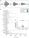
Fig. 1.
Fig. 1.
Quantitative mass spectrometry analysis identified 13 potential MAF1 host binding partners. (A) Selection criteria for candidate proteins included identifying proteins with two or more peptide hits and proteins significantly higher in abundance (greater than twofold change) in type II:MAF1b IP samples in comparison with the type II:MAF1a and type II:MAF1b mutant samples and must have an ANOVA P value ≤0.001. Nine of the final 13 candidates have a mitochondrial function based on DAVID 6.8 analysis. (B) Peak intensity plots for the cumulative ionized peptides from the 13 candidate proteins. All identified peptides for each IP sample (n = 7) are graphed against their peak ionization intensity. Error bars indicate SD. (C) Normalization of peak intensities for TOM70 cumulative peptide ionizations. Ionization values for each peptide were normalized to overall mean peptide ionization. Each peptide is a different color and each dot represents one of the seven quantitative mass spectrometry replicates. Error bars indicate SD.
Fig. 2.
Fig. 2.
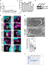
TOM70 and HSPA9 are required for MAF1b-mediated HMA. (A) U2OS cells were treated with 25 nM esiRNA targeting the candidate gene of interest for 48 h followed by type II:MAF1b infection for 24 h (MOI 2). RNA was collected at 48 h and qPCR analysis was performed. **P = 0.0021, ****P < 0.0001, unpaired two-tailed t test between cognate vehicle-treated control ΔCT values. Error bars indicate SD. (B) U2OS cells were treated as in A. Following 48 h of esiRNA treatment, cells were infected for 24 h with type II:MAF1b parasites. Cells were fixed and visualized using fluorescence microscopy. Immunofluorescence staining was performed with antibodies against the HA epitope tag and mitochondria. (Scale bars, 5 μm.) (C) HMA+ and HMA− vacuoles were counted (n = 3 coverslips per treatment group; 50 vacuoles per coverslip) using fluorescence microscopy. **P = 0.0013, ****P < 0.0001, one-way ANOVA followed by multiple comparisons. Error bars indicate SD. (D) U2OS cells were treated as in A with 25 nM esiRNA targeting TOM70, HIBADH, or vehicle control, and TOM70 protein levels were measured by Western blot analysis. β-Actin was used as a loading control. (E) U2OS cells were treated as in A with 25 nM esiRNA targeting TOM70 or HIBADH. After 48 h of treatment, cells were infected with type II:MAF1b parasites for 24 h followed by fixation in 2.5% glutaraldehyde and processed for transmission electron microscopy. For each esiRNA treatment, a minimum of 19 vacuoles were quantified. Mitochondria (M) are labeled. **P = 0.0055, ****P < 0.0001, unpaired two-tailed t test; ns, not significant. Error bars indicate SD. (F) SDS-PAGE showing results from interaction experiments between recombinant TOM70 (untagged) using 6×His-tagged TgMAF1. No significant MAF1b-specific interactions were observed with TOM70 despite robust expression of each. See text for sizes of each individual recombinant protein which is consistent with their apparent molecular masses on the stained gel.
Fig. 3.
Fig. 3.
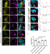
Ectopic expression of GFP-MAF1b/a confirms the requirement of TOM70 for mediating HMA. (A) U2OS cells were transfected with either GFP-MAF1b or GFP-MAF1a. Cells were fixed at either 6 or 8 h after transfection and probed with an antibody against mitochondria. Samples were visualized using epifluorescence microscopy. (B) U2OS cells were treated with 25 nM esiRNA against the indicated transcript for 48 h followed by transfection with GFP-MAF1b for 24 h. Cells were fixed and immunofluorescence staining was performed with antibodies against the mitochondria. Profile plots of both the GFP-MAF1b and mitochondria are derived from the dotted white line in the merged image (left to right) to illustrate overlap of signal intensity across each channel. (C) Overlap coefficient calculated for each of the esiRNA treatment samples (n = 4 images per treatment; representative images in B) between the GFP-MAF1b channel and the red (Alexa Fluor 594) channel used to visualize the mitochondria. **P = 0.0086, *P = 0.0101 (HIBADH), *P = 0.0127 (SAM50), one-way ANOVA followed by multiple comparisons. Error bars indicate SD. (Scale bars, 5 μm.)
Fig. 4.
Fig. 4.
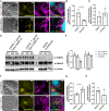
TOM70 is enriched and redistributed at the parasitophorous vacuole membrane. (A) Normal rat kidney cells expressing RFP-labeled mitochondria were infected with type II:MAF1b or type II:MAF1a parasites. Cells were fixed and visualized with epifluorescence microscopy. Immunofluorescence staining was performed with antibodies against TOM70. (B) Enrichment of either RFP-mitochondria or TOM70 at the parasitophorous vacuole was performed by quantifying pixel intensity of either the TOM70 or the mitochondria channel in a selected region on the vacuole membrane and a region off of the vacuole membrane. The ratio of these two areas was then calculated to measure enrichment of mito-RFP or TOM70 protein at the type II:MAF1b vacuole. *P = 0.0250, unpaired two-tailed t test. (C) Similar to B, enrichment ratios were calculated in cells infected with type II:MAF1a parasites. (D) U2OS cells were infected with type II:MAF1b or type II:MAF1a parasites for 24 h (MOI 2) in triplicate. Cells were lysed at 24 h in IP lysis buffer and boiled in lithium dodecyl sulfate sample buffer. Western blot analysis was performed with the listed primary antibodies and HRP-conjugated secondary antibodies. (E) Densitometric actin/TOM70 quantification of Western blots depicted in D using FIJI ImageJ software (NIH). (F) MitoRFP NRK cells were treated as in A and immunofluorescence staining was performed with antibodies against VDAC2. (G) VDAC2 raw pixel intensity values were measured as previously described in B and C. *P = 0.0470, unpaired two-tailed t test. (H) Similar to G, enrichment ratios were calculated in cells infected with type II:MAF1a parasites. Error bars in B, C, G, and H indicate SD. (Scale bars, 5 μm.)
Fig. 5.
Fig. 5.
MAF1b preferentially binds mitochondria and excludes the ER. (A) mCherry-KDEL cells were infected with either type II:MAF1b or type II:MAF1a parasites. Cells were fixed 24 hpi and visualized using fluorescence microscopy. Immunofluorescence staining was performed with antibodies against the mitochondria and HA epitope tag. Profile plots of HA-MAF1b (yellow), KDEL (cyan), and mitochondria (magenta) correlate to the arrow in the merged image. (Scale bars, 5 μm.) (B) TEM micrograph of HFFs infected with type III (HMA+) parasites. Arrowheads indicate the ER and asterisks indicate mitochondria. (B, Right) Image is a magnified region of Left (dashed square).

References

    1. Horwitz M. A., Formation of a novel phagosome by the Legionnaires’ disease bacterium (Legionella pneumophila) in human monocytes. J. Exp. Med. 158, 1319–1331 (1983). - PMC - PubMed
    1. Matsumoto A., Bessho H., Uehira K., Suda T., Morphological studies of the association of mitochondria with chlamydial inclusions and the fusion of chlamydial inclusions. J. Electron Microsc. (Tokyo) 40, 356–363 (1991). - PubMed
    1. Matsumoto A., Isolation and electron microscopic observations of intracytoplasmic inclusions containing Chlamydia psittaci. J. Bacteriol. 145, 605–612 (1981). - PMC - PubMed
    1. Adomako-Ankomah Y., et al. ., Host mitochondrial association evolved in the human parasite Toxoplasma gondii via neofunctionalization of a gene duplicate. Genetics 203, 283–298 (2016). - PMC - PubMed
    1. Dubey J. P., Sreekumar C., Redescription of Hammondia hammondi and its differentiation from Toxoplasma gondii. Int. J. Parasitol. 33, 1437–1453 (2003). - PubMed

MeSH terms

LinkOut - more resources