. 2021 Mar 23;118(12):e2023069118.
doi: 10.1073/pnas.2023069118.

Evidence supporting a time-limited hippocampal role in retrieving autobiographical memories

Affiliations

Evidence supporting a time-limited hippocampal role in retrieving autobiographical memories

Adrian W Gilmore et al. Proc Natl Acad Sci U S A. .

Abstract

The necessity of the human hippocampus for remote autobiographical recall remains fiercely debated. The standard model of consolidation predicts a time-limited role for the hippocampus, but the competing multiple trace/trace transformation theories posit indefinite involvement. Lesion evidence remains inconclusive, and the inferences one can draw from functional MRI (fMRI) have been limited by reliance on covert (silent) recall, which obscures dynamic, moment-to-moment content of retrieved memories. Here, we capitalized on advances in fMRI denoising to employ overtly spoken recall. Forty participants retrieved recent and remote memories, describing each for approximately 2 min. Details associated with each memory were identified and modeled in the fMRI time-series data using a variant of the Autobiographical Interview procedure, and activity associated with the recall of recent and remote memories was then compared. Posterior hippocampal regions exhibited temporally graded activity patterns (recent events > remote events), as did several regions of frontal and parietal cortex. Consistent with predictions of the standard model, recall-related hippocampal activity differed from a non-autobiographical control task only for recent, and not remote, events. Task-based connectivity between posterior hippocampal regions and others associated with mental scene construction also exhibited a temporal gradient, with greater connectivity accompanying the recall of recent events. These findings support predictions of the standard model of consolidation and demonstrate the potential benefits of overt recall in neuroimaging experiments.

Keywords: autobiographical memory; fMRI; hippocampus; parietal cortex; spoken recall.


Conflict of interest statement

The authors declare no competing interest.

Figures

Fig. 1.
Fig. 1.
Trial structure and approach. (A) Autobiographical memories were cued from three different recall periods (same-day, 6–18 mo ago, and 5–10 y ago), and participants selected their preferred picture cue for each trial. After viewing an enlarged version of their selected cue, participants described the memory as vividly as possible. A stop cue signaled the end of each trial. (B) Transcripts of each description were labeled and scored for content. (C and D) Transcript text was realigned with the original audio recording (C) and resynchronized with the BOLD time series (D) so that each detail could be converted to an event-related regressor. This allowed separate modeling of details and recall period effects.
Fig. 2.
Fig. 2.
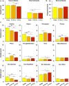
Internal and External (Ext.) details generated across temporal distances. (A) Events from 5–10 y ago had fewer Internal details than more recent events, whereas events from the Today condition had more External details than did other Recall Periods. (B) However, the overall proportion of External details did not significantly differ across Recall Periods. (C) Considerable variability was present across subtypes of details, suggesting that a simple “semanticizing account” of older memories may not accurately depict changes over time. Error bars denote within-subject SE (76). *P < 0.05; **P < 0.01; ***P < 0.001; ∼P < 0.07 (nonsignificant). For detail counts related to the Picture Description control task, see SI Appendix, Fig. S1.
Fig. 3.
Fig. 3.
Analysis of a priori, subject-specific hippocampal ROIs. (A) Anterior and posterior hippocampal ROIs were manually segmented for each subject using the uncal apex as a division landmark (27). (A, Insets) Example anterior and posterior boundary slices. (B) Posterior, but not anterior, hippocampal subregions exhibited a temporal gradient across the overt recall period, with more recent memories eliciting stronger activations than remote memories. Effects are plotted relative to the Picture Description baseline. Activity significantly differed from the baseline control task for the Today and 6–18 mo ago, but not 5–10 y ago, conditions in posterior HC regions, but anterior effects were largely indistinguishable from the control baseline (SI Appendix, Fig. S3). Error bars denote within-subject SE. *P < 0.05; Significant one-sample test vs. baseline Picture Description task (FDR, q < 0.05). AB, autobiographical; HC, hippocampus; L, left; R, right.
Fig. 4.
Fig. 4.
Voxel-wise whole-brain analysis of temporal distance effects. (A) Regions exhibiting significant main effects of temporal distance were identified in medial and lateral parietal cortex and the left superior frontal gyrus. (B) Subsequent pairwise contrasts revealed that events from earlier in the same day always elicited greater activity than did more distant events, and in all cases a monotonic reduction in activity accompanied increasing temporal distance. Results are depicted on a partially inflated human brain surface (77), and effects are plotted relative to the Picture Description baseline. Coordinates are listed in MNI152 space and refer to centers of mass for each region. Error bars denote within-subject SE. *P < 0.05; **P < 0.01; ***P < 0.001. AB, autobiographical; IPS, intraparietal sulcus; L, left; MCC, midcingulate cortex; MFG, middle frontal gyrus; PCU, precuneus; pIPL, posterior inferior parietal lobule; R, right.
Fig. 5.
Fig. 5.
Temporally graded connectivity was observed across seed regions. (A) Regions identified in the univariate whole-brain analysis served as seeds whose connectivity was compared for recent (Today) and remote (5–10 y ago) events. Green nodes depict centers of mass for each seed. (B) Maps were binarized and summed across the whole brain. Effects were consistently observed (present in four or more seed maps) in the right angular gyrus and retrosplenial cortex/parieto-occipital sulcus, parahippocampal cortex, and superior frontal cortex bilaterally. L, left; MCC, midcingulate cortex; PCU, precuneus; pIPL, posterior inferior parietal lobule; R, right.
Fig. 6.
Fig. 6.
Scene-selective cortex exhibits a temporal gradient in task-based connectivity to the posterior hippocampus. (A) Independent localizer data were used to define scene-selective regions. (B) A significant difference in task-based connectivity in the right posterior hippocampus (R post HC) and a nonsignificant tendency in the left posterior hippocampus (L post HC) were observed across Recall Periods. Error bars reflect SEM. *P < 0.05. AB, autobiographical.

References

    1. Tulving E., Elements of Episodic Memory (Oxford University Press, New York, NY, 1983).
    1. Tulving E., Memory and consciousness. Can. Psychol. 26, 1–12 (1985).
    1. Svoboda E., McKinnon M. C., Levine B., The functional neuroanatomy of autobiographical memory: A meta-analysis. Neuropsychologia 44, 2189–2208 (2006). - PMC - PubMed
    1. Hayama H. R., Vilberg K. L., Rugg M. D., Overlap between the neural correlates of cued recall and source memory: Evidence for a generic recollection network? J. Cogn. Neurosci. 24, 1127–1137 (2012). - PMC - PubMed
    1. Boccia M., Teghil A., Guariglia C., Looking into recent and remote past: Meta-analytic evidence for cortical re-organization of episodic autobiographical memories. Neurosci. Biobehav. Rev. 107, 84–95 (2019). - PubMed

Publication types

LinkOut - more resources