. 2021 Mar 23;118(12):e2025830118.
doi: 10.1073/pnas.2025830118.

Analysis of chronic inflammatory lesions of the colon for BMMF Rep antigen expression and CD68 macrophage interactions

Affiliations

Analysis of chronic inflammatory lesions of the colon for BMMF Rep antigen expression and CD68 macrophage interactions

Timo Bund et al. Proc Natl Acad Sci U S A. .

Abstract

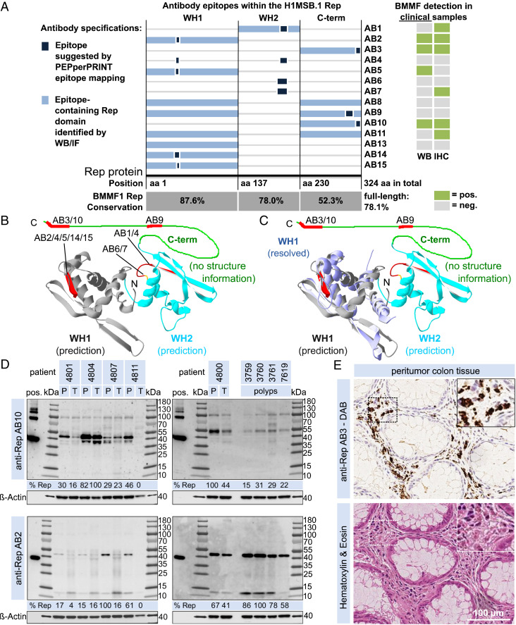
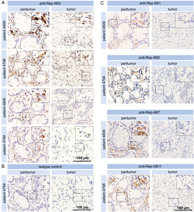
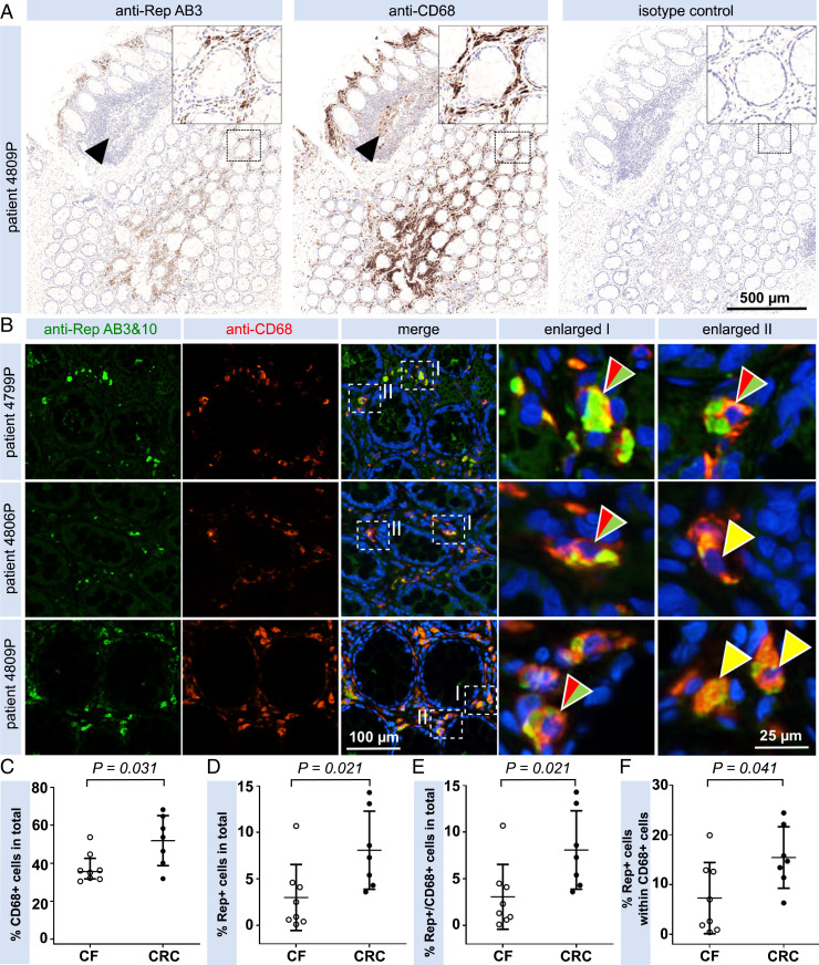
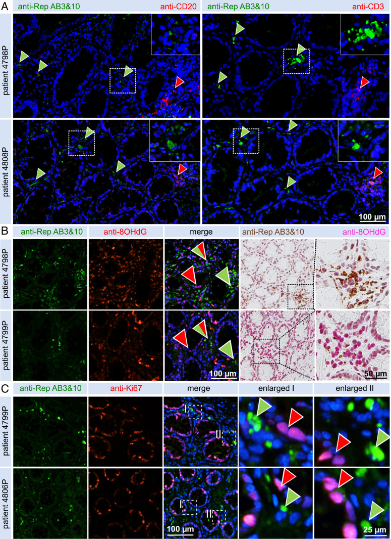
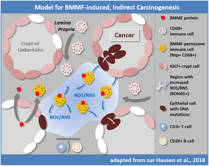
Consumption of Eurasian bovine meat and milk has been associated with cancer development, in particular with colorectal cancer (CRC). In addition, zoonotic infectious agents from bovine products were proposed to cause colon cancer (zur Hausen et al., 2009). Bovine meat and milk factors (BMMF) are small episomal DNA molecules frequently isolated from bovine sera and milk products, and recently, also from colon cancer (de Villiers et al., 2019). BMMF are bioactive in human cells and were proposed to induce chronic inflammation in precancerous tissue leading to increased radical formation: for example, reactive oxygen and reactive nitrogen species and elevated levels of DNA mutations in replicating cells, such as cancer progenitor cells (zur Hausen et al., 2018). Mouse monoclonal antibodies against the replication (Rep) protein of H1MSB.1 (BMMF1) were used to analyze BMMF presence in different cohorts of CRC peritumor and tumor tissues and cancer-free individuals by immunohistochemistry and Western blot. BMMF DNA was isolated by laser microdissection from immunohistochemistry-positive tissue regions. We found BMMF Rep protein present specifically in close vicinity of CD68+ macrophages in the interstitial lamina propria adjacent to CRC tissues, suggesting the presence of local chronic inflammation. BMMF1 (modified H1MSB.1) DNA was isolated from the same tissue regions. Rep and CD68+ detection increased significantly in peritumor cancer tissues when compared to tissues of cancer-free individuals. This strengthens previous postulations that BMMF function as indirect carcinogens by inducing chronic inflammation and DNA damage in replicating cells, which represent progress to progenitor cells for adenoma (polyps) formation and cancer.

Keywords: BMMF; antigen; chronic inflammation; colon cancer.


Conflict of interest statement

The authors declare no competing interest.

Figures

Fig. 1.
Fig. 1.
Localization and identification of antibody epitopes together with clinical information of BMMF detection by WB and IHC. (A) Epitopes identified for individual anti-Rep ABs by PEPperPRINT linear epitope mapping (dark blue box) and by WB/IF (light blue box) on a linear representation of the H1MSB.1 Rep protein (including Rep domains WH1, WH2, and the C-terminal domain) together with a summary of clinical staining results (WB and IHC, table on the right). The sequence similarity of the individual Rep domains is given for a set of BMMF1 Rep proteins (C1MI.1–4, H1MSBI.1+2, C1HB.3–6, Sphinx1.76). (B) Localization of antibody epitopes (red) on a structure prediction (16, 47) of the H1MSB.1 Rep WH1 (gray) and WH2 domains (cyan) (Swiss PDB-Viewer) (16, 47) and (C) good agreement of the predicted WH1 (gray) versus experimentally resolved H1MSB.1 WH1 structure (blue, PDB ID code 6H24) (16). (D) WB results for AB10 and AB2 for peritumor (P) and tumor (T) tissues of five CRC patients and four polyps tissues (densitometrical quantification of the target bands at the size region of about 38 to 55 kDa). (E) IHC (DAB) staining of consecutive peritumor colon tissue sections of a CRC patient with AB3 together with H&E staining. (Magnification: E, Insets, 2x.)
Fig. 2.
Fig. 2.
Immunohistochemical detection of BMMF Rep. (A) Pairs of peritumor and tumor tissue sections from four individual CRC patients were tested by IHC with anti-Rep AB3 (Magnification: A, Insets, 2x.) together with (B) isotype control. (Magnification: B, Insets 2x.) (C) IHC detection with antibodies AB1/2/7/11 respectively, of peritumor and tumor tissues from CRC patients. (Magnification: C, Insets, 2x.)
Fig. 3.
Fig. 3.
Coimmunodetection and quantification of Rep+ and CD68+ cells in peritumor CRC tissue. (A) Anti-Rep and anti-CD68 DAB IHC staining showing antibody staining in the lamina propria. No Rep staining is observed in CD68+ cells in follicular areas (arrowheads). (Magnification: A, Insets, 3.2x.) (B) Coimmunofluorescence microscopy of peritumor CRC formalin-fixed paraffin-embedded tissues showing a cytoplasmic localization of Rep (green). The majority of Rep+ cells also stained positive by anti-CD68 staining (red). Yellow arrowheads indicate colocalization of cytoplasmic Rep/CD68 signals (see enlargements), split red/green arrowheads indicate nuclei with complementation of cytoplasmic Rep/CD68 staining. (CF) Quantification of Rep+/CD68+ interstitial cells based on IF for seven peritumor tissues from CRC patients and eight colon biopsies from young donors (cancer-free, CF). About 5,000 nuclei were analyzed showing significantly increased levels of Rep+ (C), CD68+ (D), and Rep+/CD68+ cells (E) in the interstitial peritumor of colon cancer patients, when compared with interstitial regions of cancer-free individuals (medians illustrated as horizontal lines). An increased fraction of Rep+ macrophages up to 25% of all macrophages (F) is observed in peritumor colon cancer tissues versus cancer-free tissues.
Fig. 4.
Fig. 4.
Coimmunodetection of Rep together with T cell/B cell markers and markers for oxidative stress and proliferation in peritumor CRC tissue. (A) Coimmunofluorescence imaging shows no infiltration of CD20+ B cells (Left) into the vicinity of Rep+ foci (green arrowheads) in two representative tissues, although accumulations of B cells are readily visible in lymphoid follicles of tissue (red arrowheads). No pronounced infiltration of CD3+ T cells (Right) in the vicinity of Rep+ foci were observed, although distinct accumulation of T cells were noted in lymphoid follicles (red arrowheads). (Magnification: A, Insets, 2x.) (B) Detection, in consecutive sections, of increased levels of 8-OHdG in Rep+ regions; coimmunofluorescence (Left) and brightfield DAB staining (Right). (C) Coimmunofluorescence of Rep+ and Ki67+ cells demonstrating no overlap between Rep-producing cells and proliferating epithelial crypt cells.
Fig. 5.
Fig. 5.
Previously proposed hypothesis for indirect induction of colorectal cancer by BMMF (14). BMMFs are taken up by Eurasian milk and meat consumption and accumulate in CD68+ macrophages. BMMF replication and further recruitment of macrophages contribute to chronic inflammation and production of ROS and RNS, resulting in oxidative stress (represented by 8-OHdG detection) and an increased risk for DNA mutation, particularly in DNA replicating cells. T and B cells are not observed in the affected tissue regions. Replication-competent Ki67+ epithelial cells in the crypts are exposed to diffusing ROS/RNS and acquire random mutations over time, occasionally targeting cancer driver genes. The latter cells may convert into colon polyps and with additional mutations, may result in colon cancer. Modified with permission from ref. .

References

    1. Saunders K. E., et al., Novel intestinal Helicobacter species isolated from cotton-top tamarins (Saguinus oedipus) with chronic colitis. J. Clin. Microbiol. 37, 146–151 (1999). - PMC - PubMed
    1. Zur Hausen H., The search for infectious causes of human cancers: Where and why. Virology 392, 1–10 (2009). - PubMed
    1. zur Hausen H., Red meat consumption and cancer: Reasons to suspect involvement of bovine infectious factors in colorectal cancer. Int. J. Cancer 130, 2475–2483 (2012). - PubMed
    1. zur Hausen H., de Villiers E. M., Dairy cattle serum and milk factors contributing to the risk of colon and breast cancers. Int. J. Cancer 137, 959–967 (2015). - PubMed
    1. Ramos A., Hemann M. T., Drugs, bugs, and cancer: Fusobacterium nucleatum promotes chemoresistance in colorectal cancer. Cell 170, 411–413 (2017). - PubMed

Publication types