. 2021 Mar 23;118(12):e2026650118.
doi: 10.1073/pnas.2026650118.

The intrinsic instability of the hydrolase domain of lipoprotein lipase facilitates its inactivation by ANGPTL4-catalyzed unfolding

Affiliations

The intrinsic instability of the hydrolase domain of lipoprotein lipase facilitates its inactivation by ANGPTL4-catalyzed unfolding

Katrine Z Leth-Espensen et al. Proc Natl Acad Sci U S A. .

Abstract

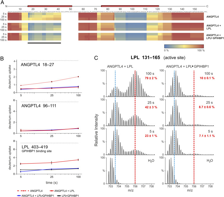
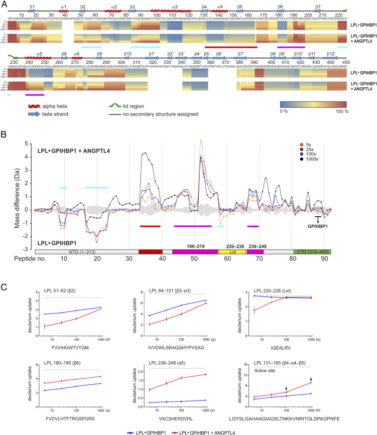
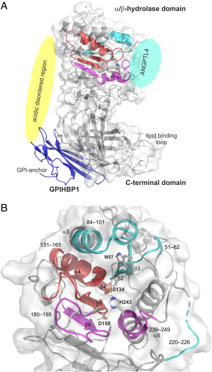
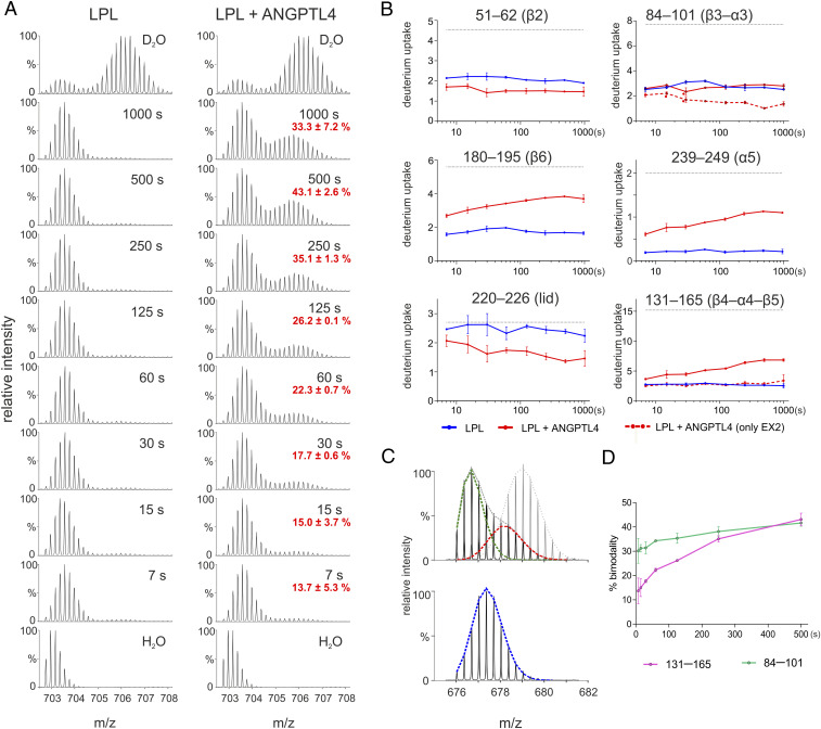
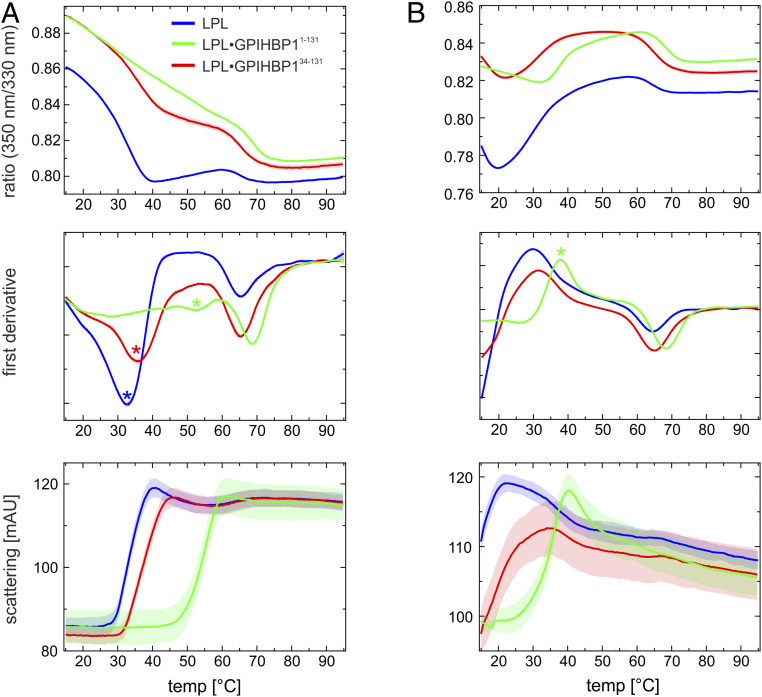
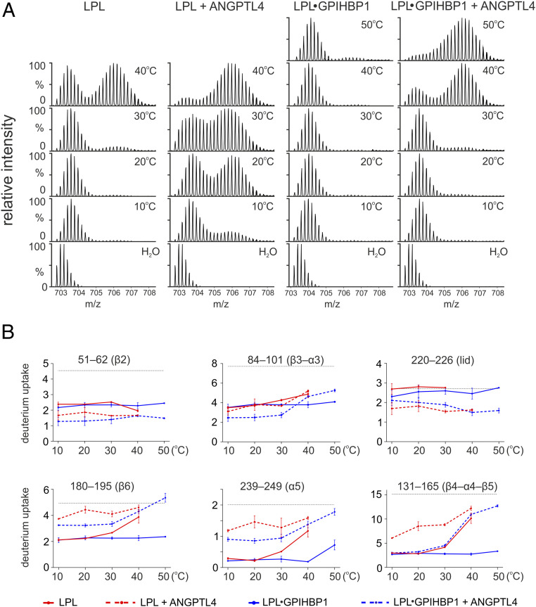
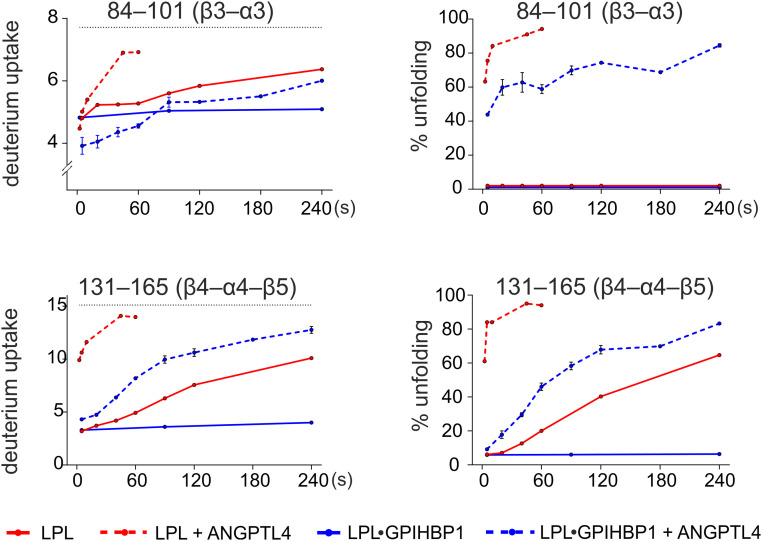
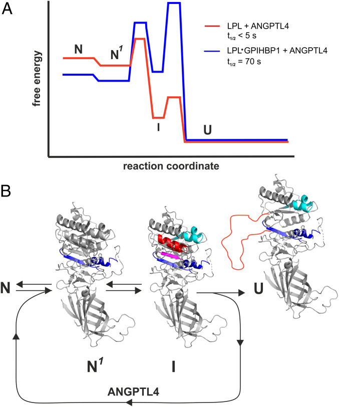
The complex between lipoprotein lipase (LPL) and its endothelial receptor (GPIHBP1) is responsible for the lipolytic processing of triglyceride-rich lipoproteins (TRLs) along the capillary lumen, a physiologic process that releases lipid nutrients for vital organs such as heart and skeletal muscle. LPL activity is regulated in a tissue-specific manner by endogenous inhibitors (angiopoietin-like [ANGPTL] proteins 3, 4, and 8), but the molecular mechanisms are incompletely understood. ANGPTL4 catalyzes the inactivation of LPL monomers by triggering the irreversible unfolding of LPL's α/β-hydrolase domain. Here, we show that this unfolding is initiated by the binding of ANGPTL4 to sequences near LPL's catalytic site, including β2, β3-α3, and the lid. Using pulse-labeling hydrogen‒deuterium exchange mass spectrometry, we found that ANGPTL4 binding initiates conformational changes that are nucleated on β3-α3 and progress to β5 and β4-α4, ultimately leading to the irreversible unfolding of regions that form LPL's catalytic pocket. LPL unfolding is context dependent and varies with the thermal stability of LPL's α/β-hydrolase domain (Tm of 34.8 °C). GPIHBP1 binding dramatically increases LPL stability (Tm of 57.6 °C), while ANGPTL4 lowers the onset of LPL unfolding by ∼20 °C, both for LPL and LPL•GPIHBP1 complexes. These observations explain why the binding of GPIHBP1 to LPL retards the kinetics of ANGPTL4-mediated LPL inactivation at 37 °C but does not fully suppress inactivation. The allosteric mechanism by which ANGPTL4 catalyzes the irreversible unfolding and inactivation of LPL is an unprecedented pathway for regulating intravascular lipid metabolism.

Keywords: GPIHBP1; HDX-MS; hypertriglyceridemia; intravascular lipolysis; intrinsic disorder.


Conflict of interest statement

The authors declare no competing interest.

Figures

Fig. 1.
Fig. 1.
The GPIHBP1•LPL complex binds to ANGPTL4, but LPL unfolding is blunted. (A) Heat maps depicting deuterium content in 40 peptic peptides from ANGPTL4 (89% sequence coverage), revealing that both unbound LPL and GPIHBP1•LPL complexes bind to ANGPTL4. Deuterium content was measured in triplicate by MS (SI Appendix, Fig. S1) and is shown relative to a fully exchanged control. Regions in blue and red show low and high deuterium uptake, respectively. Data were obtained by incubating 7 µM ANGPTL4 alone (Top) or in the presence of either 10 µM LPL (Middle) or 10 µM LPL + 20 µM GPIHBP1 (Bottom) in deuterium oxide for 5, 25, or 100 s at 25 °C. Predicted α-helices are shown above the primary sequence. The LPL binding site on ANGPTL4 was evident from ANGPTL4 peptides with reduced deuterium uptake (black bar). (B) Time-dependent deuterium uptake in peptic peptides corresponding to the first and second α-helices of ANGPTL4 (residues 18–24 and 96–111, respectively) and in the GPIHBP1-binding region of LPL (residue 403–419). The dotted black lines show fully labeled controls (determined experimentally). The dotted red line is ANGPTL4 alone; the solid red line is ANGPTL4 + LPL. The blue line is ANGPTL4 + LPL•GPIHBP1 complexes; the solid black line is LPL•GPIHBP1 complexes (dataset is from Fig. 2). (C) The unfolding of LPL by ANGPTL4 was reduced when LPL was complexed to GPIHBP1. Shown are isotope envelopes for an LPL peptic peptide covering LPL’s catalytic triad (residue 131–165) when LPL or LPL•GPIHBP1 complexes were incubated with ANGPTL4. The vertical dashed blue line shows isotope envelopes for LPL peptide 131–165 after undergoing uncorrelated EX2 exchange (a measure of local dynamics), whereas the vertical dashed red line shows LPL molecules that had undergone correlated deuterium EX1 exchange (indicative of cooperative unfolding of the hydrolase domain). Red numbers show the fractional unfolding of LPL at different incubation times.
Fig. 2.
Fig. 2.
Localizing the ANGPTL4 binding site on LPL and defining the allosteric changes in LPL triggered by ANGPTL4 binding. (A) Heat maps depicting deuterium incorporation into LPL peptides relative to a fully labeled control. The data were obtained by incubating 10 µM LPL•GPIHBP1 complexes (formed by 10 µM LPL and 30 µM GPIHBP1) alone (Upper) or with 20 µM ANGPTL4 (Lower) for 5, 25, 100, or 1,000 s in the presence of deuterium oxide at 25 °C. Online pepsin digestion generated 92 LPL peptides corresponding to 88.9% sequence coverage; 79 peptides were used to generate the heat maps (SI Appendix, Fig. S2). Secondary structures, shown above the primary sequence, are from the LPL crystal structure (37). Cyan bars indicate areas of protection (i.e., binding sites for ANGPTL4 on LPL); purple bars indicate areas of increased flexibility, and the red bar indicates global changes (bimodal isotope envelopes). (B) Butterfly plot depicting ANGPTL4-induced changes in deuterium uptake into the LPL in GPIHBP1•LPLcomplexes at four different incubation times: 5 s (orange), 25 s (red), 100 s (blue), and 1,000 s (black). Highlighted are regions with reduced deuterium uptake (residues 51–62, 84–101, and 220–226; cyan), increased deuterium uptake by uncorrelated exchange with unimodal isotope envelopes (i.e., increased flexibility; residues 180–219 and 239–249; purple), and increased deuterium uptake by correlated exchange with bimodal isotope envelopes (i.e., unfolding of residue 131–165; red). Peptic LPL peptides from GPIHBP1’s binding site are highlighted by the black bar. The shaded gray area corresponds to the largest SD in the dataset for each peptide (n = 3). (C) Deuterium uptake into selected peptic LPL peptides with reduced deuterium uptake (residues 51–62, 84–101, and 220–226), increased deuterium uptake without isotope bimodality (residues 180−195 and 239–249), and increased uptake with bimodal isotope envelopes (residue 131–165, which contains Ser134 and Asp158 of LPL’s catalytic triad). The arrows indicate correlated exchange as observed by the emergence of bimodal isotope envelopes.
Fig. 3.
Fig. 3.
Structural elements in LPL that are affected by ANGPTL4 binding. (A) Cartoon representation of the human LPL•GPIHBP1 complex with the molecular surface of LPL shown as a transparent light gray envelope (generated with PyMol using coordinates from the LPL•GPIHBP1 crystal structure; Protein Data Bank ID code 6E7K). The Trp-rich lipid-binding loop is modeled because it was not visualized in the electron density map (37). LPL elements implicated in ANGPTL4 binding are highlighted in cyan, and those undergoing allosteric changes are highlighted in red (correlated exchange) and purple (noncorrelated exchange). The position of ANGPTL4 binding is indicated with a cyan oval. GPIHBP1 (blue) binds to the C-terminal domain of LPL, and the location of GPIHBP1’s membrane-tethering site (GPI anchor) is indicated. The location of GPIHBP1’s intrinsically disordered acidic domain is depicted with a yellow oval; the acidic domain was not visualized in the crystal structure but is assumed to project over and interact with a large basic patch on the surface of LPL (37). The acidic domain stabilizes LPL’s α/β-hydrolase domain (17, 18). (B) Cartoon representation of the structural elements in LPL surrounding the catalytic triad (Ser134, Asp158, and His243). LPL regions affected by ANGPTL4 binding are color-coded: Sequences bound by ANGPTL4 are cyan; sequences that respond in an allosteric fashion to ANGPTL4 binding are shown in purple and red. The peptide segments and relevant secondary elements are identified by numbers.
Fig. 4.
Fig. 4.
Analyzing ANGPTL4 binding to LPL at 15 °C by pulse-labeled HDX-MS. (A) Impact of ANGPTL4 on the irreversible unfolding of LPL’s hydrolase domain, as judged by the emergence of a bimodal isotope envelope in the LPL peptic peptide 131–165. LPL alone (and LPL + ANGPTL4) was incubated in a protiated solvent for multiple time points (ranging from 7 to 1,000 s) and then pulse labeled for 10 s in a deuterated solvent. Incubations were performed at 15 °C to retard ANGPTL4-mediated unfolding of LPL’s α/β-hydrolase domain. A fully labeled control is shown as the first spectrum in each column. (B) Deuterium uptake in peptides representing 1) the ANGPTL4 binding site (51–62, 84–101, and 220–226), 2) regions where ANGPTL4 induces increased allosteric fluctuation (180–195 and 239–249), and 3) peptide 131–165 where bimodal isotope envelopes in pulse-labeling HDX-MS serve as a proxy for irreversible inactivation of LPL. In this experiment, two regions exhibited time-dependent ANGPTL4-induced bimodality (84–101 and 131–165). In those regions, the reduced deuterium uptake calculated for the low-mass population representing noncorrelated EX2 exchange (shown as dashed red lines) shows that region 84–101 forms part of the ANGPTL4-binding site and region 131–165 does not. (C) Isotope envelopes for LPL 84–101 after a 250-s incubation in protiated solvent at 15 °C with and without ANGPTL4 followed by a 10-s incubation at 15 °C in deuterated solvent. The fitted dashed lines represent an ANGPTL4-bound LPL peptide (green; centroid mass = 2,193.14 Da), an unfolded peptide (red; centroid mass = 2,198.67 Da), and an unoccupied, folded peptide (blue; centroid mass = 2,195.46 Da). A fully labeled control is shown by the light gray isotope envelope in the upper spectrum. (D) Time-dependent appearance of bimodal isotope envelopes in peptides 84–101 and 131–165 induced by ANGPTL4 at 15 °C.
Fig. 5.
Fig. 5.
Temperature-induced unfolding of unbound LPL and LPL•GPIHBP1 complexes in the presence and absence of ANGPTL4. (A) Thermal unfolding profiles of 10 µM LPL (blue line) in the presence of 10 µM GPIHBP11−131 (green line) or 10 µM GPIHBP134−131 (red line). Shown is the ratio of emissions at a 330 and 350 nm as a function of temperature (Upper), the first derivative of these profiles (Middle), and the change in back scattering (i.e., aggregation) (Lower). The apparent Tm for the α/β-hydrolase is highlighted by a colored asterisk. (B) Corresponding unfolding profiles recorded in the presence of 10 µM ANGPTL4. Note that the unfolding of LPL alone and in the presence of GPIHBP134−131 has already occurred (to a large extent) before the first measurement at 15 °C.
Fig. 6.
Fig. 6.
Temperature-dependent deuterium exchange in unbound LPL and LPL•GPIHBP1 complexes in both the presence and absence of ANGPTL4. (A) ANGPTL4-induced unfolding of LPL, assessed by the emergence of bimodality in the LPL peptide 131–165. The 10 µM LPL or 10 µM LPL•GPIHBP1 complexes were incubated alone or in the presence of 12 µM ANGPTL4 in protiated solvent for 180 s at the indicated temperatures, followed by 10-s pulse labeling at 25 °C in deuterated solvent. When LPL was incubated in the presence of ANGPTL4, the bimodal isotope envelope in peptide 131–165 appeared at much lower temperatures (10–20 °C) than LPL incubated without ANGPTL4 (40 °C). Similarly, when LPL•GPIHBP1 complexes were incubated in the presence of ANGPTL4, the bimodal isotope envelope in peptide 131–165 emerged at 40 °C, whereas it was absent at 50 °C when LPL•GPIHBP1 complexes were incubated without ANGPTL4. (B) Deuterium uptake in LPL peptides corresponding to the ANGPTL4 binding site (Upper) and in LPL peptides from LPL segments where ANGPTL4 binding had triggered allosteric conformational changes (Lower). Due to low peak intensity, the uptake in the lid peptide 220–226 is not shown for LPL at 40 °C.
Fig. 7.
Fig. 7.
Deuterium incorporation into LPL and LPL•GPIHBP1 complexes by pulse labeling in the presence or absence of ANGPTL4 at 37 °C. Left shows deuterium uptake in LPL peptides 84–101 and 131–165 by pulse labeling for 10 s as a function of incubation time in protiated solvent at 37 °C. Right shows the time-dependent progression in LPL unfolding as defined by the relative fractions of peptides 131–165 and 84–101 that had undergone correlated exchange (i.e., irreversibly unfolded).
Fig. 8.
Fig. 8.
Energy landscape for ANGPTL4-catalyzed unfolding of LPL and LPL•GPIHBP1 complexes. (A) Proposed model for ANGPTL4-catalyzed LPL unfolding based on continuous and pulse-labeling HDX-MS studies performed at 15, 25, or 37 °C. Half-lives (t1/2) are calculated from pulse labeling of 10 µM LPL or 10 µM LPL•GPIHBP1 complexes incubated at 37 °C in 10 mM Hepes and 150 mM NaCl (pH 7.4) in the presence of 12 µM ANGPTL4. (B) Allostery in LPL induced by ANGPTL4 binding. N is the native structure of unbound LPL or LPL•GPIHBP1 complexes; N1 is the native structure of LPL or LPL•GPIHBP1 complexes in the setting of ANGPTL4 with increased dynamics of α5 and β6 (EX2 exchange kinetics) highlighted by blue. I is the intermediate conformation with increased dynamics in β3–α3 (cyan) and with reversible unfolding (EX1) of β5 (purple) and β4–α4 (red). U is inactivated LPL with irreversibly unfolded β4–α4–β5 (red line).

References

    1. Meng X., Zeng W., Young S. G., Fong L. G., GPIHBP1, a partner protein for lipoprotein lipase, is expressed only in capillary endothelial cells. J. Lipid Res. 61, 591 (2020). - PMC - PubMed
    1. Goulbourne C. N., et al. ., The GPIHBP1-LPL complex is responsible for the margination of triglyceride-rich lipoproteins in capillaries. Cell Metab. 19, 849–860 (2014). - PMC - PubMed
    1. Young S. G., et al. ., GPIHBP1 and lipoprotein lipase, partners in plasma triglyceride metabolism. Cell Metab. 30, 51–65 (2019). - PMC - PubMed
    1. Beigneux A. P., et al. ., Autoantibodies against GPIHBP1 as a cause of hypertriglyceridemia. N. Engl. J. Med. 376, 1647–1658 (2017). - PMC - PubMed
    1. Beigneux A. P., et al. ., GPIHBP1 missense mutations often cause multimerization of GPIHBP1 and thereby prevent lipoprotein lipase binding. Circ. Res. 116, 624–632 (2015). - PMC - PubMed

Publication types