. 2021 Mar 15;6(1):123.
doi: 10.1038/s41392-021-00515-5.

Unique and complementary suppression of cGAS-STING and RNA sensing- triggered innate immune responses by SARS-CoV-2 proteins

Affiliations

Unique and complementary suppression of cGAS-STING and RNA sensing- triggered innate immune responses by SARS-CoV-2 proteins

Yajuan Rui et al. Signal Transduct Target Ther. .

Abstract

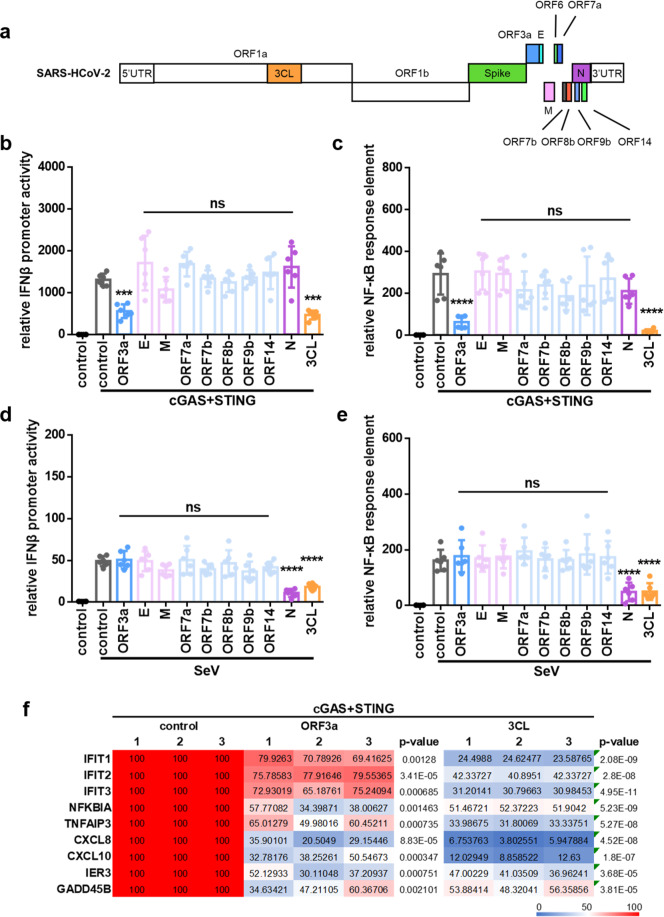
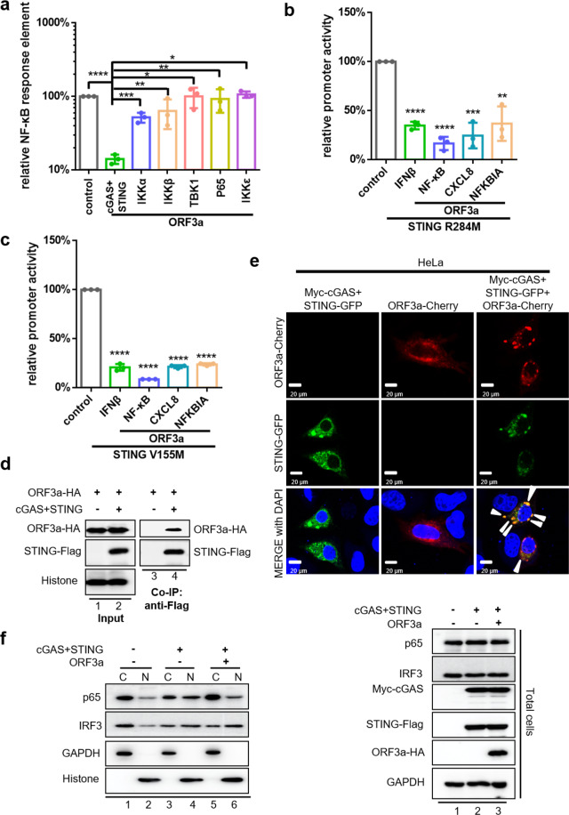
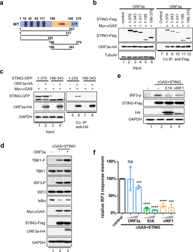
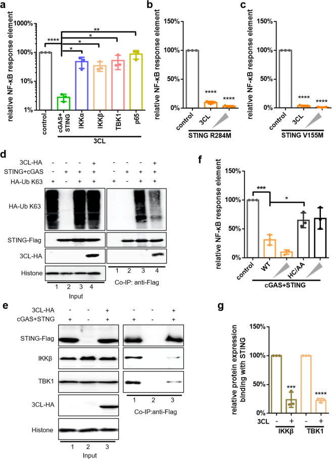
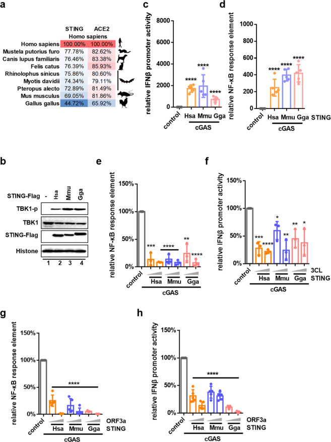
The emergence of SARS-CoV-2 has resulted in the COVID-19 pandemic, leading to millions of infections and hundreds of thousands of human deaths. The efficient replication and population spread of SARS-CoV-2 indicates an effective evasion of human innate immune responses, although the viral proteins responsible for this immune evasion are not clear. In this study, we identified SARS-CoV-2 structural proteins, accessory proteins, and the main viral protease as potent inhibitors of host innate immune responses of distinct pathways. In particular, the main viral protease was a potent inhibitor of both the RLR and cGAS-STING pathways. Viral accessory protein ORF3a had the unique ability to inhibit STING, but not the RLR response. On the other hand, structural protein N was a unique RLR inhibitor. ORF3a bound STING in a unique fashion and blocked the nuclear accumulation of p65 to inhibit nuclear factor-κB signaling. 3CL of SARS-CoV-2 inhibited K63-ubiquitin modification of STING to disrupt the assembly of the STING functional complex and downstream signaling. Diverse vertebrate STINGs, including those from humans, mice, and chickens, could be inhibited by ORF3a and 3CL of SARS-CoV-2. The existence of more effective innate immune suppressors in pathogenic coronaviruses may allow them to replicate more efficiently in vivo. Since evasion of host innate immune responses is essential for the survival of all viruses, our study provides insights into the design of therapeutic agents against SARS-CoV-2.


Conflict of interest statement

The authors declare no competing interests.

Figures

Fig. 1
Fig. 1
Identification of SARS-CoV-2 proteins involved in innate immune suppression. a Schematic diagram of the genomic organization and encoded proteins of SARS-2019 Wuhan-Hu-1 strain (MN908947.3). b, c cGAS-STING-stimulated IFNβ promoter (b) and NF-κB response element (c) activation is inhibited by ORF3a and 3CL. HEK293T cells were co-transfected with IFNβ-Luc (b), NF-κB-Luc (c), and the cGAS and STING expression vectors in the presence or absence of vectors expressing various SARS-CoV-2 proteins as indicated. d, e SeV-induced IFNβ promoter (d) and NF-κB response element (e) activation is inhibited by protein N and 3CL. HEK293T cells were co-transfected with IFNβ-Luc (d) or NF-κB-Luc (e) in the presence or absence of vectors expressing various SARS-CoV-2 proteins as indicated. SeV was added to the culture medium 12 h after transfection. pRL-TK Renilla was used as an internal control (be). Transactivation of the luciferase reporter was determined 24 h after transfection (b, c) or 12h after SeV infection (d, e) (n = 6 independent biological experiments). Means and standard deviations are presented. Statistical significance was determined by two-sided unpaired t test, ***p < 0.001; ****p < 0.0001; NS, no significance (b–e). f Inhibition of cGAS-STING-stimulated human genes by ORF3a and 3CL of SARS-CoV-2. HEK293T cells were co-transfected with the cGAS-STING expression vectors in the presence or absence of the ORF3a or 3CL expression vectors as indicated. Human genes activated by cGAS-STING alone served as a positive control and were set to 100%. Total RNA was prepared from HEK293T cells 24 h after transfection and analyzed for the transcriptional level of the indicated genes by RT-qPCR (n = 3 independent biological experiments). Statistical significance was determined by two-sided unpaired t test
Fig. 2
Fig. 2
Characterization of ORF3a-mediated cGAS-STING inhibition. a Comparison of ORF3a-mediated inhibition of cGAS-STING-, IKKα-, IKKβ-, TBK1-, p65-, and IKKε-induced NF-κB signaling. HEK293T cells were co-transfected with NF-κB-Luc and the cGAS-STING, IKKα, IKKβ, TBK1, p65, or IKKε expression vectors in the presence or absence of the ORF3a expression vector. cGAS-STING, IKKα, IKKβ, TBK1, p65, or IKKε alone was set to 100%, as appropriate. b, c ORF3a inhibits the function of STING R284M (b) and STING V155M (c). HEK293T cells were transfected with IFNβ-Luc, NF-κB-Luc, CXCL8-Luc, or NFKBIA-Luc, together with STING R284M-Flag or STING V155M and empty vector or the ORF3a expression vector. Luciferase activity induced by STING R284M or STING V155M alone served as a control and was set to 100%. pRL-TK Renilla was used as an internal control (a–c). Transactivation of the luciferase reporter was determined 24 h after transfection (n = 3 independent biological experiments). Means and standard deviations are presented. Statistical significance was determined by two-sided unpaired t test, *p < 0.05; **p < 0.01; ***p < 0.001; ****p < 0.0001 (ac). d Identification of the ORF3a–STING interaction by co-immunoprecipitation. HEK293T cells were transfected with ORF3a as well as Myc-cGAS and STING-Flag or control vector as indicated. Cell lysates were prepared and immunoprecipitated using anti-Flag beads 24 h after transfection. Precipitated samples were separated by SDS-PAGE, transferred to nitrocellulose membranes, and reacted with anti-HA antibody to detect ORF3a-HA and anti-Flag antibody to detect STING-Flag. Representative immunoblotting results are shown (n = 2 independent biological experiments). e Co-localization of intracellular ORF3a and STING. HeLa cells were transfected with Myc-cGAS and STING-GFP alone, ORF3a-Cherry alone, or Myc-cGAS and STING-GFP plus ORF3a-Cherry as shown. DAPI staining was performed to show the nucleus. Representative images from n = 3 independent biological experiments are shown. The arrowheads show representative co-localization of STING and ORF3a. f ORF3a inhibits nuclear translocation of p65, but not IRF3. HEK293T cells were transfected with the control vector or with STING-Flag and Myc-cGAS or with STING-Flag and Myc-cGAS plus ORF3a-HA, as indicated. Cells were harvested, the total cell lysates were prepared, and the nuclear (N) and cytoplasmic fractions (C) were separated 24 h after transfection. The indicated proteins were analyzed by immunoblotting using anti-p65 and anti-IRF3. GAPDH and histone were used as controls and detected using anti-GAPDH or anti-histone antibodies, respectively. Representative immunoblotting results are shown (n = 3 independent biological experiments)
Fig. 3
Fig. 3
ORF3a interacts with both N- and C-terminal fragments of human STING, and has no effect on cGAS-STING triggered IRF3 activation. a Schematic of the STING functional domains and various STING truncation constructs. b Identification of the regions in STING that are important for the ORF3a interaction. HEK293T cells were co-transfected with the ORF3a expression vector and various STING mutant constructs or control vector. Cell lysates were prepared and immunoprecipitated using anti-Flag beads 24 h after transfection. Precipitated samples were separated by SDS-PAGE, transferred to nitrocellulose membranes, and reacted with anti-HA antibody to detect ORF3a-HA and anti-Flag antibody to detect STING truncation constructs. Tubulin was used as the loading control. c The N- and C-terminal of STING are required for the interaction with ORF3a. HEK293T cells were co-transfected with ORF3a expression vector and the wild-type or truncated form of STING. Cell lysates were prepared and immunoprecipitated using anti-HA beads 24 h after transfection. Precipitated samples were separated by SDS-PAGE, transferred to nitrocellulose membranes, and reacted with anti-GFP antibody to detect full-length STING or its truncation constructs and anti-HA antibody to detect ORF3a-HA. GAPDH was used as the loading control. The bands of STING and its truncation were labeled with an asterisk. d ORF3a impairs IκBα degradation induced by cGAS-STING, but has no effect on cGAS-STING-triggered IRF3 and TBK1 phosphorylation. HEK293T cells were co-transfected with STING-Flag, Myc-cGAS, and ORF3a-HA or control vector. After 24 h, total cells were harvested, and the protein expression levels were analyzed by immunoblotting with anti-TBK1-p, anti-TBK1, anti-IRF3-p, anti-IRF3, anti-IκBα, anti-Flag, anti-Myc, anti-HA, or anti-GAPDH antibodies. e E1A and vIRF1 inhibit the expression of cGAS-STING-induced IRF3 phosphorylation. HEK293T cells were co-transfected with STING-Flag, Myc-cGAS, E1A-HA, and vIRF1-HA or control vector. After 24 h, total cells were harvested, and the protein expression levels were analyzed by immunoblotting with anti-IRF3-p, anti-Flag, anti-HA, or anti-GAPDH antibodies. f E1A and vIRF1, but not ORF3a, inhibit IRF3 response element activity induced by cGAS-STING. HEK293T cells were co-transfected with IRF3-Luc, cGAS, and STING expression vectors, and increasing amounts of ORF3a, E1A, or vIRF1 expression vectors. Transactivation of the luciferase reporter was determined as described before (n = 3 independent biological experiments). Luciferase activity stimulated by cGAS-STING alone was used as a positive control and set to 100%. Means and standard deviations are presented. Statistical significance was determined by two-sided unpaired t test, ***p < 0.001; ****p < 0.0001; NS, no significance
Fig. 4
Fig. 4
SARS-CoV-2 3CL selectively targets STING function and inhibits K63-Ub modification of STING. a SARS-CoV-2 3CL specifically antagonizes cGAS-STING-mediated NF-κB signaling. HEK293T cells were co-transfection with NF-κB-Luc and expression plasmids for cGAS and STING, IKKβ, IKKα, TBK1, or p65 with or without the expression vector for SARS-CoV-2 3CL. Luciferase activity stimulated by cGAS and STING, IKKβ, IKKα, TBK1, or p65 in the absence of SARS-CoV-2 3CL was set to 100%. b, c SARS-CoV-2 3CL inhibits STING R284M- (b) or V155M (c)-stimulated NF-κB signaling. HEK293T cells were transfected with NF-κB-Luc, pRL-TK Renilla, and the STING R284M (b) or V155M (c) expression vectors with increasing amounts of 3CL. d 3CL inhibits K63-Ub modification of STING. HEK293T cells were transfected with Ub K63 alone, STING-cGAS alone, or co-transfected with STING, cGAS, and Ub K63 in the presence or absence of 3CL. MG132 (20 µM) was added 12 h later. Cell lysates were prepared and immunoprecipitated with anti-Flag beads 24 h after transfection. Precipitated samples were prepared and reacted with anti-Flag antibody to detect Flag-STING and anti-HA antibody to detect HA-Ub-K63 and 3CL-HA. Histone was used as the loading control. e 3CL disrupts the interaction between STING and endogenous TBK1 or IKKβ. HEK293T cells were transfected with STING-cGAS in the presence or absence of 3CL. Cell lysates were prepared and immunoprecipitated using anti-Flag beads 24 h after transfection. Precipitated samples were prepared and reacted with anti-Flag antibody to detect STING-Flag and anti-TBK1 and anti-IKKβ antibody to detect endogenous proteins. Histone was used as the loading control. f The 3CL mutant HC/AA has a reduced ability to inhibit cGAS-STING-stimulated NF-κB response element activation. HEK293T cells were transfected with NF-κB-Luc, pRL-TK Renilla, and the cGAS-STING expression vector with increasing amounts of 3CL or 3CL HC/AA. cGAS-STING alone served as a positive control and was set to 100%. Transactivation of the luciferase reporter was determined 24 h after transfection (n ≥ 3 independent biological experiments) (ac, f). g The binding ability of IKKβ or TBK1 with STING was downregulated in the presence of 3CL in (e) and was quantified using the ImageJ software. Means and standard deviations are presented. Statistical significance was determined by two-sided unpaired t test, *p < 0.05; **p < 0.01; ***p < 0.001; ****p < 0.0001 (ac, fg)
Fig. 5
Fig. 5
Human, mouse, and chicken STING function can be suppressed by SARS-CoV-2 3CL. a Percentage of amino acid identity of representative vertebrate STING molecules as compared to Homo sapiens STING [Homo sapiens (NP_938023.1), Felis catus (AMD40259.1), Canis lupus familiaris (XP_022263973.1), Mustela putorius furo (XP_012907883.1), Myotis davidii (XP_006772500.1), Pteropus alecto (XP_006923104.1), Rhinolophus sinicus (XP_019595754.1), Mus musculus (NP_082537.1), and Gallus gallus (NP_001292081.1)]. Identities of human ACE2 with representative vertebrate ACE2 are shown for comparison. b Homo sapiens, Mus musculus, and Gallus gallus STING molecules can promote TBK1 phosphorylation in human cells. HEK293T cells were co-transfected with the cGAS and Homo sapiens, Mus musculus, or Gallus gallus STING expression vectors, respectively. Cells were harvested 24 h after transfection, and the protein expression levels were analyzed by immunoblotting with anti-Flag, anti-TBK1, anti-TBK1-p, or anti-Histone antibody. c, d Homo sapiens, Mus musculus, and Gallus gallus STING can all stimulate IFNβ (c) promoter and NF-κB- (d) response element activity. HEK293T cells were transfected with IFNβ-Luc (c) or NF-κB-Luc (d) and pRL-TK Renilla, together with Myc-cGAS and each of the three vertebrate STING molecules described previously. e–h SARS-CoV-2 3CL (e, f) and ORF3a (g, h) inhibit vertebrate STING function in a dose-dependent manner. HEK293T cells were transfected with NF-κB-Luc (e, g) or IFNβ-Luc (f, h) and pRL-TK Renilla, together with STING-Flag and Myc-cGAS and increasing amounts of the 3CL (e, f) or ORF3a (g, h) expression vectors. Luciferase activity induced by cGAS-STING alone was used as a positive control and set to 100%. Transactivation of the luciferase reporter was determined 24 h after transfection (n ≥ 3 independent biological experiments). Means and standard deviations are presented. The statistical significance analyses were performed using a two-sided unpaired t test, *p < 0.05; **p < 0.01; ***p < 0.001; ****p < 0.0001

Similar articles

Cited by

References

    1. Zhu N, et al. A novel coronavirus from patients with pneumonia in China, 2019. N. Engl. J. Med. 2020;382:727–733. doi: 10.1056/NEJMoa2001017. - DOI - PMC - PubMed
    1. Zhou P, et al. A pneumonia outbreak associated with a new coronavirus of probable bat origin. Nature. 2020;579:270–273. doi: 10.1038/s41586-020-2012-7. - DOI - PMC - PubMed
    1. Wu F, et al. A new coronavirus associated with human respiratory disease in China. Nature. 2020;579:265–269. doi: 10.1038/s41586-020-2008-3. - DOI - PMC - PubMed
    1. Huang C, et al. Clinical features of patients infected with 2019 novel coronavirus in Wuhan, China. Lancet. 2020;395:497–506. doi: 10.1016/S0140-6736(20)30183-5. - DOI - PMC - PubMed
    1. Li Q, et al. Early transmission dynamics in Wuhan, China, of novel coronavirus–infected pneumonia. N. Engl. J. Med. 2020;382:1199–1207. doi: 10.1056/NEJMoa2001316. - DOI - PMC - PubMed

Publication types