. 2021 Mar 15;7(1):55.
doi: 10.1038/s41420-021-00429-9.

Plasma IL-6 levels following corticosteroid therapy as an indicator of ICU length of stay in critically ill COVID-19 patients

Affiliations

Plasma IL-6 levels following corticosteroid therapy as an indicator of ICU length of stay in critically ill COVID-19 patients

Samir Awasthi et al. Cell Death Discov. .

Abstract

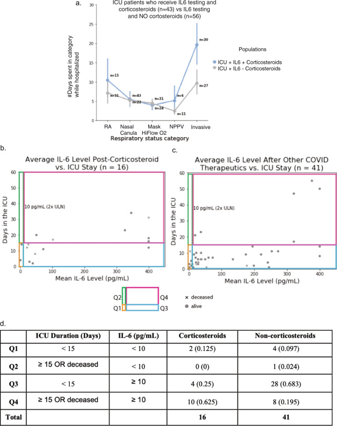
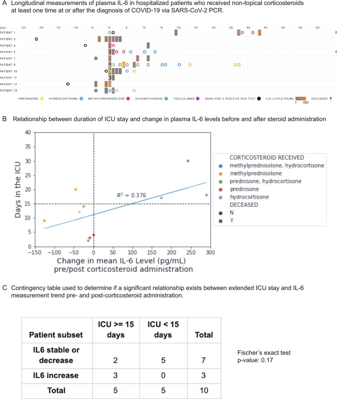
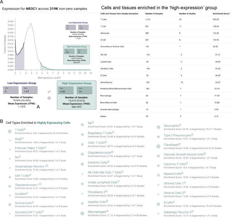
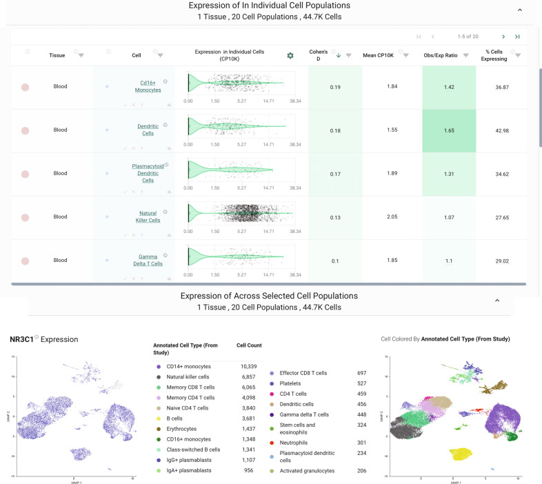
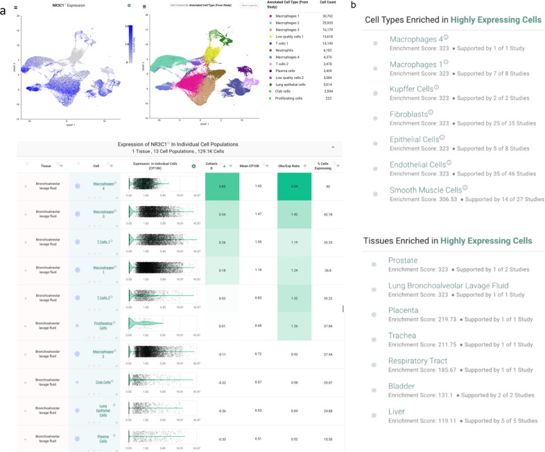
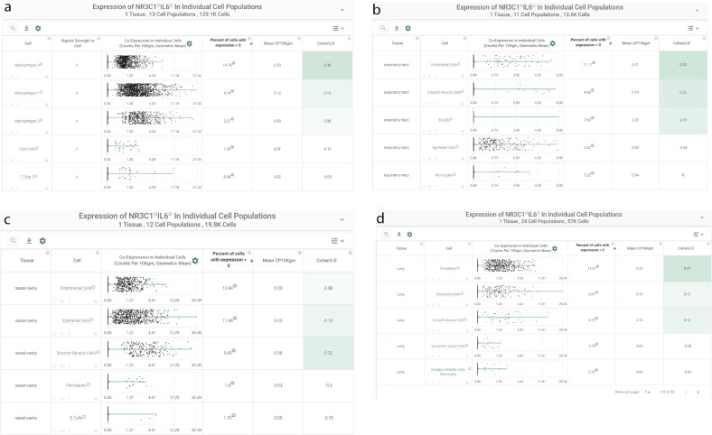
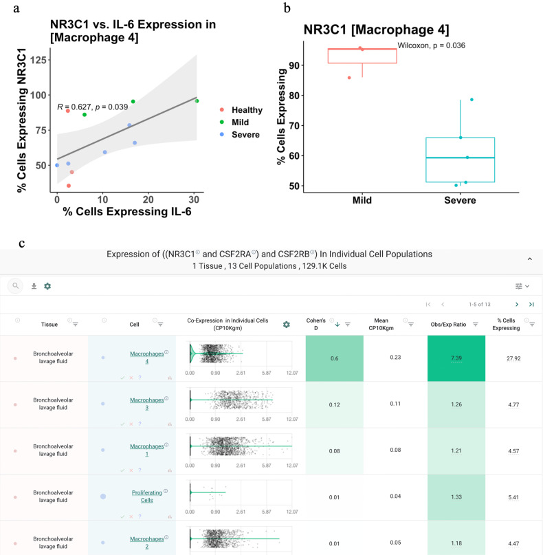
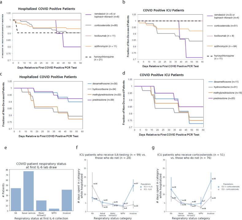
Intensive care unit (ICU) admissions and mortality in severe COVID-19 patients are driven by "cytokine storms" and acute respiratory distress syndrome (ARDS). Interim clinical trial results suggest that the corticosteroid dexamethasone displays better 28-day survival in severe COVID-19 patients requiring ventilation or oxygen. In this study, 10 out of 16 patients (62.5%) that had an average plasma IL-6 value over 10 pg/mL post administration of corticosteroids also had worse outcomes (i.e., ICU stay >15 days or death), compared to 8 out of 41 patients (19.5%) who did not receive corticosteroids (p-value = 0.0024). Given this potential association between post-corticosteroid IL-6 levels and COVID-19 severity, we hypothesized that the glucocorticoid receptor (GR or NR3C1) may be coupled to IL-6 expression in specific cell types that govern cytokine release syndrome (CRS). Examining single-cell RNA-seq data from BALF of severe COVID-19 patients and nearly 2 million cells from a pan-tissue scan shows that alveolar macrophages, smooth muscle cells, and endothelial cells co-express NR3C1 and IL-6, motivating future studies on the links between the regulation of NR3C1 function and IL-6 levels.


Conflict of interest statement

ADB is supported by Grants AI110173 and AI120698 from NIAID, 109593-62-RGRL from Amfar, and the HH Sheikh Khalifa Bin Zayed Al-Nahyan named professorship from Mayo Clinic. ADB is a consultant for Abbvie, is on scientific advisory boards for Nference and Zentalis, and is founder and President of Splissen therapeutics. WK reports grants from AstraZeneca, grants from Biogen, grants from Roche, outside the submitted work. One or more of the investigators from the Mayo Clinic associated with this project and Mayo Clinic have a Financial Conflict of Interest in the technology used in the research and that the investigator(s) and Mayo Clinic may stand to gain financially from the successful outcome of the research. This research has been reviewed by the Mayo Clinic Conflict of Interest Review Board and is being conducted in compliance with Mayo Clinic Conflict of Interest policies. The authors on this manuscript from nference have equity in nference and have a financial interest in nference.

Figures

Fig. 1
Fig. 1. Relationship between IL-6 levels and ICU duration for COVID-19 ICU patients who had at least one plasma IL-6 measurement after administration of either corticosteroids or other classes of drugs.
a Among the COVID19 patients that were admitted to ICU at some point during their hospital stay and had undergone an IL6 test, the patients that were on corticosteroids (n = 43) experienced longer durations on higher levels of respiratory support compared to the patients that were not on corticosteroids (n = 56). b Relationship between ICU duration and IL-6 levels following the administration of non-topical corticosteroids for n = 21 ICU patients who received corticosteroids. c Relationship between ICU duration and IL-6 levels following the administration of one or more of the following drugs: tocilizumab, azithromycin, hydroxychloroquine, or antivirals (n = 64 patients); these patients did not receive corticosteroids. d Summarizing the number of patients in each of the four quadrants (Q1, Q2, Q3, and Q4) in (b) and (c) above. A Fisher exact test of proportions suggests that a higher proportion of patients who receive corticosteroids and have an IL-6 value over 10 pg/mL have poor outcomes (ICU stay ≥15 days or death) when compared to the equivalent proportion of those treated with non-corticosteroid drugs including antivirals, tocilizumab, azithromycin, and hydroxychloroquine (p = 0.0006).
Fig. 2
Fig. 2. Constraining cohort of COVID-19 ICU patients to those who had at least one plasma IL-6 measurement before and after corticosteroid administration.
A Longitudinal measurements of plasma IL-6 in hospitalized patients who received non-topical corticosteroids at least one time at or after the diagnosis of COVID-19 via SARS-CoV-2 PCR. B Relationship between duration of ICU stay and change in plasma IL-6 levels before and after steroid administration (patient numbers are mapped onto Fig. 8a). If multiple pre/post-corticosteroid administration levels are available, the mean is used. C Contingency table used to determine if a significant relationship exists between extended ICU stay and IL-6 measurement trend pre- and post-corticosteroid administration.
Fig. 3
Fig. 3. Analysis of bulk tissue RNA sequencing data from gene expression omnibus reveals increased NR3C1 expression in human immune cells.
A Expression profile of NR3C1 across ~319,000 non-zero expressing bulk RNA-sequencing samples from the gene expression omnibus. Cells of the immune system are enriched among the highest expressing samples, including both lymphocytes and myeloid cells. B Single-cell RNA-sequencing expression profile of NR3C1 across ~1.9 million individual human-derived cells. Cells of the immune system are enriched among the highest expressing samples, including both lymphocytes and myeloid cells.
Fig. 4
Fig. 4. nferX single-cell platform analysis of NR3C1 expression.
In a study of peripheral blood samples from healthy donors (n = 6) and COVID-19 patients between days 2 and 16 of symptom onset (n = 7), NR3C1 was appreciably expressed in diverse immune cell types including T cells, B cells, dendritic cells, monocytes, and plasmacytoid dendritic cells.
Fig. 5
Fig. 5. Analysis of available single-cell RNA sequencing data from cells recovered from bronchoalveolar lavage fluid of COVID-19 patients.
A Expression of NR3C1 in cells from bronchoalveolar lavage fluid (BALF) of COVID-19 patients (n = 9; 3 mild, 6 severe) by single-cell RNA-sequencing. B Summary of IL-6/NR3C1 co-expression across 1.9 million human cells from single-cell RNA-sequencing datasets. Shown are cell and tissue types most highly enriched for detection of both NR3C1 and IL-6 in the same individual cells.
Fig. 6
Fig. 6. IL-6/NR3C1 co-expression by single-cell RNA-sequencing.
A Bronchoalveolar lavage fluid of COVID-19 patients; and (B)–(D) nasal cavity, respiratory tract, lungs.
Fig. 7
Fig. 7. Relationship of NR3C1 and IL-6 expression to COVID-19 status and clinical severity.
A Correlation between IL-6 and NR3C1 expression in the macrophage population found to most robustly co-express these two genes (“Macrophages 4”). Each dot represents the percentage of cells from this cluster (i.e., cell population) in which IL-6 and NR3C1 were detected, and dots are colored by COVID-19 status and clinical severity. Values shown are the Pearson correlation coefficient (“R”) and corresponding p-value (“p”). B Comparison of NR3C1 expression in the “Macrophage 4” cell population from patients with mild versus severe COVID-19. C Coexpression of NR3C1 with the two subunits of the GM-CSF receptor (CSF2RA and CSF2RB) in the bronchoalveolar lavage fluid of COVID-19 patients (n = 9; 3 mild, 6 severe). Top coexpressing populations, all of which are macrophages, are shown.
Fig. 8
Fig. 8. Survival curves and respiratory status for the study cohort.
Survival curves for (a) hospitalized and (b) ICU-COVID-19 patients who received corticosteroids, tocilizumab, azithromycin, hydroxychloroquine, or antivirals (remdesivir or lopinavir–ritonavir) following their SARS-CoV-2-positive PCR test. c Survival curves for each corticosteroid administered patient who is hospitalized, split up by the exact corticosteroid drug. d Survival curves for each corticosteroid administered patient who is in the ICU, split up by the exact corticosteroid drug, i.e., dexamethasone, hydrocortisone, methylprednisolone, or prednisone. e Bar plot of COVID-19 patient respiratory status at first IL-6 lab draw, showing that ICU patients who receive IL-6 testing are typically initially tested early in the course of respiratory failure (panel A), (f) yet spend a long time in critical respiratory failure, indicating that physicians may be using IL-6 as part of their assessment of patients who are rapidly progressing towards critical respiratory failure. Here, RA indicates that the patient is breathing room air. The nasal cannula includes all nasal cannula with oxygen flow rates up to 15 L/min. Mask/HiFlowO2 includes devices designed to increase the percentage of oxygen inspired with each breath, such as a non-rebreather, Oxymizer, OxyMask, high flow nasal cannula, etc. NPPV includes all forms of non-invasive positive pressure ventilation, such as CPAP (continuous positive airway pressure), BiPAP (bilevel positive airway pressure), and oxygen-supplemented CPAP/BiPAP. Invasive ventilation includes intubation and mechanical ventilation. g Across every respiratory status, COVID-19 patients who received corticosteroids generally are worse from a respiratory standpoint than patients who did not receive corticosteroids.

References

    1. Wagner, T. et al. Augmented curation of clinical notes from a massive EHR system reveals symptoms of impending COVID-19 diagnosis. 10.1101/2020.04.19.20067660. - PMC - PubMed
    1. Zhou F, et al. Clinical course and risk factors for mortality of adult inpatients with COVID-19 in Wuhan, China: a retrospective cohort study. Lancet. 2020;395:1054–1062. doi: 10.1016/S0140-6736(20)30566-3. - DOI - PMC - PubMed
    1. Goyal P, et al. Clinical characteristics of Covid-19 in New York city. N. Engl. J. Med. 2020;382:2372–2374. doi: 10.1056/NEJMc2010419. - DOI - PMC - PubMed
    1. COVID-19 Studies from the World Health Organization Database—ClinicalTrials.gov. https://clinicaltrials.gov/ct2/who_table.
    1. Ulhaq ZS, Soraya GV. Interleukin-6 as a potential biomarker of COVID-19 progression. Med. Mal. Infect. 2020;50:382–383. doi: 10.1016/j.medmal.2020.04.002. - DOI - PMC - PubMed