Review
. 2021 Jun 16;117(7):1645-1661.
doi: 10.1093/cvr/cvab080.

New aspects of endocrine control of atrial fibrillation and possibilities for clinical translation

Affiliations
Review

New aspects of endocrine control of atrial fibrillation and possibilities for clinical translation

Martin Aguilar et al. Cardiovasc Res. .

Abstract

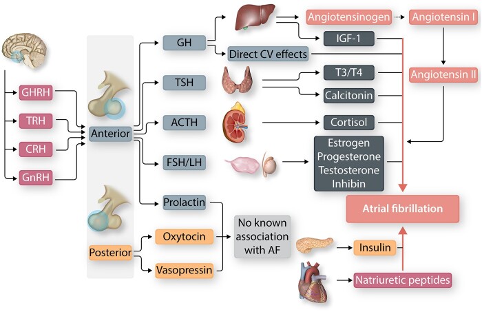
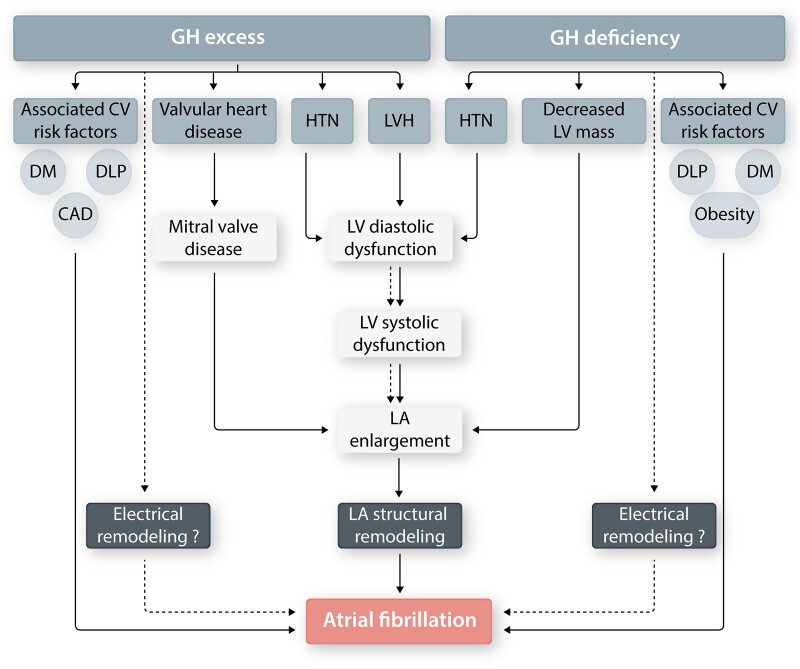
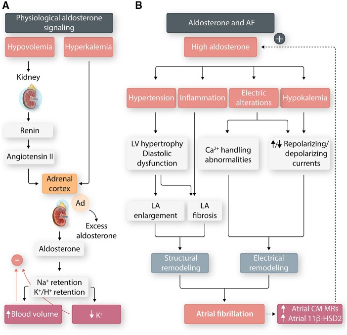
Hormones are potent endo-, para-, and autocrine endogenous regulators of the function of multiple organs, including the heart. Endocrine dysfunction promotes a number of cardiovascular diseases, including atrial fibrillation (AF). While the heart is a target for endocrine regulation, it is also an active endocrine organ itself, secreting a number of important bioactive hormones that convey significant endocrine effects, but also through para-/autocrine actions, actively participate in cardiac self-regulation. The hormones regulating heart-function work in concert to support myocardial performance. AF is a serious clinical problem associated with increased morbidity and mortality, mainly due to stroke and heart failure. Current therapies for AF remain inadequate. AF is characterized by altered atrial function and structure, including electrical and profibrotic remodelling in the atria and ventricles, which facilitates AF progression and hampers its treatment. Although features of this remodelling are well-established and its mechanisms are partly understood, important pathways pertinent to AF arrhythmogenesis are still unidentified. The discovery of these missing pathways has the potential to lead to therapeutic breakthroughs. Endocrine dysfunction is well-recognized to lead to AF. In this review, we discuss endocrine and cardiocrine signalling systems that directly, or as a consequence of an underlying cardiac pathology, contribute to AF pathogenesis. More specifically, we consider the roles of products from the hypothalamic-pituitary axis, the adrenal glands, adipose tissue, the renin-angiotensin system, atrial cardiomyocytes, and the thyroid gland in controlling atrial electrical and structural properties. The influence of endocrine/paracrine dysfunction on AF risk and mechanisms is evaluated and discussed. We focus on the most recent findings and reflect on the potential of translating them into clinical application.

Keywords: Arrhythmia; Atrial fibrillation; Endocrine system; Heart.


Figures

Figure 1
Figure 1
Summary of the endocrine glands and hormones associated with atrial fibrillation (AF). The anterior pituitary secretes tropic hormones in response to hypothalamic stimulation. Tropic hormones stimulate the release of physiologically active hormones from their target organ(s). The posterior pituitary hormones do not have known direct electrophysiological effects. ACTH, adrenocorticotropic hormone; CRH, corticotropin-releasing hormone; CV, cardiovascular; FSH, follicle-stimulating hormone; GH, growth hormone; GHRH, GH releasing-hormone; GnRH, gonadotropin-releasing hormone; IGF-1, insulin-like growth factor-I; LH, luteinizing hormone; T3, triiodothyronine; T4, tetraiodothyronine; TRH, thyrotropin-releasing hormone.
Figure 2
Figure 2
Growth hormone (GH) and AF. GH excess and deficiency have both been associated with an increased risk of AF. Chronic GH excess leads to left-ventricular (LV) diastolic dysfunction and left-atrial (LA) enlargement, contributing to pro-arrhythmic LA structural remodelling. In addition, GH deficiency has been associated with decreased LV mass and LV systolic dysfunction. GH dysregulation also often co-exists with pro-AF cardiovascular (CV) risk factors [i.e. hypertension (HTN), diabetes (DM), dyslipidaemia (DLP), coronary artery disease (CAD)]. The effect of GH excess and/or deficiency, if any, on LA electrophysiology (electrical remodelling) is not known.
Figure 3
Figure 3
Hyperaldosteronism and AF. (A) Hypovolemia and hyperkalaemia are the primary physiological stimuli for adrenal aldosterone secretion, which acts on the nephron distal tubule and collecting duct to retain Na+ and excrete K+. (B) Mechanism of aldosterone-related AF. Hyperaldosteronism causes angiotensin-independent hypertension and left atrial (LA) inflammation, leading to pro-fibrillatory LA remodelling. It also produces pro-AF electrical remodelling in the form of LA action potential-shortening, increased sarcoplasmic reticulum Ca2+ sparks, and delayed afterdepolarizations. Sustained AF may potentiate the effects of hyperaldosteronism by upregulating of the mineralocorticoid receptor (MR) on AF atrial cardiomyocytes (CMs). Furthermore, AF increases 11b-hydroxysteroid dehydrogenase type 2 (11b-HSD2), which metabolizes cortisol, thereby increasing MR occupancy by aldosterone.
Figure 4
Figure 4
Current understanding of the mechanistic links between obesity and AF. The corporal load model states that excess body mass (adipose and/or lean) poses a haemodynamic load culminating in pro-AF left-atrial (LA) remodelling. Obesity has also been associated with pro-fibrillatory electrical remodelling in the form of shorter effective refractory period (ERP), slower conduction velocity (CV) and increased conduction heterogeneity. Adipocytes have a pro-inflammatory secretome which can affect LA electrophysiology indirectly (systemic adiposity) or directly (epicardial adiposity). Finally, obesity often co-exists with a number of pro-AF cardiovascular risk factors.
Figure 5
Figure 5
Atrial electrical and structural remodelling in angiotensin-II mediated hypertension and in mouse models of type-1 (T1DM) and type-2 (T2DM) diabetes mellitus. (A) Angiotensin-II infusion in mice causes distinct patterns of ion channel remodelling and changes in action potential morphology in left and right atrial myocytes. Angiotensin-II also causes right and LA fibrosis. These alterations lead to conduction abnormalities and increased susceptibility to AF. Loss of NPR-C leads to worsened ion channel remodelling and atrial fibrosis, as well as enhanced AF susceptibility, while NPR-C activation prevents some ion channel remodelling, reduces right and LA fibrosis, and decreases AF burden. (B) T1DM (Akita mice) is associated with reductions in AP Vmax due to reduction in atrial INa as well as increases in AP duration due to reduction in IKur. T2DM (db/db mice) show increases in AP duration due to reduction in both Ito and IKur while INa amplitude and AP Vmax are not altered. Both T1DM and T2DM are associated with increased atrial fibrosis. These alterations lead to conduction abnormalities and increased susceptibility to AF. Insulin treatment in T1DM prevents reductions in atrial INa and reduces atrial fibrosis leading to improved conduction and reduced AF susceptibility.
Figure 6
Figure 6
Thyroid dysfunction and AF. (A) The hypothalamic-pituitary-thyroid axis forms a closed negative-feedback system. The thyroid secretes primarily T4, which is converted to T3, the more metabolically active thyroid hormone, by the enzyme 5′-ionidase. Thyroid hormone effects can be genomic or non-genomic. Genomic effects are mediated by binding of T3 to the nuclear thyroid α-receptor-1 (TRα1), which interacts with the thyroid release elements (TREs) to promote/suppress thyroid hormone-regulated genes. Non-genomic effects are mediated by T3 and T4 as they interact with extra-nuclear receptors, which may or may not be structurally related to the thyroid receptor. (B) Hyperthyroidism leads to high-output heart failure (HF), causing left atrial enlargement (LAE), activation of the renin–angiotensin–aldosterone system (RAAS), and increased adrenergic stimulation. Altered intracellular Ca2+ promotes the formation of early (EADs) and delayed afterdepolarizations (DADs) from the pulmonary veins (PVs). Action potential duration (APD) shortening promotes re-entry. Finally, hypertension (HTN) also contributes to pro-fibrillatory left-atrial structural remodelling. TRH, thyrotropin-releasing hormone; TSH, thyroid-stimulating hormone; DNA, deoxyribonucleic acid.
Figure 7
Figure 7
Calcitonin signalling and AF-induced remodelling. (A) In healthy hearts, atrial cardiomyocytes secrete calcitonin, which binds to the calcitonin-receptors (CTRs) on atrial fibroblasts, controlling extracellular matrix deposition and helping to maintain normal sinus rhythm. In AF, calcitonin signalling is disordered by reduced secretion of calcitonin by atrial cardiomyocytes and reduced calcitonin-receptor responsiveness; these changes impede the calcitonin-mediated brake on fibrogenesis causing atrial fibrosis and increased arrhythmogenesis (created with Biorender.com). (B) CTRs (green) in atrial fibroblasts co-stained with filamin A (red) and 4′,6-diamidino-2-phenylindole (DAPI) (blue). Relocalization of CTRs from the cell membrane to the nucleus explains CTR hyporesponsiveness. Scale bar = 50 μm; adapted from Moreira et al.

References

    1. Carpenter PC. Diagnostic evaluation of Cushing's syndrome. Endocrinol Metab Clin North Am 1988;17:445–472. - PubMed
    1. van der Hooft CS, Heeringa J, Brusselle GG, Hofman A, Witteman JC, Kingma JH, Sturkenboom MC, Stricker BH. Corticosteroids and the risk of atrial fibrillation. Arch Intern Med 2006;166:1016–1020. - PubMed
    1. Di Dalmazi G, Vicennati V, Pizzi C, Mosconi C, Tucci L, Balacchi C, Cosentino ER, Paolisso P, Fanelli F, Gambineri A, Pelusi C, Repaci A, Garelli S, Galie N, Borghi C, Golfieri R, Pagotto U. Prevalence and incidence of atrial fibrillation in a large cohort of adrenal incidentalomas: a long-term study. J Clin Endocrinol Metab 2020;105. - PubMed
    1. Neary NM, Booker OJ, Abel BS, Matta JR, Muldoon N, Sinaii N, Pettigrew RI, Nieman LK, Gharib AM. Hypercortisolism is associated with increased coronary arterial atherosclerosis: analysis of noninvasive coronary angiography using multidetector computerized tomography. J Clin Endocrinol Metab 2013;98:2045–2052. - PMC - PubMed
    1. Mancini T, Kola B, Mantero F, Boscaro M, Arnaldi G. High cardiovascular risk in patients with Cushing's syndrome according to 1999 WHO/ISH guidelines. Clin Endocrinol 2004;61:768–777. - PubMed

Publication types

MeSH terms