. 2022 Apr;38(2):203-222.
doi: 10.1007/s10565-021-09592-2. Epub 2021 Mar 15.

Dimethyl sulfoxide stimulates the AhR-Jdp2 axis to control ROS accumulation in mouse embryonic fibroblasts

Affiliations

Dimethyl sulfoxide stimulates the AhR-Jdp2 axis to control ROS accumulation in mouse embryonic fibroblasts

Kenly Wuputra et al. Cell Biol Toxicol. 2022 Apr.

Abstract

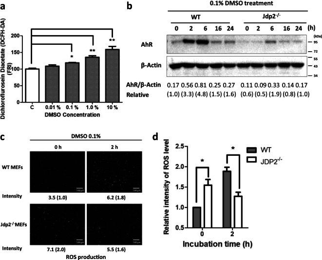
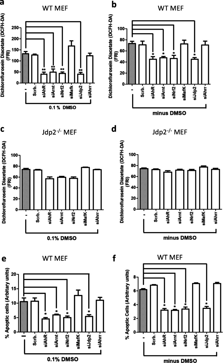
The aryl hydrocarbon receptor (AhR) is a ligand-binding protein that responds to environmental aromatic hydrocarbons and stimulates the transcription of downstream phase I enzyme-related genes by binding the cis element of dioxin-responsive elements (DREs)/xenobiotic-responsive elements. Dimethyl sulfoxide (DMSO) is a well-known organic solvent that is often used to dissolve phase I reagents in toxicology and oxidative stress research experiments. In the current study, we discovered that 0.1% DMSO significantly induced the activation of the AhR promoter via DREs and produced reactive oxygen species, which induced apoptosis in mouse embryonic fibroblasts (MEFs). Moreover, Jun dimerization protein 2 (Jdp2) was found to be required for activation of the AhR promoter in response to DMSO. Coimmunoprecipitation and chromatin immunoprecipitation studies demonstrated that the phase I-dependent transcription factors, AhR and the AhR nuclear translocator, and phase II-dependent transcription factors such as nuclear factor (erythroid-derived 2)-like 2 (Nrf2) integrated into DRE sites together with Jdp2 to form an activation complex to increase AhR promoter activity in response to DMSO in MEFs. Our findings provide evidence for the functional role of Jdp2 in controlling the AhR gene via Nrf2 and provide insights into how Jdp2 contributes to the regulation of ROS production and the cell spreading and apoptosis produced by the ligand DMSO in MEFs.

Keywords: AhR; Dimethyl sulfoxide; Jun dimerization protein 2; Mouse embryonic fibroblasts; Nrf2; Reactive oxygen species.


Conflict of interest statement

The authors declare no competing interests.

Figures

Fig. 1
Fig. 1
Extension of actin stress fibers and cell spreading of WT and AhR−/− MEFs after exposure to DMSO. a Cells were starved of serum overnight before DMSO treatment for 24 h and exposed to 0.1% DMSO for 2 h. At the end of treatment, cells were rinsed with PBS, fixed with 4% formaldehyde, and then processed for F-actin staining and phosphorylated myosin light chain staining as described in the “Materials and methods” section. Nuclei were stained with DAPI, as indicated by blue fluorescence and the images with each of the three fluorescent colors merged. b Quantification of signaling of F-actin fibers. Five fields were examined, and the fluorescence intensity was quantified. WT MEFs and AhR−/− MEFs in the presence and absence of 0.1% DMSO for 2-h treatment. Cell areas with actin fibers were calculated as described in the “Materials and methods” section. Data represent the mean ± SEM (n = 5), **P < 0.01. c Quantification of signaling of pMLC2. Five fields were examined, and the fluorescence intensity was quantified. WT MEFs and AhR−/− MEFs in the presence and absence of 0.1% DMSO for 2-h treatment. Cell areas with pMLC2 were calculated as described in the “Materials and methods” section. Data represent the mean ± SEM (n = 5), ** P < 0.01. Representative immunoblots d and quantitative results e for cells harvested 2 h after 0.1% DMSO treatment. Data are presented as mean ± SEM (n = 5). All statistical analysis was performed by Student’s t-test (** P < 0.01)
Fig. 2
Fig. 2
Measurement of ROS activity in WT MEFs in response to DMSO. a MEFs incubated with 0 or 0.01, 0.1, 1.0, and 10.0% DMSO for 2 h were stained with 25 micro M CM-H2-DCFDA and examined by flow cytometry as described in the “Materials and methods” section. Data are presented as mean ± SEM (n = 5, in triplicate). ROS levels in the control incubation without DMSO were arbitrarily set to 100. (* P < 0.05, ** P < 0.01 ). b Comparison of the levels of AhR protein in WT and Jdp2−/− MEFs after incubation with 0.1% DMSO for 0, 2, 6, 16, and 24 h. c Comparative ROS activity was measured in WT and Jdp2−/− MEFs in response to 0.1% DMSO for 2 h. ROS production was detected using CM-H2DCFDA as described in the “Materials and methods” section. Representative fluorescence images of ROS generation in WT (top) and Jdp2−/− (bottom) MEFs are shown. d Collective fluorescence images of ROS levels detected using CM-H2DCFDA after treatment with DMSO were analyzed using ImageJ software. The fluorescence of WT MEFs without DMSO exposure is taken as 1.0. Values represent mean ± SEM (n = 6). Statistical analysis was performed by two-way ANOVA with Bonferroni posttests (*P < 0.05)
Fig. 3
Fig. 3
Characterization of AhR promoter activity. a Schematic representation of the positions of each DRE and ARE in the AhR promoter region. Each element is shown at the position from the putative transcription start site, which was mutated to generate the mutants of DRE and ARE. b Relative activity of pGL4.1-AhR-luciferase in WT and Jdp2−/− MEFs treated with 0.1% DMSO for 0, 2, 6, and 16 h. Luciferase activities were calculated as the ratio of the AhR-luciferase activity to that of the control pGL4.1 and expressed as relative luciferase activity. Values represent the mean ± SEM of five independent measurements. Statistical analysis was done by two-way ANOVA with Bonferroni posttests (* P < 0.05, ** P < 0.01). c–f Effects of the mutation of each cis element, i.e., ARE1, ARE2, DRE1, DRE2, and DRE3, on the AhR promoter region. Luciferase activity was measured in WT MEFs (c, e) and Jdp2−/− MEFs (d, f) in the absence (c, d) or presence of 0.1% DMSO (e, f) as described in the “Materials and methods” section. The luciferase activity of full length (FL) AhR-luciferase was arbitrarily set to 1.0. Values represent the mean ± SEM (n = 5). Statistical analysis was performed by one-way ANOVA with Tukey’s test (** P < 0.01). g, h Effect of siRNAs against each representative transcription factor (AhR, Arnt, Nrf2, MafK, Jdp2, and Ahrr) on pGL4.1-AhR-luciferase activity in WT MEFs in the absence (g) or presence (h) of 0.1% DMSO. The luciferase activity of FL AhR-luciferase was arbitrarily set to 1.0. Values represent the mean ± SEM (n = 3). Statistical analysis was performed by one-way ANOVA with Tukey’s test (** P < 0.01)
Fig. 4
Fig. 4
Regulation of production of ROS and apoptotic activity in WT and Jdp2−/− MEFs by siRNA against AhR, Arnt, Ahrr, Nrf2, Jdp2, and MafK. a–d WT (a, b) and Jdp2−/− MEFs (c, d) incubated with various siRNAs against AhR, Arnt, Nrf2, Ahrr, Jdp2, and MafK without (b, d) and with 0.1% DMSO (a, c) for 2 h were stained with 0.25 M CM-H2DCFDA and examined by flow cytometry as described in the “Materials and methods” section. Data are presented as mean ± SEM (n = 3, in triplicate). Statistical analysis was done by one-way ANOVA with Tukey’s test (* P < 0.05, ** P < 0.01 ). e, f Effect of siRNA against each representative transcription factor (AhR, Arnt, Nrf2, MafK, Jdp2, and Ahrr) on the apoptotic activity in WT MEFs in the presence (e) and absence (f) of 0.1% DMSO. Values represent the mean ± SEM (n = 3). Statistical analysis was performed by one-way ANOVA with Tukey’s test (*P < 0.05)
Fig. 5
Fig. 5
Differential recruitments of transcription factors such as AhR, Arnt, Nrf2, and MafK to ARE and DRE sites in WT and Jdp2−/− MEFs. a Schematic representation of the mouse AhR promoter and the position of cis elements, such as ARE1, ARE2, DRE1, and DRE2/3, which were detected in the ChIP assay. a–d; f–h The regions that were amplified by PCR with the specific corresponding primers (ARE1, ARE2 and DRE1) and (e, i) with the primers that contained the DRE 2 and 3 cis elements were indicated in WT MEFs. The ChIP-qPCR analyses were performed using chromatin extracts from WT (b–e) and Jdp2−/− MEFs (f–i) with the indicated antibodies and normal IgG (as a negative control). The probes of ARE1 (b, f), ARE2 (c, g), DRE1 (d, h), and DRE2/3 (e, i) are shown in the presence of 0.1% DMSO, respectively. The values in the absence of DMSO are detailed in Fig. S6. Values represent mean ± SEM (n = 5). Statistical analysis was performed by one-way ANOVA with Tukey’s test (** P < 0.01)
Fig. 6
Fig. 6
Effect of Jdp2, MafK, and Nrf2 on AhR-luciferase activity via DRE2 and DRE3 in Jdp2−/− MEFs. a AhR-luciferase, DRE2 mutant-luciferase, and DRE3 mutant-luciferase (50 ng, respectively) plus 0–200 ng of pcDNA-Jdp2 were transfected into Jdp2−/− MEFs. One day after transfection, 0.1% DMSO was added for 2 h, cells were collected, and luciferase activity was measured as described in the “Materials and methods” section. b AhR-luciferase, DRE2 mutant-luciferase, and DRE3 mutant-luciferase (50 ng, respectively) plus 0–200 ng of pcDNA-Nrf2 were transfected into Jdp2−/− MEFs. One day after transfection, 0.1% DMSO was added for 2 h, cells were harvested, and luciferase activity was measured as described in the “Materials and methods” section. Data represent the mean ± SEM (n = 5). c AhR WT-luciferase, DRE2 mutant-luciferase, and DRE3 mutant-luciferase (50 ng, respectively) plus 0–200 ng of pcDNA-MafK were transfected into Jdp2−/− MEFs. One day after transfection, 0.1% DMSO was added for 2 h, cells were collected, and luciferase activity was measured as described in the “Materials and methods” section. All statistical analysis was performed by two-way ANOVA with Bonferroni posttests (* P < 0.05, ** P < 0.01)
Fig. 7
Fig. 7
Jdp2 interacts with AhR and Nrf2 in nuclei and rescue of JdP2−/− MEFs by AhR. a Cell lysates (300 μg) from cytosolic and nuclear fractions of WT and Jdp2−/− MEFs were immunoprecipitated with antibodies against AhR, and the bound proteins were blotted with antibodies against Nrf2, Arnt, AhR, and Jdp2, as described in the “Materials and methods” section. IgG, as a negative control. b Cell lysates (300 μg) from cytosolic and nuclear fractions from WT MEFs were immunoprecipitated with anti-Jdp2 antibodies, and the bound proteins were blotted with anti-Nrf2, anti-AhR, and anti-Jdp2 antibodies, as described in the “Materials and methods” section. IgG was used as a negative control. c Rescue of DMSO-induced cell spreading by overexpression of Jdp2, its mutant, and AhR. Rescue of 2-h DMSO-induced cell spreading (c) and apoptosis (e) by increasing doses (50, 100, and 200 ng) of pcDNA-Flag-Jdp2, pcDNA-FlagJdp2-FL34R, and pcDNA-HA-AhR in Jdp2−/− MEFs. The fluorescence intensity in Jdp2−/− MEFs without DMSO exposure was set as 1.0. Data represent the mean ± SEM (n = 5; ** P < 0.01). d Representative results of western blot for the tagged proteins of Jdp2, Jdp2FL34R mutant, and AhR in Jdp2−/− MEFs transformants by pcDNA-Flag-Jdp2, pcDNA-FlagJdp2-FL34R, and pcDNA-HA-AhR, respectively. f Schematic representation of DMSO-induced AhR activation through the complex of AhR, Nrf2, and Jdp2 to increase ROS production, cell spreading, and apoptosis in WT MEFs. In Jdp2−/− MEFs, only a residual amount of AhR-Arnt was recruited to the DRE2 and DRE3 elements of the AhR promoter.

Similar articles

Cited by

References

    1. Aronheim A, Zandi E, Hennemann H, Elledge SJ, Karin M. Isolation of an AP-1 repressor by a novel method for detecting protein-protein interactions. Mol Cell Biol. 1997;17(6):3094–3102. doi: 10.1128/mcb.17.6.3094. - DOI - PMC - PubMed
    1. Barbarov Y, Timaner M, Alishekevitz D, Hai T, Yokoyama KK, Shaked Y, et al. Host JDP2 expression in the bone marrow contributes to metastatic spread. Oncotarget. 2015;6(35):37737–37749. doi: 10.18632/oncotarget.5648. - DOI - PMC - PubMed
    1. Barouki R, Coumoul X, Fernandez-Salguero PM. The aryl hydrocarbon receptor, more than a xenobiotic-interacting protein. FEBS Lett. 2007;581(19):3608–3615. doi: 10.1016/j.febslet.2007.03.046. - DOI - PubMed
    1. Beg AA, Sha WC, Bronson RT, Ghosh S, Baltimore D. Embryonic lethality and liver degeneration in mice lacking the RelA component of NF-kappa B. Nature. 1995;376(6536):167–170. doi: 10.1038/376167a0. - DOI - PubMed
    1. Birnbaum MRE, Harris MW. Teratogenic effects of 2,3,7,8-tetrabromodibenzo-p-dioxin and three polybrominated dibenzofurans in C57BL/6N mice. Toxicol Appl Pharmacol. 1991;107(1):141–152. doi: 10.1016/0041-008x(91)90338-f. - DOI - PubMed

Publication types

MeSH terms

LinkOut - more resources