. 2021 Oct;37(5):751-771.
doi: 10.1007/s10565-021-09596-y. Epub 2021 Mar 15.

The DNA methylation inhibitor RG108 protects against noise-induced hearing loss

Affiliations

The DNA methylation inhibitor RG108 protects against noise-induced hearing loss

Zhiwei Zheng et al. Cell Biol Toxicol. 2021 Oct.

Abstract

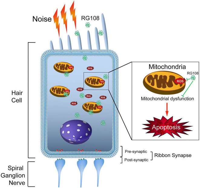
Background: Noise-induced hearing loss represents a commonly diagnosed type of hearing disability, severely impacting the quality of life of individuals. The current work is aimed at assessing the effects of DNA methylation on noise-induced hearing loss.

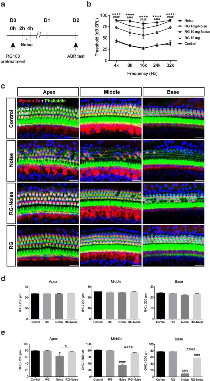
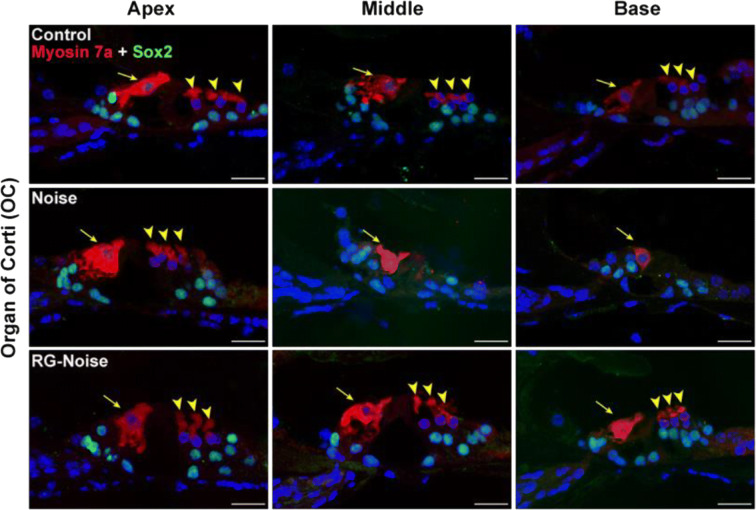
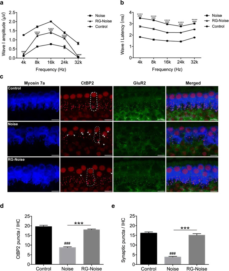
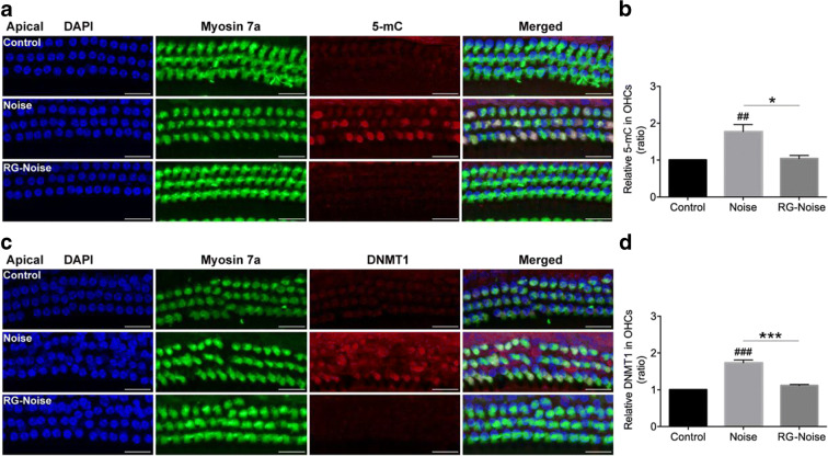
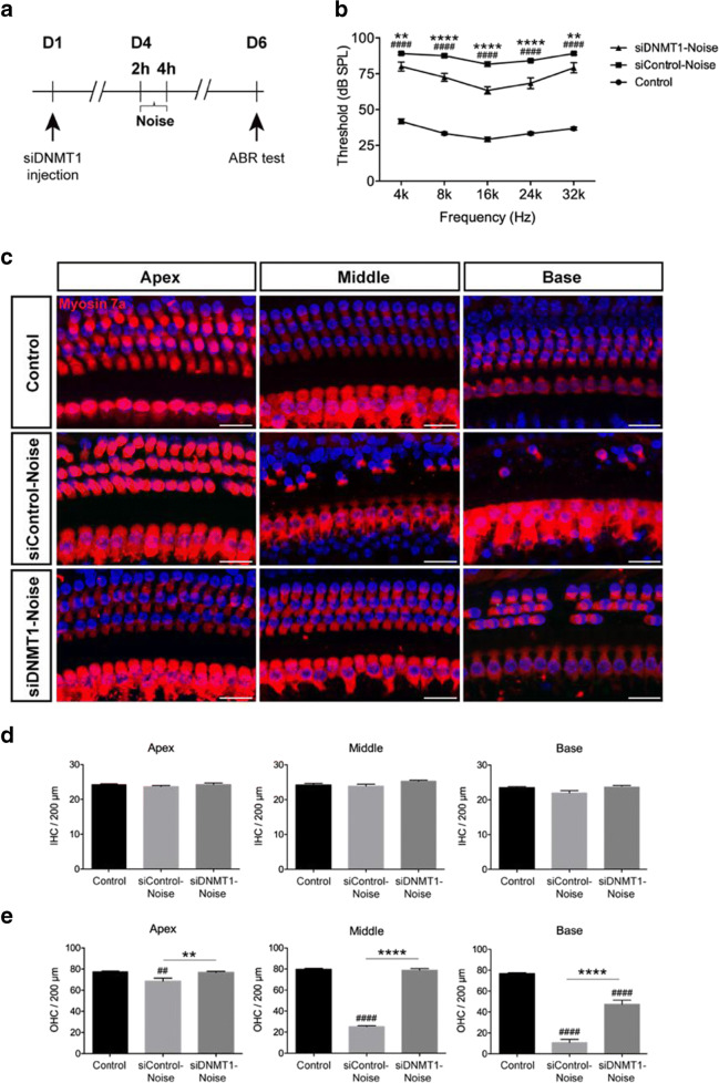
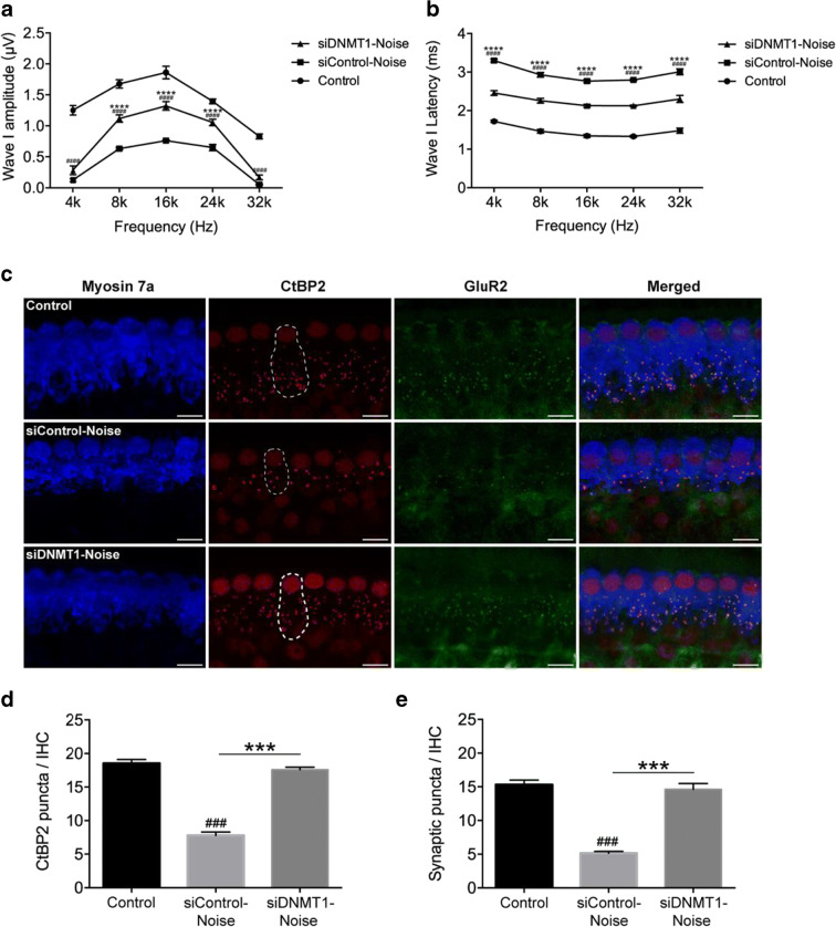
Methods: Blocking DNA methyltransferase 1 (DNMT1) activity with a selective inhibitor RG108 or silencing DNMT1 with siRNA was used in this study. Auditory brainstem responses were measured at baseline and 2 days after trauma in mice to assess auditory functions. Whole-mount immunofluorescent staining and confocal microcopy of mouse inner ear specimens were performed to analyze noise-induced damage in cochleae and the auditory nerve at 2 days after noise exposure.

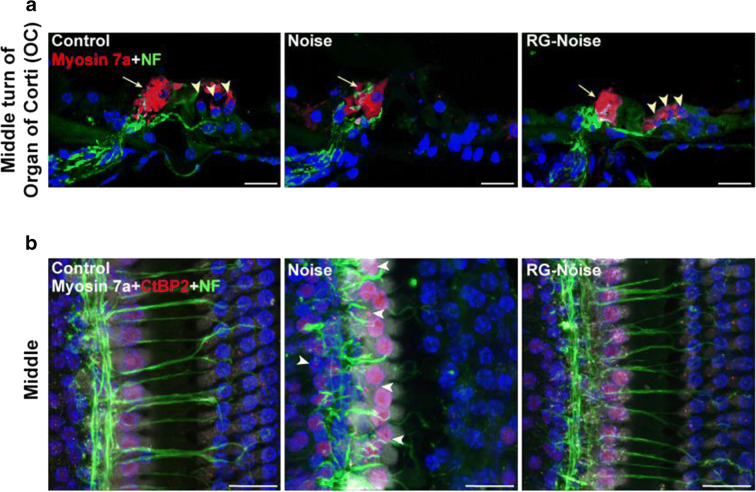
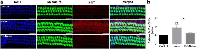
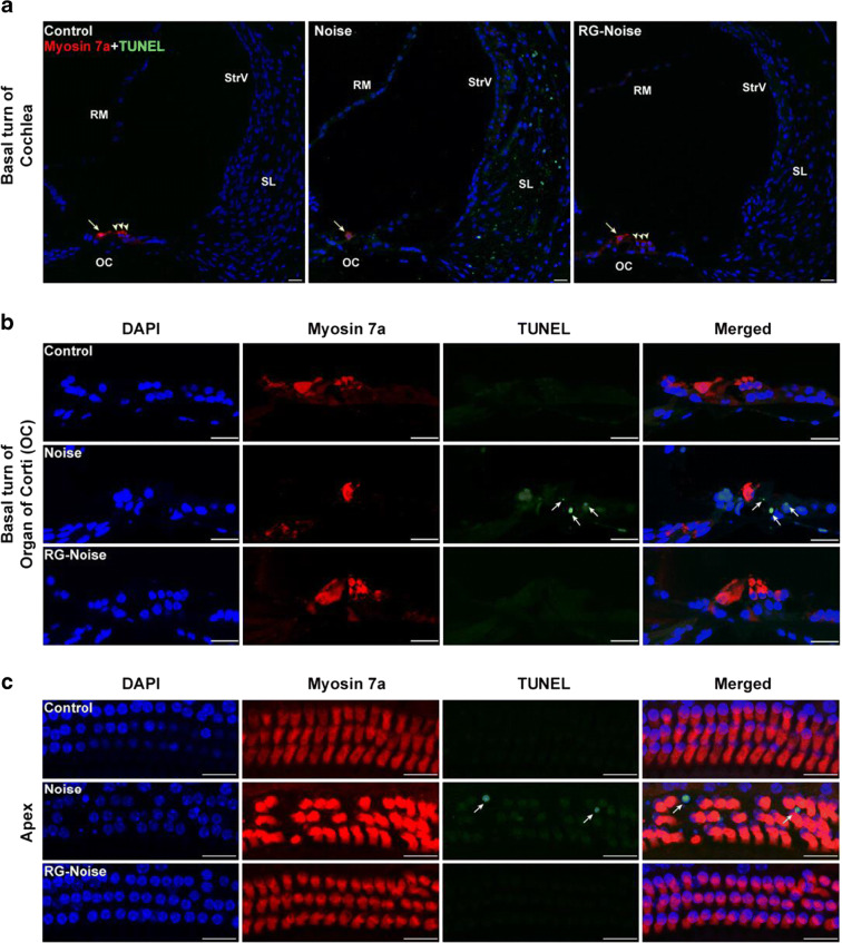
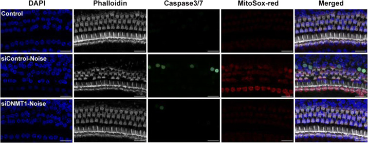
Results: The results showed that noise exposure caused threshold elevation of auditory brainstem responses and cochlear hair cell loss. Whole-mount cochlea staining revealed a reduction in the density of auditory ribbon synapses between inner hair cells and spiral ganglion neurons. Inhibition of DNA methyltransferase activity via a non-nucleoside specific pharmacological inhibitor, RG108, or silencing of DNA methyltransferase-1 with siRNA significantly attenuated ABR threshold elevation, hair cell damage, and the loss of auditory synapses.

Conclusions: This study suggests that inhibition of DNMT1 ameliorates noise-induced hearing loss and indicates that DNMT1 may be a promising therapeutic target. Graphical Headlights • RG108 protected against noise-induced hearing loss • RG108 administration protected against noise-induced hair cell loss and auditory neural damage. • RG108 administration attenuated oxidative stress-induced DNA damage and subsequent apoptosis-mediated cell loss in the cochlea after noise exposure.

Keywords: Cochlea; DNA methylation; Hair cells; Noise-induced hearing loss.


Conflict of interest statement

The authors declare no competing interests.

Figures

None
Graphical Headlights • RG108 protected against noise-induced hearing loss • RG108 administration protected against noise-induced hair cell loss and auditory neural damage. • RG108 administration attenuated oxidative stress-induced DNA damage and subsequent apoptosis-mediated cell loss in the cochlea after noise exposure.
Fig. 1
Fig. 1
Assessment of ABR threshold shifts and hair cell (HC) loss at 2 days after noise exposure. (a) Experimental schedule. Mice pre-injected with saline or RG108 were exposed to 120-dB noise for 2 h, and the ABR thresholds were measured at 4, 8, 16, 24, and 32 kHz at 2 days after noise exposure. (b) Noise exposure caused elevation of ABR thresholds across tested frequencies, while pretreatment with 10 mg RG108 significantly attenuated the noise-induced ABR threshold shifts at each tested frequency. n = 6 mice for each condition. Data are mean ± SEM. Statistical analysis was performed using a two-way ANOVA, ####p < 0.0001 for noise vs. control, ****p < 0.0001 for noise vs. 10 mg RG108-Noise. (c) Representative images of myosin 7a and phalloidin staining of cochlear turns from different groups at 2 days after noise exposure. OHC1, OHC2, OHC3: 1st, 2nd, and 3rd rows of outer hair cells (OHCs); inner hair cell (IHC). Scale bar = 20 μm. (d, e) Quantitative analysis of noise-induced losses of IHC (d) and OHC (e) along the cochlear explants. Total IHC or OHC count per 200 μm was compared among no noise (control), 10 mg RG108 (RG), noise, and 10 mg RG108-Noise (RG-Noise) groups. A significant loss of OHC was observed at both base and middle turns in noise-exposed mice compared with no noise control mice. In contrast, OHC loss in the RG108 pretreatment group was significantly lower than that in the noise exposure group, but still higher than in the no noise control group (except middle and apex turns). No significant differences were observed in IHCs loss among these groups. n = 6 mice for each condition. Data are mean ± SEM. Statistical analysis was performed using a one-way ANOVA, #p < 0.05, ####p < 0.0001 for noise vs. control; *p < 0.05 and ****p < 0.0001 for noise vs. RG-Noise. IHC, inner hair cell; OHCs, outer hair cells
Fig. 2
Fig. 2
Assessment of hair cell (HC) loss at 2 days after noise exposure. Immunostaining was performed in the inner ear sections of mice from different groups. Representative images showed reduction in immunoreactivity for myosin 7a (red) and Sox2 (supporting cell marker, green) in the organ of Corti (OC) after noise exposure compared to controls without exposure. Pretreatment with RG108 significantly attenuated noise-induced decrease of OHCs and supporting cells. Images were taken from the apical, middle, and basal turns of cochlea in the inner ear sections. The yellow arrowheads point to three rows of outer hair cells, and the yellow arrow indicates an inner hair cell. Scale bar =20 μm
Fig. 3
Fig. 3
Assessment of noise-induced losses of synapses. (a, b) Representative ABR wave I amplitudes and latencies at 4, 8, 16, 24, and 32 kHz in mice for each condition. ABR wave I amplitudes in RG108 pretreated mice were significantly increased compared to the noise exposed mice. Threshold adjusted ABR wave I latency measurements demonstrated that RG108 pretreatment prevented increase of wave I latency induced by noise when tested 2 days after the noise exposure. n = 6 mice for each condition. Data are mean ± SEM. Statistical analysis was performed using a two-way ANOVA, ####p < 0.0001 for noise vs. control, ****p < 0.0001 for noise vs. RG-Noise. (c) Representative confocal images of immunolabeling for presynaptic ribbons (CtBP2-red), postsynaptic receptor patches (GluR2-green), and IHCs (myosin 7a-blue) in the middle turn corresponding to 16 kHz in each group (no noise-exposed cochlea; noise-exposed cochlea; RG108-treated cochlea at 2 days post-noise exposure). The white dash lines indicate the outline of IHCs. The CtBP2 antibody also stains hair cell nuclei. The white arrowheads point to ribbons that are far from the basolateral surface of the IHC toward the perinuclear positions. The white arrow indicates abnormally large ribbons in the exposed ear. Scale bar = 10 μm. (d, e) Quantitative data showed that noise reduced, and RG108 pretreatment increased, the number of CtBP2-immunolabeled ribbons (d) and paired ribbon puncta, defined as juxtaposed CtBP2- and GluR2-positive puncta (e) at 16 kHz cochlear region; n = 24 IHCs from four cochleae of each group. Data are mean ± SEM. ###p < 0.001 for noise vs. control; ***p < 0.001 for noise vs. RG108-Noise
Fig. 4
Fig. 4
Assessment of noise-induced synaptic degeneration. (a) Analysis for cochlear nerve terminals (neurofilament, NF-green) and HCs (myosin 7a-red) in mice for each condition. Images were taken from the middle turn of cochlea in the inner ear sections. The yellow arrowheads point to three rows of outer hair cells, and the yellow arrow indicates an inner hair cell. Scale bar = 20 μm. (b) Assessment of synaptic ribbons (CtBP2-red), cochlear nerve terminals (neurofilament, NF-green), and HCs (myosin 7a-white) in mice for each condition. Images were obtained from the middle cochlear turn. The white arrowheads in noise image point to ribbons are not paired with auditory nerve terminals, and some appear far from the basolateral surface of the IHC. Scale bar = 20 μm
Fig. 5
Fig. 5
Immunofluorescence analyses of oxidative stress-associated 3-nitrotyrosine (3-NT) expression in OHCs. (a) Representative images of myosin 7a (green) and 3-NT (red) staining of apical cochlear turns from different groups at 2 days after noise exposure. RG108 treatment reduced the noise-increased 3-NT level in OHCs. Scale bar = 20 μm. (b) Quantification of 3-NT staining in OHCs confirmed a significant reduction with RG108 administration. n = 3 per group with one cochlea used per mouse. Data are mean ± SEM. ##p < 0.01 for noise vs. control; *p < 0.05 for noise vs. RG-Noise
Fig. 6
Fig. 6
Detection of apoptotic cell death by TUNEL assay in the cochlea. (a, b) Immunostaining was performed in the inner ear sections of mice from different groups. Representative images showed an increase in immunoreactivity for TUNEL (green) in the spiral ligament (SL) and organ of Corti (OC) after noise exposure compared to controls without exposure. RG108 pretreatment significantly attenuated noise-induced apoptosis. Images were obtained from the basal turn of cochlea. The yellow arrowheads in (a) point to three rows of outer hair cells, and the yellow arrow indicates an inner hair cell. The white arrows in (b) point to TUNEL-positive cells. Scale bar = 20 μm. (c) Representative images of TUNEL staining of cochlea from different groups at 2 days after noise exposure. Images were obtained from the apical cochlear region. Scale bar = 20 μm. OC: organ of Corti; RM: Reissner’s membrane; SL: spiral ligament; StrV: stria vascularis
Fig. 7
Fig. 7
Immunofluorescence analyses of 5-mC and DNMT1 expression in OHCs. (a) Representative images showed an increase in immunoreactivity for 5-mC (red) in OHCs stained with myosin 7a (green) after noise exposure compared to controls without exposure. RG108 pretreatment significantly attenuated noise-induced 5-mC in OHCs. Images were taken from the apical turn. Scale bar =20 μm. (b) Quantification of 5-mC staining in OHCs confirmed a significant reduction with RG108 administration. n = 4 per group with one cochlea used per mouse. Data are mean ± SEM. ##p < 0.01 for noise vs. control; *p < 0.05 for noise vs. RG-Noise. (c) Representative images showed an increase in immunoreactivity for DNMT1 (red) in OHCs stained with myosin 7a (green) after noise exposure compared to controls without exposure. RG108 pretreatment significantly attenuated noise-induced DNMT1 in OHCs. Images were taken from the apical turn. Scale bar =20 μm. (d) Quantification of DNMT1 staining in OHCs confirmed a significant reduction with RG108 administration. n = 4 per group with one cochlea used per mouse. Data are mean ± SEM. ###p < 0.001 for noise vs. control; ***p < 0.001 for noise vs. RG-Noise
Fig. 8
Fig. 8
Evaluation of protective effect of siDNMT1 on noise-induced hearing loss. (a) Experimental schedule. Mice pre-injected with siControl or siDNMT1 were exposed to 120-dB noise for 2 h, and the ABR thresholds were measured at 4, 8, 16, 24, and 32 kHz at 2 days after noise exposure. (b) Noise exposure caused elevation of ABR thresholds across tested frequencies, while pretreatment with siDNMT1 significantly attenuated the noise-induced ABR threshold shifts at each tested frequency. n = 6 mice for each condition. Data are mean ± SEM. Statistical analysis was performed using a two-way ANOVA, ####p < 0.0001 for siControl-noise vs. control, **p < 0.01 and ****p < 0.0001 for siControl-noise vs. siDNMT1-Noise. (c) Representative images of myosin 7a staining of cochlear turns from different groups. Scale bar = 20 μm. (d, e) Quantitative analysis of noise-induced losses of IHC (d) and OHC (e) along the cochlear explants. Total IHC or OHC count per 200 μm was compared among no noise (control), siControl-noise, and siDNMT1-Noise groups. A significant loss of OHC was observed at the both base and middle turns in siControl noise-exposed mice compared with control mice. In contrast, OHC loss in the siDNMT1 pretreatment group was significantly lower than that in the siControl-noise group. No significant differences were observed in IHCs loss among the groups. n = 6 mice for each condition. Data are mean ± SEM. Statistical analysis was performed using a one-way ANOVA, ##p < 0.01, ####p < 0.0001 for siControl-noise vs. control; **p < 0.01 and ****p < 0.0001 for siControl-noise vs. siDNMT1-noise
Fig. 9
Fig. 9
Evaluation of protective effect of siDNMT1 on noise-induced losses of synapses. (a) ABR wave I amplitudes in siDNMT1 pretreated mice were significantly increased compared to the noise exposed mice. (b) Threshold adjusted ABR wave I latency measurements demonstrated that siDNMT1 pretreatment prevented the increase of wave I latency induced by noise when tested 2 days after the noise exposure. n = 6 mice for each condition. Data are mean ± SEM. Statistical analysis was performed using a two-way ANOVA, ####p < 0.0001 for siControl-noise vs. control, ****p < 0.0001 for siControl-noise vs. siDNMT1-noise. (c) Representative confocal images of immunolabeling for presynaptic ribbons (CtBP2-red), postsynaptic receptor patches (GluR2-green), and IHCs (myosin 7a-blue) in the middle turn corresponding to 16 kHz in each group. The white dash lines indicate the outline of IHCs. Scale bar = 10 μm. (d, e) Quantitative data showed that noise reduced, and siDNMT1 pretreatment increased, the number of CtBP2-immunolabeled ribbons (d) and paired ribbon puncta, defined as juxtaposed CtBP2- and GluR2-positive puncta (e) at 16 kHz cochlear region; n = 24 IHCs from four cochleae of each group. Data are mean ± SEM. ###p < 0.001 for siControl-noise vs. control, ***p < 0.001 for siControl-noise vs. siDNMT1-noise
Fig. 10
Fig. 10
Detection of apoptotic cell death and mitochondrial ROS accumulation in the cochlea. Representative images showed an increase in immunoreactivity for caspase 3/7 (green) and MitoSox-red (red) in OHCs stained with phalloidin (white) after noise exposure compared to controls without exposure. Pretreatment with siDNMT1 significantly attenuated noise-induced caspase 3/7 and MitoSox-red in OHCs. Images were taken from the apical turn; Scale bar = 20 μm

Similar articles

Cited by

References

    1. Akil O, Seal RP, Burke K, Wang CS, Alemi A, During M, Edwards RH, Lustig LR. Restoration of hearing in the VGLUT3 knockout mouse using virally mediated gene therapy. Neuron. 2012;75(2):283–293. doi: 10.1016/j.neuron.2012.05.019. - DOI - PMC - PubMed
    1. Akil O, Blits B, Lustig LR, Leake PA. Virally mediated overexpression of glial-derived neurotrophic factor elicits age- and dose-dependent neuronal toxicity and hearing loss. Hum Gene Ther. 2019;30(1):88–105. doi: 10.1089/hum.2018.028. - DOI - PMC - PubMed
    1. Al Shoyaib A, Archie SR, Karamyan VT. Intraperitoneal route of drug administration: should it be used in experimental animal studies? Pharm Res. 2019;37(1):12. doi: 10.1007/s11095-019-2745-x. - DOI - PMC - PubMed
    1. Auclair G, Weber M. Mechanisms of DNA methylation and demethylation in mammals. Biochimie. 2012;94(11):2202–2211. doi: 10.1016/j.biochi.2012.05.016. - DOI - PubMed
    1. Bird A. DNA methylation patterns and epigenetic memory. Genes Dev. 2002;16(1):6–21. doi: 10.1101/gad.947102. - DOI - PubMed

Publication types

LinkOut - more resources