

Uses of Human MR and PET Imaging in Research of Neurodegenerative Brain Diseases
- PMID: 33723751
- PMCID: PMC8423895
- DOI: 10.1007/s13311-021-01030-9
Uses of Human MR and PET Imaging in Research of Neurodegenerative Brain Diseases
Abstract
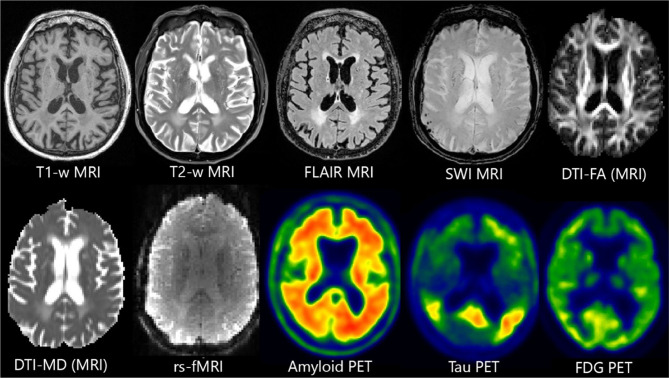
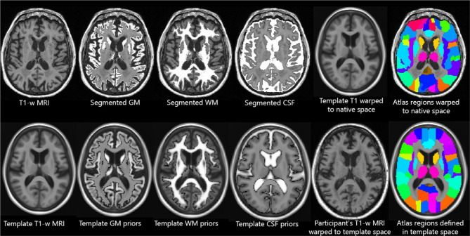
In the past decades, many neuroimaging studies have aimed to improve the scientific understanding of human neurodegenerative diseases using MRI and PET. This article is designed to provide an overview of the major classes of brain imaging and how/why they are used in this line of research. It is intended as a primer for individuals who are relatively unfamiliar with the methods of neuroimaging research to gain a better understanding of the vocabulary and overall methodologies. It is not intended to describe or review any research findings for any disease or biology, but rather to broadly describe the imaging methodologies that are used in conducting this neurodegeneration research. We will also review challenges and strategies for analyzing neuroimaging data across multiple sites and studies, i.e., harmonization and standardization of imaging data for multi-site and meta-analyses.
Keywords: Alzheimer's disease; Image analyses; Imaging; Neurodegeneration; Research; Review.
© 2021. The American Society for Experimental NeuroTherapeutics, Inc.
Figures
References
-
- Andersson J, Smith S, Jenkinson M. “FNIRT-FMRIB’s non-linear image registration tool,” in Annual Meeting of the Organization for Human Brain Mapping (OHBM), 2008.
Publication types
MeSH terms
LinkOut - more resources
Full Text Sources
Other Literature Sources
Medical
Miscellaneous
