Review
. 2021 Mar;15(1):1-15.
doi: 10.1007/s12105-021-01301-z. Epub 2021 Mar 15.

Craniofacial Development: Neural Crest in Molecular Embryology

Affiliations
Review

Craniofacial Development: Neural Crest in Molecular Embryology

Daniela Marta Roth et al. Head Neck Pathol. 2021 Mar.

Abstract

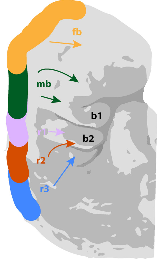
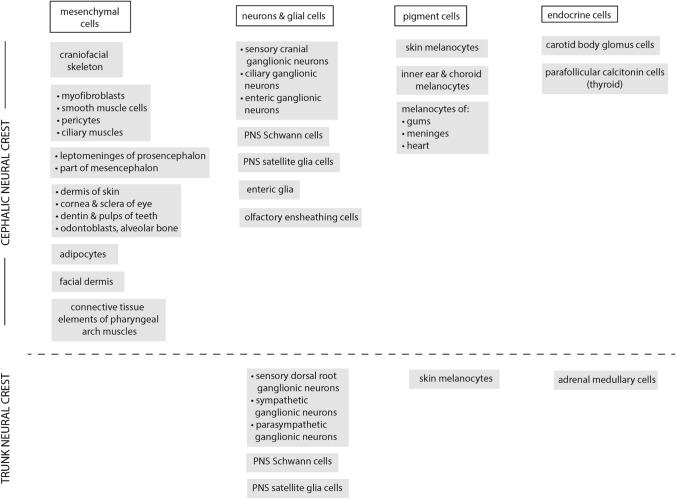
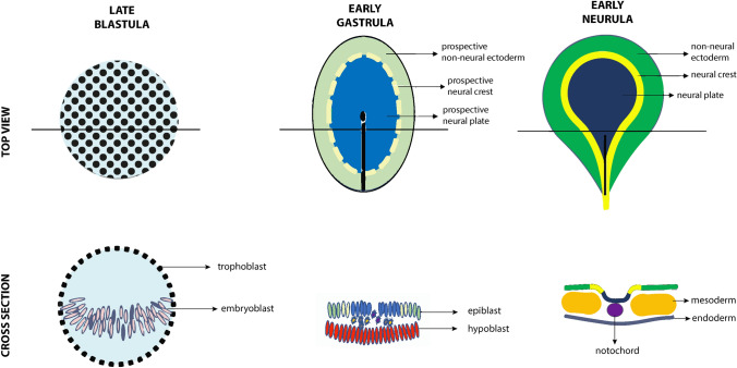
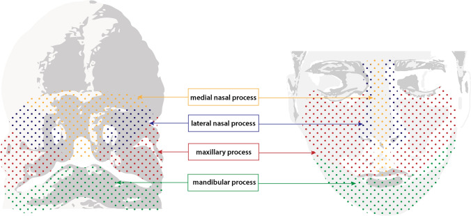
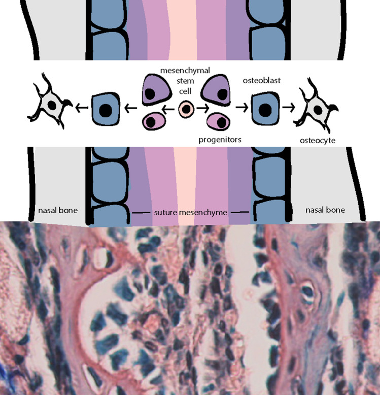
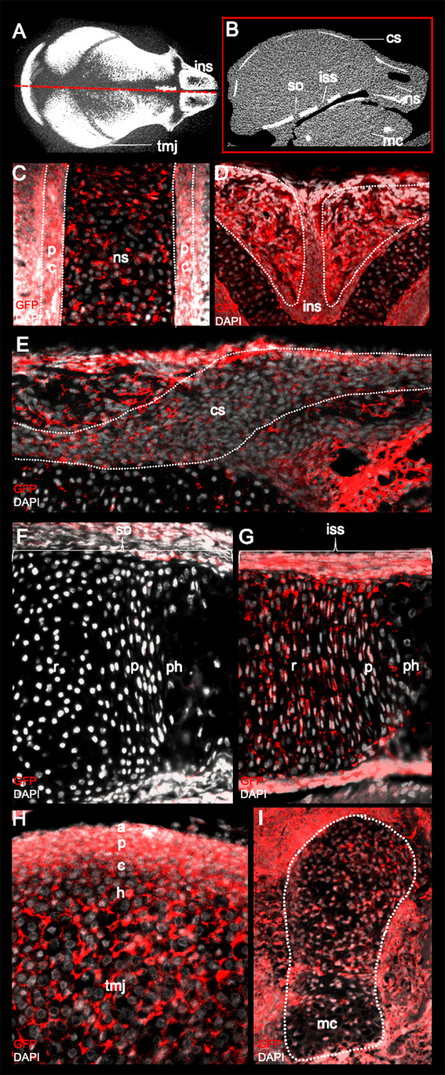
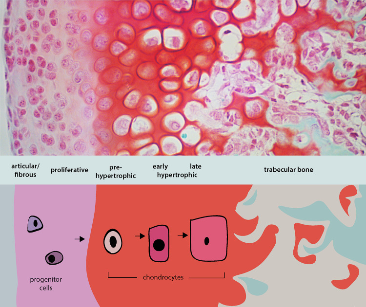
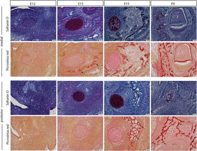
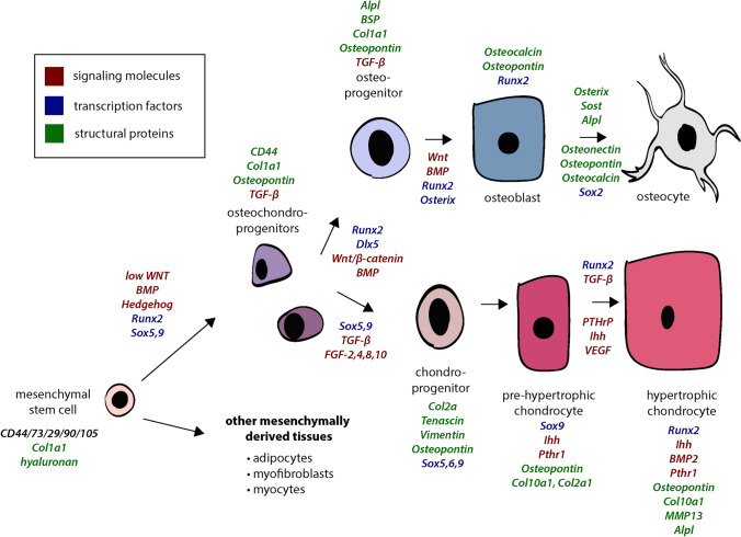
Craniofacial development, one of the most complex sequences of developmental events in embryology, features a uniquely transient, pluripotent stem cell-like population known as the neural crest (NC). Neural crest cells (NCCs) originate from the dorsal aspect of the neural tube and migrate along pre-determined routes into the developing branchial arches and frontonasal plate. The exceptional rates of proliferation and migration of NCCs enable their diverse contribution to a wide variety of craniofacial structures. Subsequent differentiation of these cells gives rise to cartilage, bones, and a number of mesenchymally-derived tissues. Deficiencies in any stage of differentiation can result in facial clefts and abnormalities associated with craniofacial syndromes. A small number of conserved signaling pathways are involved in controlling NC differentiation and craniofacial development. They are used in a reiterated fashion to help define precise temporospatial cell and tissue formation. Although many aspects of their cellular and molecular control have yet to be described, it is clear that together they form intricately integrated signaling networks required for spatial orientation and developmental stability and plasticity, which are hallmarks of craniofacial development. Mutations that affect the functions of these signaling pathways are often directly or indirectly identified in congenital syndromes. Clinical applications of NC-derived mesenchymal stem/progenitor cells, persistent into adulthood, hold great promise for tissue repair and regeneration. Realization of NCC potential for regenerative therapies motivates understanding of the intricacies of cell communication and differentiation that underlie the complexities of NC-derived tissues.

Keywords: Bone; Cartilage; Neural crest; Orofacial development; Signalling.


Conflict of interest statement

The authors declare no conflict of interest.

Figures

Fig. 1
Fig. 1
Migration pattern of cell populations from embryonic neural crest. Graphic representation of stage 12, week 4 human embryo. fb: forebrain; mb: midbrain; r1: rhombomere 1; r2: rhombomere 2; r3: rhombomere 3; b1: branchial arch 1; b2: branchial arch 2
Fig. 2
Fig. 2
Neural crest derivatives. Adapted from Sperber, 2018 [56] and Etchevers et al. [57]
Fig. 3
Fig. 3
Establishment of neural crest population in early embryogenesis. Graphic representation of chick embryogenesis from late blastula to early neurula. Initially, cells of the embryoblast within the trophoblast are interspersed with no apparent specification. At the stage of the early gastrula, these cells form the epiblast and hypoblast, further segregated into the prospective non-neural ectoderm, neural crest, and neural plate. The onset of neurulation in the early neurula brings distinction of these already-designated cell populations into the non-neural ectoderm, neural crest, neural plate, mesoderm, and endoderm grouped around the notochord
Fig. 4
Fig. 4
Structures derived from facial prominences of the early embryo. Graphic representation of 5 week human embryo (left) with labelled facial prominences: medial nasal process (yellow), lateral nasal process (blue), maxillary process (red), and mandibular process (green). Structures derived from these processes are indicated in their respective colors on an adult face (right)
Fig. 5
Fig. 5
Differentiation of cells within cranial sutures. Graphic representation (top) of cranial suture cell populations and their presumptive differentiation pathway. Note: the indicated intramembranous bone stage overtop each region of the suture is not the only composing cell type, however, would likely be found within. In reality, the suture mesenchyme is a heterogeneous population of cells ranging in differentiation from stem cells to committed osteoprogenitors. Corresponding orcein/methylene blue stain (bottom) of the internasal suture of a 4-week old mouse portrays suture mesenchyme in red/brown studded with blue nuclei between two layers of blue cuboidal osteoblasts lining the red/brown bone. Within the lacunae of the bone reside dark blue osteocytes
Fig. 6
Fig. 6
Identification of lineage-traced neural crest cells (NCCs) in the craniofacial complex using Green fluorescent protein (GFP) staining. a & b Annotation of various craniofacial structures on a midsagittal cross-section (red box) of a P0 micro-computed tomography mouse scan. Paraffin-embedded frontal section of the P0 mice skulls stained with GFP and counterstained with DAPI indicates the presence of NC-derived cells in the (c) cartilage and perichondrium (pc) of the nasal septum (ns) and the (d) internasal suture (ins). Sagittal sections of P0 skulls demonstrate the presence of NC-derived cells in the (e) periosteum and osteogenic front of the coronal suture. NCCs also contribute to the resting (r), proliferative (p), pre-hypertrophic (ph) zone of the (G) intersphenoidal (iss) and not (f) sphenooccipital (so) synchondroses of the cranial base. (h) The articular (a), proliferative (p), chondrogenic (c) and hypertrophic (h) layers of the temporomandibular joint (TMJ) are also neural crest-derived along with the (i) chondrocytes of the Meckel's cartilage (mc)
Fig. 7
Fig. 7
Illustration of the chondrocyte organization and differentiation during endochondral ossification. (Top panel) Safranin O staining (red) of paraffin-embedded temporomandibular joint (TMJ) section counterstained with fast green indicating the organization of the TMJ into articular (outermost/surface), proliferative, pre-hypertrophic, early and late hypertrophic chondrocytes followed by bone. (Bottom panel) Chondrocyte progenitors undergo differentiation into proliferative chondrocytes which then mature to become hypertrophic chondrocytes. The hypertrophic chondrocytes either undergo apoptosis or transdifferentiate into bone. This process of progenitors undergoing an intermediate cartilage phase to form bone is called endochondral ossification
Fig. 8
Fig. 8
Cartilage and intramembranous bone formation throughout mandibular development. Paraffin histology demonstrating mandibular development via intramembranous bone formation throughout mouse development, from embryonic day 12 post-conception (E12) to postnatal day 0 (P0). As Meckel’s cartilage (deep orange on Safranin O stain, indicated by white dotted lines and arrows) is degraded, bone forms in the surrounding space (red stain on Picrosirius red stain, indicated by black arrows). Note that the stage of intramembranous bone formation differs along the length of the mandible, with the medial mandible ossifying sooner than the posterior aspect
Fig. 9
Fig. 9
Differentiation of mesenchymal stem cells. Gene names listed in italics representing activity of signaling molecules, transcription factors, and structural proteins at each stage of the differentiation pathway for bone and cartilage. Red text references genes encoding for signaling molecules, blue text references genes encoding for transcription factors, and green text references genes encoding for structural proteins

Similar articles

Cited by

References

    1. Dooley CM, Wali N, Sealy IM, White RJ, Stemple DL, Collins JE, et al. The gene regulatory basis of genetic compensation during neural crest induction. PLOS Genet. Public Lib Sci. 2019;15:1008213. doi: 10.1371/journal.pgen.1008213. - DOI - PMC - PubMed
    1. Saint-Jeannet J-P, editor. Neural Crest Induction and Differentiation [Internet]. Springer US; 2006 [cited 2020 Oct 26]. https://www.springer.com/gp/book/9780387351360
    1. Donoghue PCJ, Graham A, Kelsh RN. The origin and evolution of the neural crest. Bioessays. 2008;30:530–41. doi: 10.1002/bies.20767. - DOI - PMC - PubMed
    1. Le Douarin NM, Ziller C, Couly GF. Patterning of Neural Crest Derivatives in the Avian Embryo: in Vivo and in Vitro Studies. Developmental Biology. 1993;159:24–49. doi: 10.1006/dbio.1993.1219. - DOI - PubMed
    1. Rothstein M, Bhattacharya D, Simoes-Costa M. The molecular basis of neural crest axial identity. Dev Biol. 2018;444(Suppl 1):S170–80. doi: 10.1016/j.ydbio.2018.07.026. - DOI - PMC - PubMed

LinkOut - more resources