. 2021 Feb 9;217(2):iyaa030.
doi: 10.1093/genetics/iyaa030.

A fish with no sex: gonadal and adrenal functions partition between zebrafish NR5A1 co-orthologs

Affiliations

A fish with no sex: gonadal and adrenal functions partition between zebrafish NR5A1 co-orthologs

Yi-Lin Yan et al. Genetics. .

Erratum in

Abstract

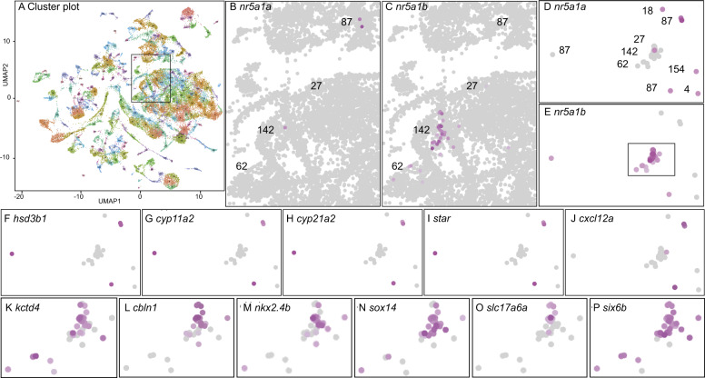
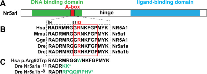
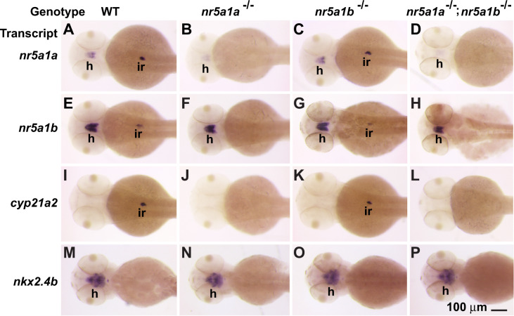
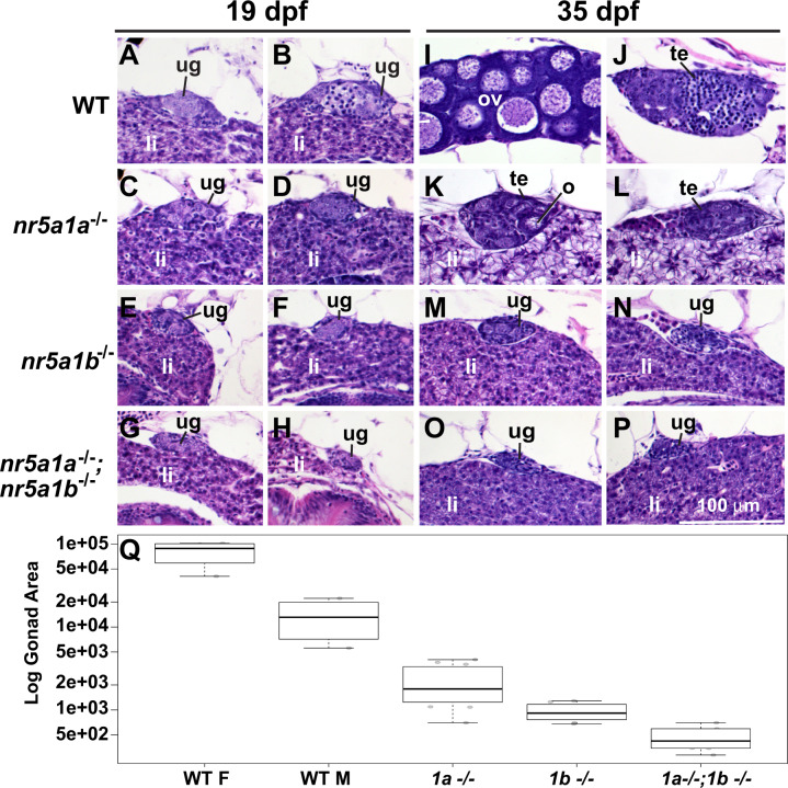
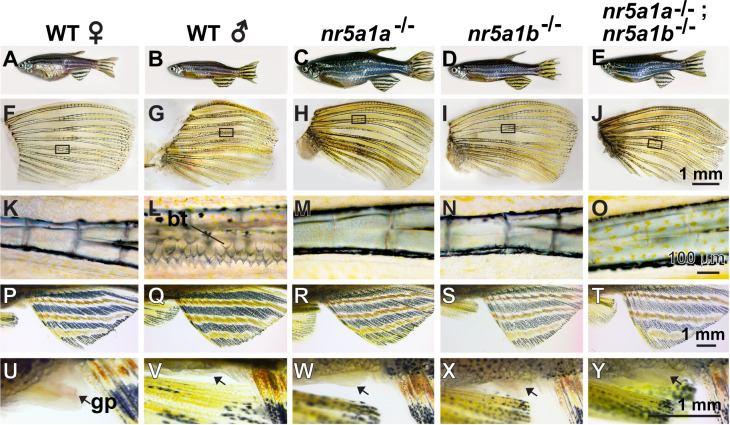
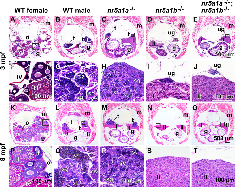
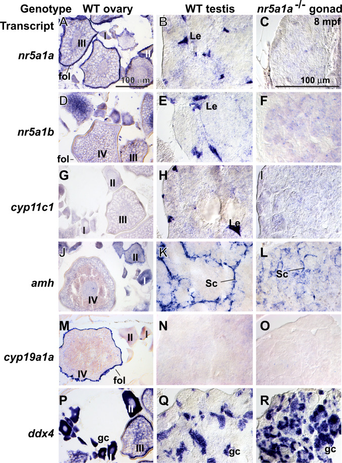
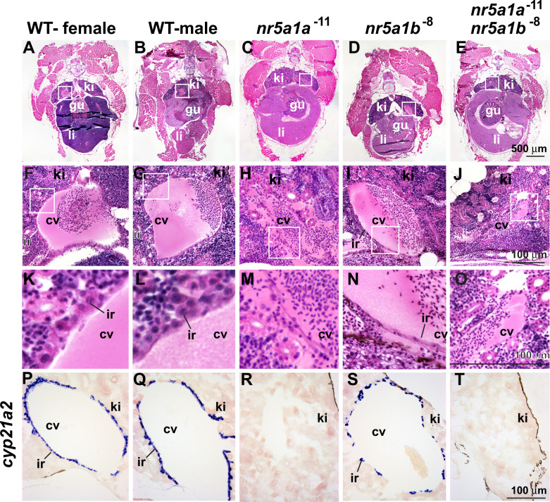
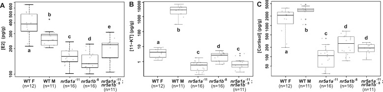
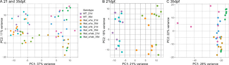
People with NR5A1 mutations experience testicular dysgenesis, ovotestes, or adrenal insufficiency, but we do not completely understand the origin of this phenotypic diversity. NR5A1 is expressed in gonadal soma precursor cells before expression of the sex-determining gene SRY. Many fish have two co-orthologs of NR5A1 that likely partitioned ancestral gene subfunctions between them. To explore ancestral roles of NR5A1, we knocked out nr5a1a and nr5a1b in zebrafish. Single-cell RNA-seq identified nr5a1a-expressing cells that co-expressed genes for steroid biosynthesis and the chemokine receptor Cxcl12a in 1-day postfertilization (dpf) embryos, as does the mammalian adrenal-gonadal (interrenal-gonadal) primordium. In 2dpf embryos, nr5a1a was expressed stronger in the interrenal-gonadal primordium than in the early hypothalamus but nr5a1b showed the reverse. Adult Leydig cells expressed both ohnologs and granulosa cells expressed nr5a1a stronger than nr5a1b. Mutants for nr5a1a lacked the interrenal, formed incompletely differentiated testes, had no Leydig cells, and grew far larger than normal fish. Mutants for nr5a1b formed a disorganized interrenal and their gonads completely disappeared. All homozygous mutant genotypes lacked secondary sex characteristics, including male breeding tubercles and female sex papillae, and had exceedingly low levels of estradiol, 11-ketotestosterone, and cortisol. RNA-seq showed that at 21dpf, some animals were developing as females and others were not, independent of nr5a1 genotype. By 35dpf, all mutant genotypes greatly under-expressed ovary-biased genes. Because adult nr5a1a mutants form gonads but lack an interrenal and conversely, adult nr5a1b mutants lack a gonad but have an interrenal, the adrenal, and gonadal functions of the ancestral nr5a1 gene partitioned between ohnologs after the teleost genome duplication, likely owing to reciprocal loss of ancestral tissue-specific regulatory elements. Identifying such elements could provide hints to otherwise unexplained cases of Differences in Sex Development.

Keywords: Differences in Sex Development; Genetics of Sex; SF1; adreno-gonadal primordium; disorders of sex development; scRNA-seq; subfunctionalization.


Figures

Figure 1
Figure 1
scRNA-seq identification of genes co-expressed with nr5a1a and nr5a1b. (A) In total, 220 clusters from scRNA-seq of whole animals at 24, 48, and 120 hpf (Farnsworth et al. 2020). The box indicates the portion enlarged in (B) and (C). (B) nr5a1a-expressing cells; color intensity is proportional to log expression level. (C) nr5a1b-expressing cells. (D) nr5a1a- and/or nr5a1b-expressing cells labeled only for nr5a1a-expression, with clusters numbered. (E) nr5a1a- and/or nr5a1b-expressing cells labeled only for nr5a1b-expression. The box indicates the portion enlarged in panels J–O. (F–I) Expression of the steroid biosynthesis genes hsd3b1, cyp11a2, cyp21a2, and star. (J) Cluster 87 cells labeled for the cytokine cxcl12a. (K–P) Expression of ventromedial hypothalamus genes kctd4, cbln1, nkx2.4b, sox14, slc17a6a, and six6b, respectively, in the boxed region of (E).
Figure 2
Figure 2
CRISPR/Cas9-induced nr5a1a and nr5a1b mutants. (A) Consequences of induced mutations on Nr5a1 proteins (DNA-binding domain, green; Abox, red; ligand-binding domain, blue). (B) Amino acid sequences surrounding the zebrafish CRISPR target sites for orthologs in human (Hsa), mouse (Mmu), chicken (Gga), and zebrafish (Dre). The human mutations R84H and G91S are dominant and cause 46, XY sex reversal; R92W is a dominant mutation resulting in variable testis development in 46, XX individuals; and R92Q is a recessive that causes adrenal insufficiency, 46, XX sex reversal, and 46, XY sex reversal (Lin and Achermann 2008; Bashamboo et al. 2016; Miyado et al. 2016; Werner et al. 2017). (C) The zebrafish nr5a1a−11 and nr5a1b−8 deletion alleles are predicted to result in polypeptides with short out-of-frame sequences (green) that are truncated owing to a premature stop codon (*) before or within the A-box.
Figure 3
Figure 3
Gene expression patterns in embryos at 48 hpf. (A, E, I, M) Wild-type embryos. (B, F, J, N) nr5a1a−11 mutant embryos. (C, G, K, O) nr5a1b−8 mutant embryos. (D, H, L, P) nr5a1a−11; nr5a1b−8 double mutant embryos. In situ hybridization for nr5a1a (A, B, C, D), nr5a1b (E, F, G, H), cyp21a2 (I, J, K, L), nkx2.4b (M, N, O, P). interrenal development depends on nr5a1a, but hypothalamus expression of nkx2-family marker genes is not strongly affected in either single mutant or double mutants. Based on three clutches, each containing 20–25 wild types, 42–48 heterozygotes, 19–24 nr5a1a mutants, 18–27 nr5a1b mutants, and 8–12 double mutant embryos. h, hypothalamus; ir, interrenal. Scale bar in P represents 100 μm.
Figure 4
Figure 4
Histology of gonads in 19 and 35 dpf wild-type and nr5a1-mutant fish. (A–H) 19 dpf. (I–P) 35 dpf. In 19-dpf juveniles, gonads in all (8/8 fish) wild-type fish (WT) were undifferentiated (e.g., A, B). Gonads in all nr5a1a−11 mutants (6/6 fish) (e.g., C, D), all nr5a1b−8 mutants (6/6 fish) (e.g., E, F) and in all (4/4 fish) nr5a1a−11; nr5a1b−8 double mutants (e.g., G, H) were undifferentiated like wild-type gonads but all mutant gonads were smaller than wild-type sibling gonads, especially in nr5a1b−8 mutants and nr5a1a−11; nr5a1b−8 double mutants (e.g., E–H). At 35-dpf (I–P), wild-type fish contained gonads that were clearly either ovaries (4/4 fish) (e.g., I) or testis (4/4 fish) (e.g., J). In 35-dpf nr5a1a−11 mutants, all fish examined had smaller testis (13/13 fish) (e.g., K, L, Q); In 35-dpf nr5a1b−8 mutants (8/8 fish), gonads were still undifferentiated (e.g., M, N, Q); In 35-dpf nr5a1a−11; nr5a1b−8 double mutants, gonads were still undifferentiated and were even smaller than earlier (4/4 fish) (e.g., O, P, Q). (Q), Log cross-sectional area of the gonad in arbitrary units for 35-dpf wild-type females (4/4 fish) and males (4/4 fish); for 35-dpf nr5a1a−11 mutants (13/13 fish); 35-dpf nr5a1b−8 mutants (8/8 fish), and 35-dpf nr5a1a−11; nr5a1b−8 double mutants (4/4 fish). Gonad sizes were significantly smaller in nr5a1 mutants compared to wild-type siblings. Outer bars, minimum and maximum; thick bar, median; lower box edge, first quartile; upper box edge third quartile. li, liver; ov, ovary; te, testis; ug, undifferentiated gonad. Statistical significance: different letters (a–e) signify differences at P < 0.05. Scale bar in P is 100 µm for all panels.
Figure 5
Figure 5
Secondary sex characteristics. (A–E) 8-mpf adult zebrafish. (A) Wild-type female. (B) Wild-type male. (C) nr5a1a−11 mutant. (D) nr5a1b−8 mutant. (E) nr5a1a−11; nr5a1b−8 double mutant. Among mutants, body shape was male-like but body color was not. (F–O) Pectoral fin of 8-mpf fish. (F) wild-type female, (G) wild-type male, (H) nr5a1a−11 mutant, (I) nr5a1b−8 mutant, and (J) nr5a1a−11; nr5a1b−8 double mutant. (K–O) Boxed region of pectoral fin in higher magnification, showing breeding tubercles (bt) in (I) wild-type male but not in (K) wild-type female or in any nr5a1 mutants (M–O). (P–T) Anal fins dissected from (P) wild-type female, (Q) wild-type male, (R) nr5a1a−11 mutant, (S) nr5a1b−8 mutant, and (T) nr5a1a−11; nr5a1b−8 double mutant. (U–Y) Genital papilla (gp, arrows) located anterior to the anal fin. (U) Long genital papilla in wild-type female compared to (V) wild-type male and (W–Y) nr5a1 mutants. Based on five fish of each genotype, a total of 25 fish. bt, breeding tubercles; gp, genital papilla. Scale bar in J for F–J: 1 mm; scale bar in O for K–O: 100 μm; scale bar in T for P–T; scale bar in Y for U–Y: 1 mm.
Figure 6
Figure 6
Histology of gonads in adult zebrafish. (A–J) 3-mpf adult zebrafish. (A, F) wild-type female, (B, G) wild-type male. (C, H) nr5a1a−11 mutant (n.8). (D, I) nr5a1b−8 mutant (n.5). (E, J) nr5a1a−11; nr5a1b−8 double mutant (n.4). (K–T) 8-mpf adult zebrafish. (K, P) Wild-type female, (L, Q) wild-type male, (M, R); nr5a1a−11 mutant (n.8). (N, S); nr5a1b−8 mutant (n.8). (O, T) nr5a1a−11; nr5a1b−8 double mutant (n.4). (A–E) Low magnification at 3 mpf, (F–J), high magnification of boxed region in AE at 3 mpf (K–O), low magnification at 8 mpf (P–T), high magnification of boxed region at (K–O) at 8 mpf. Crosssections of 3mpf (A, F) and 8-mpf wild-type female siblings (K, P) show maturing (stage-I and -II) and vitellogenic (stage-III and -IV) follicles. Wild-type male siblings at 3mpf (B, G) and 8 mpf (L, Q) had formed all spermatogenic stages. Mutant nr5a1a males at 3 mpf at low (C), and high magnification (H) show that older animals had developed more immature sperm and spermatocytes, compared to wild-type siblings (B, C, G, H). I, II, III, IV: ovarian follicle stages 1–4; g, gut; li, liver; o, ovary; s, Sertoli cells; sc, spermatocytes; sg, spermatogonia; st, spermatids; sz, spermatozoa; t, testis. Scale bar in (E) for (A–E): 500 μm; scale bar in (J) for (F–J): 100 mm; scale bar in (O) for (K–O): 500 mm; scale bar in (R) for (P–R): 100 μm; scale bar in (T) for (S) and (T): 100 μm.
Figure 7
Figure 7
Gene expression patterns in adult gonads at 8 mpf. (A, D, G, J, M, P) Wild-type ovaries. (B, E, H, K, N, Q) Wild-type testis. (C, F, I, L, O, R) nr5a1a−11 mutant testis. In situ hybridization using probes for (A, B, C) nr5a1a, (D, E, F) nr5a1b, (G, H, I) cyp11c1, (J, K, L) amh, (M, N, O) cyp19a1a, and (P, Q, R) ddx4. Scale bar in A (for all wild-type ovaries) and scale bar in C (for all testis panels) represents 100 μm. I, II, III, IV: ovarian follicle stages 1–4; fol, follicle cell; gc, germ cell; Le, Leydig cell; Sc, Sertoli cell.
Figure 8
Figure 8
Nr5a1 activity is required for normal interrenal morphology in adult zebrafish. (A–O) Cross-sections of 8-mpf adult zebrafish histology and (P–T) in situ hybridization for cyp21a2. (A, F, K, P) Wild-type females. (B, G, L, Q) Wild-type males. (C, H, M, R) nr5a1a−11 mutants. (D, I, N, S) nr5a1b−8 mutants. (E, J, O, T) nr5a1a−11; nr5a1b−8 double mutants. Histological sections of anterior trunk: (A–E) at low magnification; white box expanded in (F–J) medium magnification; and white box expanded in (K–O) high magnification. (P–T) In situ hybridization of cyp21a2 to interrenal cells. cv, cardinal vein; gu, gut; ir, interrenal; li, liver; ki, kidney. Scale bar in (E) for (A–E); scale bar in J for F–J; scale bar in (O) for (K–O); scale bar in (T) for (P–T). All scale bars: 100 μm. Based on five fish of each genotype for morphology (25 fish), and four fish of each genotype for gene expression (20 fish).
Figure 9
Figure 9
Loss of function of nr5a1 ohnologs disrupts whole-body concentrations of sex steroids and cortisol at 4.5 mpf (log scale). (A) Estradiol (E2, pg/g wet weight). (B) 11-Keto testosterone (11-KT) (pg/g wet weight). (C) Cortisol (pg/g wet weight). WT F (wild-type females, n.12); WT M (wild-type males, n.11); nr5a1a−11 (n.11); nr51b−8 (n.16); nr5a1a−11; nr5a1b−8 double mutants (n.11). Different letters (a–e) signify statistically different groups at P < 0.05.
Figure 10
Figure 10
Principal component analysis of RNA-seq data for juvenile and young adult wild-type and nr5a1 mutants. Each dot represents a different individual fish. (A) Analysis of all samples together. (B) Analysis of 21-dpf samples. (C) Analysis of 35-dpf samples. WT, wild type; 1a, nr5a1a mutants; 1b, nr5a1b mutants; 1ab, nr5a1a; nr5a1b double mutants; 21d, 21 dpf; 35d, 35 dpf.

Similar articles

Cited by

References

    1. Achermann JC, Ito M, Ito M, Hindmarsh PC, Jameson JL. 1999. A mutation in the gene encoding steroidogenic factor-1 causes XY sex reversal and adrenal failure in humans. Nat Genet. 22:125–126. - PubMed
    1. Aiuti A, Webb IJ, Bleul C, Springer T, Gutierrez-Ramos JC. 1997. The chemokine SDF-1 is a chemoattractant for human CD34+ hematopoietic progenitor cells and provides a new mechanism to explain the mobilization of CD34+ progenitors to peripheral blood. J Exp Med. 185:111–120. - PMC - PubMed
    1. Amores A, Catchen J, Ferrara A, Fontenot Q, Postlethwait JH. 2011. Genome evolution and meiotic maps by massively parallel DNA sequencing: spotted gar, an outgroup for the teleost genome duplication. Genetics. 188:799–808. - PMC - PubMed
    1. Amores A, Force A, Yan YL, Joly L, Amemiya C, et al. 1998. Zebrafish hox clusters and vertebrate genome evolution. Science. 282:1711–1714. - PubMed
    1. Anamthathmakula P, Miryala CSJ, Moreci RS, Kyathanahalli C, Hassan SS et al. 2019. Steroidogenic factor 1 (Nr5a1) is required for sertoli cell survival post sex determination. Sci Rep. 9:4452. - PMC - PubMed

Publication types

LinkOut - more resources