Meta-Analysis
. 2021 Mar 16;3(3):CD013639.
doi: 10.1002/14651858.CD013639.pub4.

Thoracic imaging tests for the diagnosis of COVID-19

Affiliations
Meta-Analysis

Thoracic imaging tests for the diagnosis of COVID-19

Nayaar Islam et al. Cochrane Database Syst Rev. .

Update in

  • Thoracic imaging tests for the diagnosis of COVID-19.
    Ebrahimzadeh S, Islam N, Dawit H, Salameh JP, Kazi S, Fabiano N, Treanor L, Absi M, Ahmad F, Rooprai P, Al Khalil A, Harper K, Kamra N, Leeflang MM, Hooft L, van der Pol CB, Prager R, Hare SS, Dennie C, Spijker R, Deeks JJ, Dinnes J, Jenniskens K, Korevaar DA, Cohen JF, Van den Bruel A, Takwoingi Y, van de Wijgert J, Wang J, Pena E, Sabongui S, McInnes MD; Cochrane COVID-19 Diagnostic Test Accuracy Group. Ebrahimzadeh S, et al. Cochrane Database Syst Rev. 2022 May 16;5(5):CD013639. doi: 10.1002/14651858.CD013639.pub5. Cochrane Database Syst Rev. 2022. PMID: 35575286 Free PMC article.

Abstract

Background: The respiratory illness caused by SARS-CoV-2 infection continues to present diagnostic challenges. Our 2020 edition of this review showed thoracic (chest) imaging to be sensitive and moderately specific in the diagnosis of coronavirus disease 2019 (COVID-19). In this update, we include new relevant studies, and have removed studies with case-control designs, and those not intended to be diagnostic test accuracy studies.

Objectives: To evaluate the diagnostic accuracy of thoracic imaging (computed tomography (CT), X-ray and ultrasound) in people with suspected COVID-19.

Search methods: We searched the COVID-19 Living Evidence Database from the University of Bern, the Cochrane COVID-19 Study Register, The Stephen B. Thacker CDC Library, and repositories of COVID-19 publications through to 30 September 2020. We did not apply any language restrictions.

Selection criteria: We included studies of all designs, except for case-control, that recruited participants of any age group suspected to have COVID-19 and that reported estimates of test accuracy or provided data from which we could compute estimates.

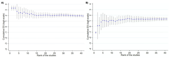
Data collection and analysis: The review authors independently and in duplicate screened articles, extracted data and assessed risk of bias and applicability concerns using the QUADAS-2 domain-list. We presented the results of estimated sensitivity and specificity using paired forest plots, and we summarised pooled estimates in tables. We used a bivariate meta-analysis model where appropriate. We presented the uncertainty of accuracy estimates using 95% confidence intervals (CIs).

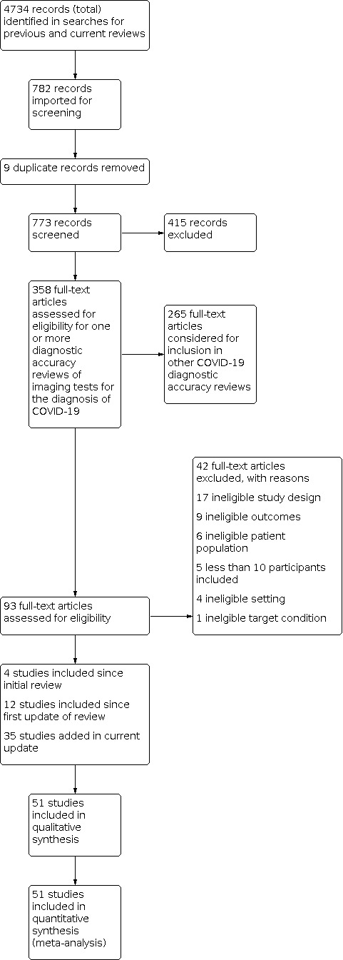
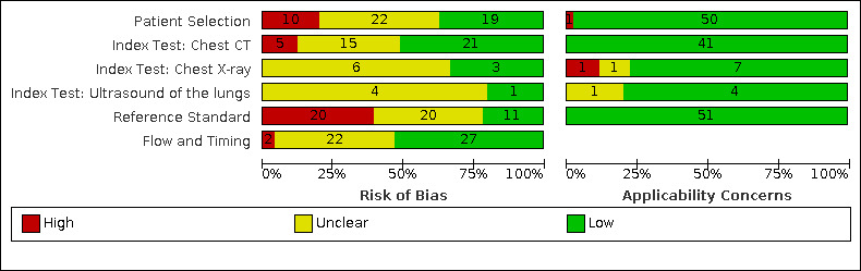
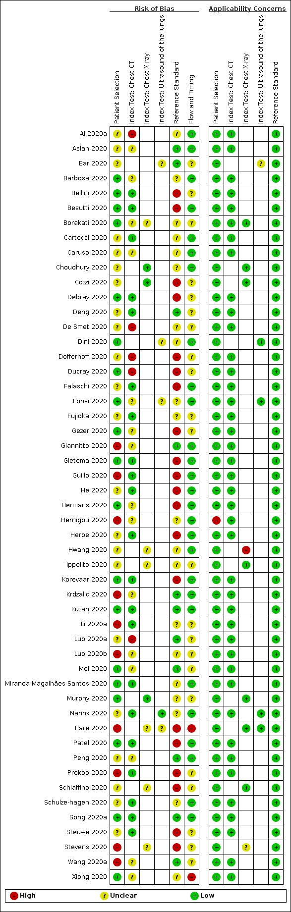
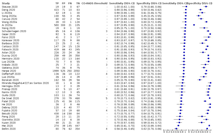
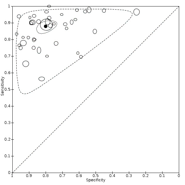
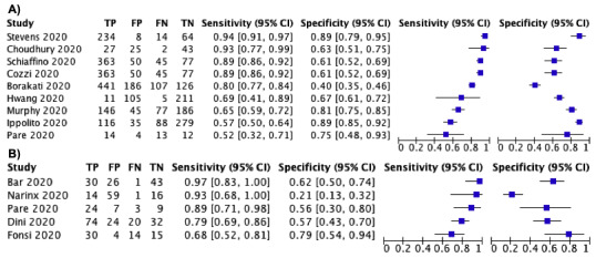
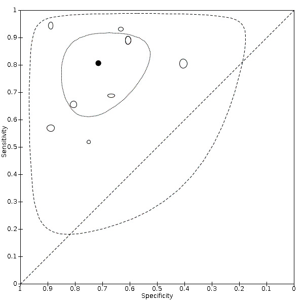
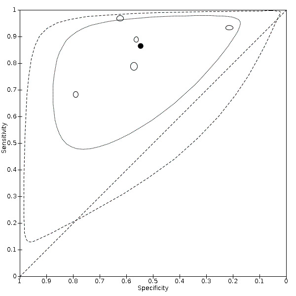
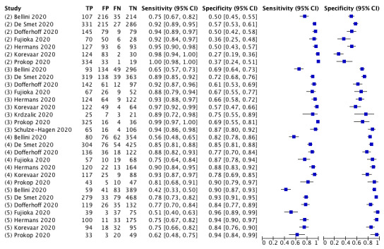
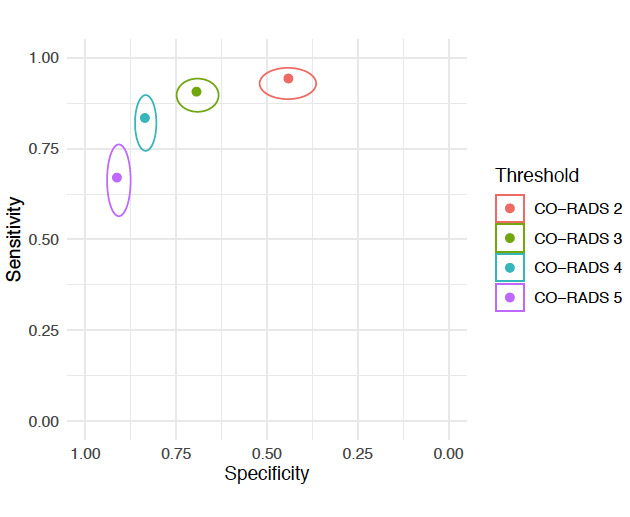
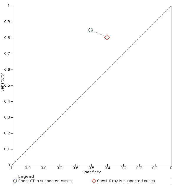
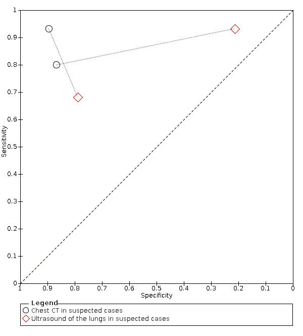
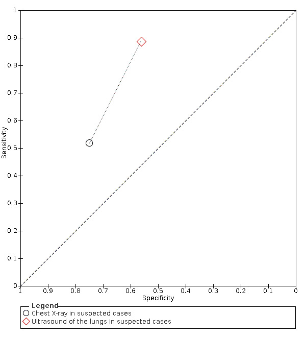
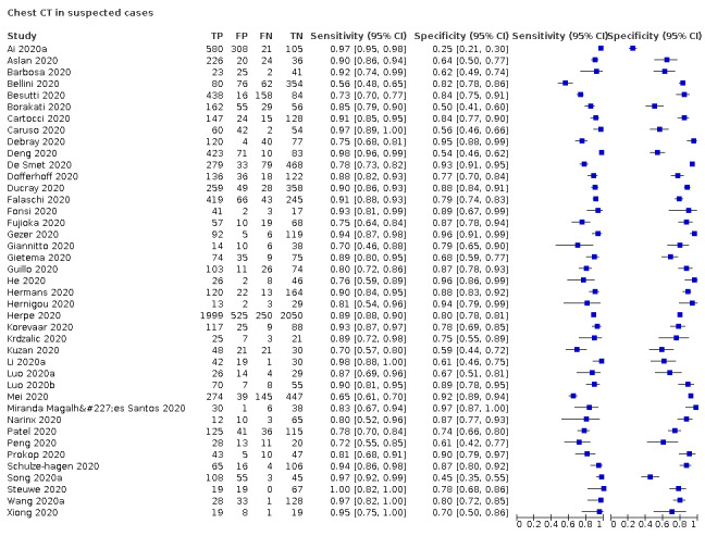
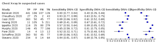
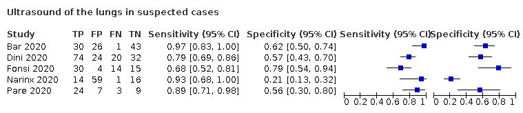
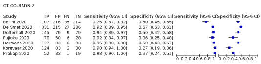
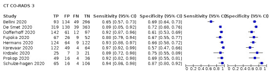
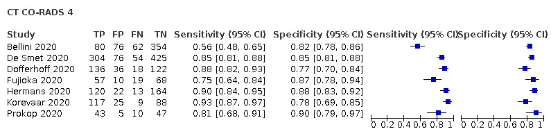
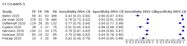
Main results: We included 51 studies with 19,775 participants suspected of having COVID-19, of whom 10,155 (51%) had a final diagnosis of COVID-19. Forty-seven studies evaluated one imaging modality each, and four studies evaluated two imaging modalities each. All studies used RT-PCR as the reference standard for the diagnosis of COVID-19, with 47 studies using only RT-PCR and four studies using a combination of RT-PCR and other criteria (such as clinical signs, imaging tests, positive contacts, and follow-up phone calls) as the reference standard. Studies were conducted in Europe (33), Asia (13), North America (3) and South America (2); including only adults (26), all ages (21), children only (1), adults over 70 years (1), and unclear (2); in inpatients (2), outpatients (32), and setting unclear (17). Risk of bias was high or unclear in thirty-two (63%) studies with respect to participant selection, 40 (78%) studies with respect to reference standard, 30 (59%) studies with respect to index test, and 24 (47%) studies with respect to participant flow. For chest CT (41 studies, 16,133 participants, 8110 (50%) cases), the sensitivity ranged from 56.3% to 100%, and specificity ranged from 25.4% to 97.4%. The pooled sensitivity of chest CT was 87.9% (95% CI 84.6 to 90.6) and the pooled specificity was 80.0% (95% CI 74.9 to 84.3). There was no statistical evidence indicating that reference standard conduct and definition for index test positivity were sources of heterogeneity for CT studies. Nine chest CT studies (2807 participants, 1139 (41%) cases) used the COVID-19 Reporting and Data System (CO-RADS) scoring system, which has five thresholds to define index test positivity. At a CO-RADS threshold of 5 (7 studies), the sensitivity ranged from 41.5% to 77.9% and the pooled sensitivity was 67.0% (95% CI 56.4 to 76.2); the specificity ranged from 83.5% to 96.2%; and the pooled specificity was 91.3% (95% CI 87.6 to 94.0). At a CO-RADS threshold of 4 (7 studies), the sensitivity ranged from 56.3% to 92.9% and the pooled sensitivity was 83.5% (95% CI 74.4 to 89.7); the specificity ranged from 77.2% to 90.4% and the pooled specificity was 83.6% (95% CI 80.5 to 86.4). For chest X-ray (9 studies, 3694 participants, 2111 (57%) cases) the sensitivity ranged from 51.9% to 94.4% and specificity ranged from 40.4% to 88.9%. The pooled sensitivity of chest X-ray was 80.6% (95% CI 69.1 to 88.6) and the pooled specificity was 71.5% (95% CI 59.8 to 80.8). For ultrasound of the lungs (5 studies, 446 participants, 211 (47%) cases) the sensitivity ranged from 68.2% to 96.8% and specificity ranged from 21.3% to 78.9%. The pooled sensitivity of ultrasound was 86.4% (95% CI 72.7 to 93.9) and the pooled specificity was 54.6% (95% CI 35.3 to 72.6). Based on an indirect comparison using all included studies, chest CT had a higher specificity than ultrasound. For indirect comparisons of chest CT and chest X-ray, or chest X-ray and ultrasound, the data did not show differences in specificity or sensitivity.

Authors' conclusions: Our findings indicate that chest CT is sensitive and moderately specific for the diagnosis of COVID-19. Chest X-ray is moderately sensitive and moderately specific for the diagnosis of COVID-19. Ultrasound is sensitive but not specific for the diagnosis of COVID-19. Thus, chest CT and ultrasound may have more utility for excluding COVID-19 than for differentiating SARS-CoV-2 infection from other causes of respiratory illness. Future diagnostic accuracy studies should pre-define positive imaging findings, include direct comparisons of the various modalities of interest in the same participant population, and implement improved reporting practices.


Conflict of interest statement

Nayaar Islam has no known conflicts of interest.

Sanam Ebrahimzadeh has no known conflicts of interest.

Jean‐Paul Salameh has no known conflicts of interest.

Sakib Kazi has no known conflicts of interest.

Nicholas Fabiano has no known conflicts of interest.

Lee Treanor has no known conflicts of interest.

Marissa Absi has no known conflicts of interest.

Zachary Hallgrimson has no known conflicts of interest.

Mariska MG Leeflang has no known conflicts of interest.

Lotty Hooft has no known conflicts of interest.

Christian B van der Pol has no known conflicts of interest.

Ross Prager has no known conflicts of interest.

Samanjit Singh Hare has no known conflicts of interest.

Carole Dennie has no known conflicts of interest.

René Spijker: the Dutch Cochrane Centre (DCC) has received grants for performing commissioned systematic reviews. In no situation did the commissioner have any influence on the results of the work.

Jonathan J Deeks has no known conflicts of interest.

Jacqueline Dinnes has no known conflicts of interest.

Kevin Jenniskens has no known conflicts of interest.

Daniel Korevaar has no known conflicts of interest.

Jérémie F Cohen has no known conflicts of interest.

Ann Van den Bruel has no known conflicts of interest.

Yemisi Takwoingi has no known conflicts of interest.

Janneke van de Wijgert has no known conflicts of interest.

Johanna AAG Damen has no known conflicts of interest.

Junfeng Wang received a consultancy fee from Biomind, an Artificial Intelligence (AI) company providing machine intelligence solutions in medical imaging. The consultancy service was about design of clinical studies, not related to this review. The company had no influence on the results of the work.

Matthew McInnes has no known conflicts of interest.

Figures

1
1
Study flow diagram.
2
2
Risk of bias and applicability concerns graph: review authors' judgements about each domain presented as percentages across included studies.
3
3
Risk of bias and applicability concerns summary: review authors' judgements about each domain for each included study.
4
4
Forest plot of chest CT in suspected cases.
5
5
Summary ROC plot of chest CT in suspected cases. The summary point is indicated by the solid black circle, individual studies are indicated by outlined circles (scale=study sample size). The dotted border and the dashed border represent 95% confidence regions and 95% prediction regions, respectively.
6
6
Forest plot of A) chest X‐ray and B) ultrasound in suspected cases.
7
7
Summary ROC plot of chest X‐ray in suspected cases. The summary point is indicated by the solid black circle, individual studies are indicated by outlined circles (scale=study sample size). The dotted border and the dashed border represent 95% confidence regions and 95% prediction regions, respectively.
8
8
Summary ROC plot of ultrasound of the lungs in suspected cases. The summary point is indicated by the solid black circle, individual studies are indicated by outlined circles (scale=study sample size). The dotted border and the dashed border represent 95% confidence regions and 95% prediction regions, respectively.
9
9
Forest plot of chest CT studies in suspected cases that used the CO‐RADS scoring system. Threshold (i.e. 2, 3 4, or 5) indicated in parentheses ahead of Study ID. Grouped by threshold.
10
10
Pooled sensitivity and specificity estimates and 95% confidence intervals at varying CO‐RADS thresholds: CO‐RADS 2 (n = 7), CO‐RADS 3 (n = 9), CO‐RADS 4 (n = 7), and CO‐RADS 5 (n = 7).
11
11
Summary ROC plot of comparative studies: chest CT versus chest X‐ray. The dotted line connects index tests pairs evaluated in the same study (i.e. a comparative study).
12
12
Summary ROC plot of comparative studies: chest CT versus ultrasound of the lungs. The dotted lines connect index tests pairs evaluated in the same study (i.e. a comparative study).
13
13
Summary ROC plot of comparative studies: chest X‐ray versus ultrasound of the lungs. The dotted line connects index tests pairs evaluated in the same study (i.e. a comparative study).
14
14
Cumulative meta‐analysis of chest CT A) logit‐sensitivity and B) logit‐specificity. Includes 41 studies in the current review. Studies are ranked according to date of publication (1 = oldest, 41 = most recent).
15
15
Pooled sensitivity and specificity estimates and 95% confidence intervals across all versions of this review.
1
1. Test
Chest CT in suspected cases
2
2. Test
Chest X‐ray in suspected cases
3
3. Test
Ultrasound of the lungs in suspected cases
4
4. Test
CT CO‐RADS 2
5
5. Test
CT CO‐RADS 3
6
6. Test
CT CO‐RADS 4
7
7. Test
CT CO‐RADS 5

Update of

Similar articles

Cited by

References

References to studies included in this review

Ai 2020a {published data only}
    1. Ai T, Yang Z, Hou H, Zhan C, Chen C, Lv W, et al. Correlation of chest CT and RT-PCR testing for coronavirus disease 2019 (COVID-19) in China: a report of 1014 cases. Radiology 2020;296(2):E32-E40. - PMC - PubMed
Aslan 2020 {published data only}
    1. Aslan S, Bekci T, Cakir IM, Ekiz M, Yavuz I, Sahin AM. Diagnostic performance of low-dose chest CT to detect COVID-19: a Turkish population study. Diagnostic and Interventional Radiology 2020. [DOI: 10.5152/dir.2020.20350] - DOI - PMC - PubMed
Bar 2020 {published data only}
    1. Bar S, Lecourtois A, Diouf M, Goldberg E, Bourbon C, Arnaud E, et al. The association of lung ultrasound images with COVID‐19 infection in an emergency room cohort. Anaesthesia 2020;75(12):1620-25. - PMC - PubMed
Barbosa 2020 {published data only}
    1. Barbosa PN, Bitencourt AG, Miranda GD, Almeida MF, Chojniak R. Chest CT accuracy in the diagnosis of SARS-CoV-2 infection: initial experience in a cancer center. Radiologia Brasileira 2020;53(4):211-5. - PMC - PubMed
Bellini 2020 {published data only}
    1. Bellini D, Panvini N, Rengo M, Vicini S, Lichtner M, Tieghi T, et al. Diagnostic accuracy and interobserver variability of CO-RADS in patients with suspected coronavirus disease-2019: a multireader validation study. European Radiology 2020. [DOI: 10.1007/s00330-020-07273-y] - DOI - PMC - PubMed
Besutti 2020 {published data only}
    1. Besutti G, Giorgi Rossi P, Iotti V, Spaggiari L, Bonacini R, Nitrosi A, et al. Accuracy of CT in a cohort of symptomatic patients with suspected COVID-19 pneumonia during the outbreak peak in Italy. European Radiology 2020 Dec;30(12):6818-27. - PMC - PubMed
Borakati 2020 {published data only}
    1. Borakati A, Perera A, Johnson J, Sood T. Chest X-ray has poor diagnostic accuracy and prognostic significance in COVID-19: a propensity matched database study. medRxiv [Preprint] 2020. [DOI: DOI: 10.1101/2020.07.07.20147934] - PubMed
Cartocci 2020 {published data only}
    1. Cartocci G, Colaiacomo MC, Lanciotti S, Andreoli C, De Cicco ML, Brachetti G, et al. Chest CT for early detection and management of coronavirus disease (COVID-19): a report of 314 patients admitted to Emergency Department with suspected pneumonia. Radiologia Medica 2020;125(10):931-42. - PMC - PubMed
Caruso 2020 {published data only}
    1. Caruso D, Zerunian M, Polici M, Pucciarelli F, Polidori T, Rucci C, et al. Chest CT features of COVID-19 in Rome, Italy. Radiology 2020;2962(2):E79-E85. - PMC - PubMed
Choudhury 2020 {published data only}
    1. Choudhury RS, Shahi PK, Sharma S, Dhar R. Utility of chest radiography on admission for initial triaging of COVID-19 in symptomatic patients. ERJ Open Research 2020;6(3):00357-2020. - PMC - PubMed
Cozzi 2020 {published data only}
    1. Cozzi A, Schiaffino S, Arpaia F, Della Pepa G, Tritella S, Bertolotti P, et al. Chest x-ray in the COVID-19 pandemic: Radiologists' real-world reader performance. European Journal of Radiology 2020 Nov;132:109272. - PMC - PubMed
Debray 2020 {published data only}
    1. Debray M-P, Tarabay H, Males L, Mahdjoub E, Pavlovsky T. Observer agreement and clinical significance of chest CT reporting in patients suspected of COVID-19. medRxiv [Preprint] 2020. [DOI: ] - PMC - PubMed
Deng 2020 {published data only}
    1. Deng Z, Zhang X, Li Y, Xu H, Gang Y, Wang H, et al. Value of chest CT screening in the early COVID-19 outbreak. Chinese Journal of Radiology 2020;5(54):430-4.
De Smet 2020 {published data only}
    1. Smet KD, Smet DD, Demedts I, Bouckaert B, Ryckaert T, Laridon E, et al. Diagnostic power of chest CT for COVID-19: to screen or not to screen. medRxiv [Preprint] 2020. [DOI: ]
Dini 2020 {published data only}
    1. Dini FL, Bergamini C, Allegrini A, Scopelliti M, Secco G, Miccoli M, et al. Bedside wireless lung ultrasound for the evaluation of COVID-19 lung injury in senior nursing home residents. Monaldi Archives for Chest Disease 2020;90(3). [DOI: 10.4081/monaldi.2020.1446] - DOI - PubMed
Dofferhoff 2020 {published data only}
    1. Dofferhoff AS, Swinkels A, Sprong T, Berk Y, Spanbroek M, Nabuurs-Franssen MH, et al. Diagnostic algorithm for COVID-19 at the ER. Nederlands Tijdschrift voor Geneeskunde 2020;164:D5042. - PubMed
Ducray 2020 {published data only}
    1. Ducray V, Vlachomitrou AS, Bouscambert-Duchamp M, Si-Mohamed S, Gouttard S, Mansuy A, et al. Chest CT for rapid triage of patients in multiple emergency departments during COVID-19 epidemic: experience report from a large French university hospital. European Radiology 2020;31(2):795-803. [DOI: 10.1007/s00330-020-07154-4] - DOI - PMC - PubMed
Falaschi 2020 {published data only}
    1. Falaschi Z, Danna PS, Arioli R, Pasché A, Zagaria D, Percivale I, et al. Chest CT accuracy in diagnosing COVID-19 during the peak of the Italian epidemic: a retrospective correlation with RT-PCR testing and analysis of discordant cases. European Journal of Radiology 2020;130:109192. - PMC - PubMed
Fonsi 2020 {published data only}
    1. Fonsi GB, Sapienza P, Brachini G, Andreoli C, De Cicco ML, Cirillo B, et al. Is lung ultrasound imaging a worthwhile procedure for severe acute respiratory syndrome coronavirus 2 pneumonia detection? Journal of Ultrasound in Medicine 2020. [DOI: 10.1002/jum.15487 ] - PubMed
Fujioka 2020 {published data only}
    1. Fujioka T, Takahashi M, Mori M, Tsuchiya J, Yamaga E, Horii T, et al. Evaluation of the usefulness of CO-RADS for chest CT in patients suspected of having COVID-19. Diagnostics 2020;10(9):608. - PMC - PubMed
Gezer 2020 {published data only}
    1. Gezer NS, Ergan B, Barış MM, Appak Ö, Sayıner AA, Balcı P, et al. COVID-19 S: a new proposal for diagnosis and structured reporting of COVID-19 on computed tomography imaging. Diagnostic and Intervional Radiology 2020;26(4):315–22. - PMC - PubMed
Giannitto 2020 {published data only}
    1. Giannitto C, Sposta FM, Repici A, Vatteroni G, Casiraghi E, Casari E, et al. Chest CT in patients with a moderate or high pretest probability of COVID-19 and negative swab. Radiologia Medica 2020;125:1260-70. - PMC - PubMed
Gietema 2020 {published data only}
    1. Gietema HA, Zelis N, Nobel JM, Lambriks LJ, Van Alphen LB, Oude Lashof AM, et al. CT in relation to RT-PCR in diagnosing COVID-19 in The Netherlands: a prospective study. PLoS ONE 2020;15(7):e0235844. [DOI: 10.1371/journal.pone.0235844] - DOI - PMC - PubMed
Guillo 2020 {published data only}
    1. Guillo E, Bedmar Gomez I, Dangeard S, Bennani S, Saab I, Tordjman M, et al. COVID-19 pneumonia: diagnostic and prognostic role of CT based on a retrospective analysis of 214 consecutive patients from Paris, France. European Journal of Radiology 2020;131:109209. - PMC - PubMed
He 2020 {published data only}
    1. He J-L, Luo L, Luo Z-D, Lyu J-X, Ng M-Y, Shen X-P, et al. Diagnostic performance between CT and initial real-time RT-PCR for clinically suspected 2019 coronavirus disease (COVID-19) patients outside Wuhan, China. Respiratory Medicine 2020;168:105980. - PMC - PubMed
Hermans 2020 {published data only}
    1. Hermans JJ, Groen J, Zwets E, Boxma-De Klerk BM, Van Werkhoven JM, Ong DS, et al. Chest CT for triage during COVID-19 on the emergency department: myth or truth? Emergency Radiology 2020;27(6):641-51. - PMC - PubMed
Hernigou 2020 {published data only}
    1. Hernigou J, Cornil F, Poignard A, El Bouchaibi S, Mani J, Naouri JF, et al. Thoracic computerised tomography scans in one hundred eighteen orthopaedic patients during the COVID-19 pandemic: identification of chest lesions; added values; help in managing patients; burden on the computerised tomography scan department. International Orthopaedics (SICOT) 2020;44(8):1571–80. - PMC - PubMed
Herpe 2020 {published data only}
    1. Herpe G, Lederlin M, Naudin M, Ohana M, Chaumoitre K, Gregory J, et al. Efficacy of chest CT for COVID-19 pneumonia in France. Radiology 2020;298(2):E81-E87. - PMC - PubMed
Hwang 2020 {published data only}
    1. Hwang EJ, Kim H, Yoon SH, Goo JM, Park CM. Implementation of a deep learning-based computer-aided detection system for the interpretation of chest radiographs in patients suspected for COVID-19. Korean Journal of Radiology 2020;21(10):1150. - PMC - PubMed
Ippolito 2020 {published data only}
    1. Ippolito D, Pecorelli A, Maino C, Capodaglio C, Mariani I, Giandola T, et al. Diagnostic impact of bedside chest X-ray features of 2019 novel coronavirus in the routine admission at the emergency department: case series from Lombardy region. European Journal of Radiology 2020;129:109092. - PMC - PubMed
Korevaar 2020 {published data only}
    1. Korevaar DA, Kootte RS, Smits LP, Van den Aardweg JG, Bonta PI, Schinkel J, et al. Added value of chest computed tomography in suspected COVID-19: an analysis of 239 patients. European Respiratory Journal 2020;56(2):2001377. - PMC - PubMed
Krdzalic 2020 {published data only}
    1. Krdzalic J, De Jaegere TM, Kwee RM. Diagnostic performance of chest CT in screening patients with suspected COVID-19 infection in a Western population. Brtish Jounal of Radiology 2020;93(1113):20200643. - PMC - PubMed
Kuzan 2020 {published data only}
    1. Kuzan TY, Murzoglu Altıntoprak K, et al. A comparison of clinical, laboratory and chest CT findings of laboratory-confirmed and clinically diagnosed COVID-19 patients at first admission. Diagnostic and Interventional Radiology [Epub ahead of print] 2020 Sep 2. [DOI: 10.5152/dir.2020.20270] - DOI - PMC - PubMed
Li 2020a {published data only}
    1. Li Z, Zeng B, Lei P, Liu J, Fan B, Shen Q, et al. Differentiating pneumonia with and without COVID-19 using chest CT images: from qualitative to quantitative. Journal of X-ray Science and Technology 2020;28(4):583-9. - PMC - PubMed
Luo 2020a {published data only}
    1. Luo L, Luo Z, Jia Y, Zhou C, He J, Lyu J, et al. CT differential diagnosis of COVID-19 and non-COVID-19 in symptomatic suspects: a practical scoring method. BMC Pulmonary Medicine 2020;20(1):129. - PMC - PubMed
Luo 2020b {published data only}
    1. Luo N, Zhang H, Zhou Y, Kong Z, Sun W, Huang N, et al. Utility of chest CT in diagnosis of COVID-19 pneumonia. Diagnostic and Interventional Radiology 2020;26(5):437–42. - PMC - PubMed
Mei 2020 {published data only}
    1. Mei X, Lee H-C, Diao K, Huang M, Lin B, Liu C, et al. Artificial intelligence-enabled rapid diagnosis of COVID-19 patients. medRxiv [Preprint] 2020. [DOI: ] - PMC - PubMed
Miranda Magalhães Santos 2020 {published data only}
    1. Miranda Magalhães Santos JM, Paula Alves Fonseca A, Pinheiro Zarattini Anastacio E, Formagio Minenelli F, Furtado de Albuquerque Cavalcanti C, Borges da Silva Teles G. Initial results of the use of a standardized diagnostic criteria for chest computed tomography findings in coronavirus disease 2019. Journal of Computer Assisted Tomography 2020;44(5):647-51. - PubMed
Murphy 2020 {published data only}
    1. Murphy K, Smits H, Knoops AJ, Korst MB, Samson T, Scholten ET, et al. COVID-19 on chest radiographs: a multireader evaluation of an artificial intelligence system. Radiology 2020;296(3):E166-72. - PMC - PubMed
Narinx 2020 {published data only}
    1. Narinx N, Smismans A, Symons R, Frans J, Demeyere A, Gillis M. Feasibility of using point-of-care lung ultrasound for early triage of COVID-19 patients in the emergency room. Emergency Radiology 2020;27(6):663-70. - PMC - PubMed
Pare 2020 {published data only}
    1. Pare J, Camelo I, Mayo K, Leo M, Dugas J, Nelson K, et al. Point-of-care lung ultrasound is more sensitive than chest radiograph for evaluation of COVID-19. Western Journal of Emergency Medicine 2020;21(4):771-8. [DOI: 10.5811/westjem.2020.5.47743] - DOI - PMC - PubMed
Patel 2020 {published data only}
    1. Patel M, Chowdhury J, Zheng M, Abramian O, Verga S, Zhao H, et al. High Resolution CHEST CT(HRCT) evaluation in patients hospitalized with COVID-19 infection. medRxiv [Preprint] 2020. [DOI: ]
Peng 2020 {published data only}
    1. Peng D, Zhang J, Xu Y, Liu Z, Wu P. Clinical analysis and early differential diagnosis of suspected pediatric patients with 2019 novel coronavirus infection. medRxiv [Preprint] 2020. [DOI: ]
Prokop 2020 {published data only}
    1. Prokop M, Van Everdingen W, Van Rees Vellinga T, Quarles van Ufford H, Stöger L, Beenen L, et al. CO-RADS: a categorical CT assessment scheme for patients suspected of having COVID-19—definition and evaluation. Radiology 2020;296(2):E97–E104. - PMC - PubMed
Schiaffino 2020 {published data only}
    1. Schiaffino S, Tritella S, Cozzi A, Carriero S, Blandi L, Ferraris L, et al. Diagnostic performance of chest X-Ray for COVID-19 pneumonia during the SARS-CoV-2 pandemic in Lombardy, Italy. Journal of Thoracic Imaging 2020;35(4):W105–6. - PubMed
Schulze‐hagen 2020 {published data only}
    1. Schulze-Hagen M, Hübel C, Meier-Schroers M, Yüksel C, Sander A, Sähn M, et al. Low-dose chest CT for the diagnosis of COVID-19—a systematic, prospective comparison with PCR. Deutsches Aerzteblatt Online 2020;117(22-23):389-95. [DOI: 10.3238/arztebl.2020.0389] - DOI - PMC - PubMed
Song 2020a {published data only}
    1. Song S, Wu F, Liu Y, Jiang H, Xiong F, Guo X, et al. Correlation between chest CT findings and clinical features of 211 COVID-19 suspected patients in Wuhan, China. Open Forum Infectious Diseases 2020;7(6):ofaa171. - PMC - PubMed
Steuwe 2020 {published data only}
    1. Steuwe A, Rademacher C, Valentin B, Köhler M-H, Appel E, Keitel V, et al. Dose-optimised chest computed tomography for diagnosis of coronavirus disease 2019 (COVID-19) - evaluation of image quality and diagnostic impact. Journal of Radiological Protection 2020;40(3):877-91. - PubMed
Stevens 2020 {published data only}
    1. Stevens BJ. Reporting radiographers' interpretation and use of the British Society of Thoracic Imaging's coding system when reporting COVID-19 chest X-rays. Radiography 2020;S1078817420300985. - PMC - PubMed
Wang 2020a {published data only}
    1. Wang T, Xiong Z, Zhou H, Luo W, Tang H, Liu J. Design, validation, and clinical practice of standardized imaging diagnostic report for COVID-19. Zhong Nan da Xue Xue Bao. Yi Xue Ban [Journal of Central South University. Medical Sciences] 2020;45(3):229-35. - PubMed
Xiong 2020 {published data only}
    1. Xiong Z, Fu L, Zhou H, Liu JK, Wang AM, Huang Y, et al. Construction and evaluation of a novel diagnosis pathway for 2019-Corona virus disease. Zhonghua Yi Xue za Zhi 2020;100(16):1223-9. - PubMed

References to studies excluded from this review

Ai 2020b {published data only}
    1. Ai J, Gong J, Xing L, He R, Tian F, Wang J, et al. Analysis of factors associated early diagnosis in coronavirus disease 2019 (COVID-19). medRxiv [Preprint] 2020. [DOI: ]
Ai 2020c {published data only}
    1. Ai J, Chen J, Wang Y, Liu X, Fan W, Qu G, et al. The cross-sectional study of hospitalized coronavirus disease 2019 patients in Xiangyang, Hubei province. medRxiv [Preprint] 2020. [DOI: 10.1101/2020.02.19.20025023] - DOI
Arentz 2020 {published data only}
    1. Arentz M, Yim E, Klaff L, Lokhandwala S, Riedo FX, Chong M, et al. Characteristics and outcomes of 21 critically ill patients with COVID-19 in Washington State. JAMA 2020;323(16):1612-4. - PMC - PubMed
Bai 2020a {published data only}
    1. Bai HX, Hsieh B, Xiong Z, Halsey K, Choi JW, Tran TM, et al. Performance of radiologists in differentiating COVID-19 from viral pneumonia on chest CT. Radiology 2020;296(2):E46-E54. - PMC - PubMed
Bai 2020b {published data only}
    1. Bai HX, Wang R, Xiong Z, Hsieh B, Chang K, Halsey K, et al. Artificial intelligence augmentation of radiologist performance in distinguishing COVID-19 from pneumonia of other origin at chest CT. Radiology 2020;296(3):E156-65. - PMC - PubMed
Chang 2020 {published data only}
    1. Chang D, Lin M, Wei L, Xie L, Zhu G, Dela Cruz CS, et al. Epidemiologic and clinical characteristics of novel coronavirus infections involving 13 patients outside Wuhan, China. JAMA 2020;323(11):1092-3. [DOI: 10.1001/jama.2020.1623 10.1001/jama.2020.1623.] - PMC - PubMed
Chen 2020a {published data only}
    1. Chen S, Wu JJ, Li MZ, Xu DZ, Zhu YZ, Wang HC, et al. Clinical features of 109 cases of novel coronavirus pneumonia. Chinese Journal of Infectious Diseases 2020;38(12):E015. [DOI: 10.3760 / cma.j.issn.1000-6680.2020.0015]
Chen 2020b {published data only}
    1. Chen X, Tang Y, Mo Y, Li S, Lin D, Yang Z, et al. A diagnostic model for coronavirus disease 2019 (COVID-19) based on radiological semantic and clinical features: a multi-center study. European Radioliology 2020;30(9):4893-902. [DOI: 10.1007/s00330-020-06829-2] - DOI - PMC - PubMed
Chen 2020c {published data only}
    1. Chen Z, Fan H, Cai J, Li Y, Wu B, Hou Y, et al. High-resolution computed tomography manifestations of COVID-19 infections in patients of different ages. European Journal of Radiology 2020;126:108972. - PMC - PubMed
Cheng 2020 {published data only}
    1. Cheng Z, Lu Y, Cao Q, Qin L, Pan Z, Yan F, et al. Clinical features and chest CT manifestations of coronavirus disease 2019 (COVID-19) in a single-center study in Shanghai, China. American Journal of Roentgenology 2020;215(1):121-6. [DOI: 10.2214/ajr.20.22959 10.2214/AJR.20.22959.] - PubMed
Çinkooğlu 2020 {published data only}
    1. Çinkooğlu A, Bayraktaroğlu S, Savaş R. Lung changes on chest CT during 2019 novel coronavirus (COVID-19) pneumonia. European Journal of Breast Health 2020;16(2):89-90. [DOI: 10.5152/ejbh.2020.010420] - DOI - PMC - PubMed
Colombi 2020 {published data only}
    1. Colombi D, Bodin FC, Petrini M, Maffi G, Morelli N, Milanese G, et al. Well-aerated lung on admitting chest CT to predict adverse outcome in COVID-19 pneumonia. Radiology 2020;296(2):E86-E96. [DOI: 10.1148/radiol.2020201433] - DOI - PMC - PubMed
Dai 2020 {published data only}
    1. Dai H, Zhang X, Xia J, Zhang T, Shang Y, Huang R, et al. High-resolution chest CT features and clinical characteristics of patients infected with COVID-19 in Jiangsu, China. International Journal of Infectious Diseases 2020;95:106-12. [DOI: ] - PMC - PubMed
Ding 2020 {published data only}
    1. Ding X, Xu J, Zhou J, Long Q. Chest CT findings of COVID-19 pneumonia by duration of symptoms. European Journal of Radiology 2020;127:109009. [DOI: 10.1016/j.ejrad.2020.109009] - DOI - PMC - PubMed
Dong 2020 {published data only}
    1. Dong J, Wu L, Jin Q, Chen J, He J. Chest CT scan of hospitalized patients with COVID-19: a case-control study. medRxiv [Preprint] 2020. [DOI: ]
Guan 2020 {published data only}
    1. Guan W, Liu J, Yu C. CT findings of coronavirus disease (COVID-19) severe pneumonia. American Journal of Roentgenology 2020;5:W85-W6. [DOI: 10.2214/AJR.20.23035] - DOI - PubMed
Hao 2020 {published data only}
    1. Hao W, Li M. Clinical diagnostic value of CT imaging in COVID-19 with multiple negative RT-PCR testing. Travel Medicine and Infectious Disease 2020;34:101627. [DOI: 10.1016/j.tmaid.2020.101627] - DOI - PMC - PubMed
Himoto 2020 {published data only}
    1. Himoto Y, Sakata A, Kirita M, Hiroi T, Kobayashi K I, Kubo K, et al. Diagnostic performance of chest CT to differentiate COVID-19 pneumonia in non-high-epidemic area in Japan. Japanese Journal of Radiology 2020;38(5):400-6. - PMC - PubMed
Huang 2020 {published data only}
    1. Huang P, Liu T, Huang L, Liu H, Lei M, Xu W, et al. Use of chest CT in combination with negative RT-PCR assay for the 2019 novel coronavirus but high clinical suspicion. Radiology 2020;295(1):22-3. [DOI: 10.1148/radiol.2020200330 10.1148/radiol.2020200330. Epub 2020 Feb 12.] - PMC - PubMed
Liang 2020 {published data only}
    1. Liang Y, Liang J, Zhou Q, Li X, Lin F, Deng Z, et al. Prevalence and clinical features of 2019 novel coronavirus disease (COVID-19) in the Fever Clinic of a teaching hospital in Beijing: a single-center, retrospective study. medRxiv [Preprint] 2020. [DOI: ]
Lu 2020 {published data only}
    1. Lu X, Zhang L, Du H, Zhang J, Li Y, Qu J, et al. SARS-CoV-2 infection in children. New England Journal of Medicine 2020;382(17):1663-5. [DOI: 10.1056/NEJMc2005073] - DOI - PMC - PubMed
Mao 2020 {published data only}
    1. Mao X, Liu X-P, Xiong M, Yang X, Jin X, Li Z, et al. Development and validation of chest CT-based imaging biomarkers for early stage COVID-19 screening. medRxiv [Preprint] 2020. [DOI: ] - PMC - PubMed
Miao 2020a {published data only}
    1. Miao C, Zhuang J, Jin M, Xiong H, Huang P, Zhao Q, et al. A comparative multi-centre study on the clinical and imaging features of confirmed and unconfirmed patients with COVID-19. medRxiv [Preprint] 2020. [DOI: ]
Miao 2020b {published data only}
    1. Miao C, Jin M, Miao L, Yang X, Huang P, Xiong H, et al. Early chest computed tomography to diagnose COVID-19 from suspected patients: a multicenter retrospective study. American Journal of Emergency Medicine 2020;S0735-6757(20):30281-3. - PMC - PubMed
Pakray 2020 {published data only}
    1. Pakray A, Walker D, Figacz A, Kilanowski S, Rhodes C, Doshi S, et al. Imaging evaluation of COVID-19 in the emergency department. Emergency Radiology 2020;27:579-88. - PMC - PubMed
Poggiali 2020 {published data only}
    1. Poggiali E, Dacrema A, Bastoni D, Tinelli V, Demichele E, Mateo Ramos P, et al. Can lung US help critical care clinicians in the early diagnosis of novel coronavirus (COVID-19) pneumonia? Radiology 2020;295(E6):200847. [DOI: 10.1148/radiol.2020200847] - DOI - PMC - PubMed
Pu 2020 {published data only}
    1. Pu J, Leader J, Bandos A, Shi J, Du P, Yu J, et al. Any unique image biomarkers associated with COVID-19? European Radiology 2020;30(NA):6221–7. - PMC - PubMed
Siegel 2020 {published data only}
    1. Siegel A, Chang PJ, Jarou ZJ, Paushter DM, Harmath CB, Arevalo JB, et al. Lung base findings of coronavirus disease (COVID-19) on abdominal CT in patients with predominant gastrointestinal symptoms. American Journal of Roentgenology 2020;215(3):607-9. [DOI: 10.2214/AJR.20.23232] - DOI - PubMed
Song 2020b {published data only}
    1. Song F, Shi N, Shan F, Zhang Z, Shen J, Lu H, et al. Emerging 2019 novel Coronavirus (2019-nCoV) pneumonia. Radiology 2020;295(1):210-7. [DOI: 10.1148/radiol.2020200274 10.1148/radiol.2020200274. Epub 2020 Feb 6.] - PMC - PubMed
Tavare 2020 {published data only}
    1. Tavare AN, Braddy A, Brill S, Jarvis H, Sivaramakrishnan A, Barnett J, et al. Managing high clinical suspicion COVID-19 inpatients with negative RT-PCR: a pragmatic and limited role for thoracic CT. Thorax 2020;75(7):537-8. [DOI: ] - PMC - PubMed
Wang 2020b {published data only}
    1. Wang Y, Dong C, Hu Y, Li C, Ren Q, Zhang X, et al. Temporal changes of CT findings in 90 patients with COVID-19 pneumonia: a longitudinal study. Radiology 2020;296(2):E55-E64. [DOI: 10.1148/radiol.2020200843 10.1148/radiol.2020200843.] - PMC - PubMed
Wu 2020a {published data only}
    1. Wu J, Feng CL, Xian XY, Qiang J, Zhang J, Mao QX, et al. Novel coronavirus pneumonia (COVID-19) CT distribution and sign features. Zhonghua Jie He He Hu Xi za Zhi [Chinese Journal of Tuberculosis and Respiratory Diseases] 2020;43(4):321-6. [DOI: 10.3760/cma.j.cn112147-20200217-00106 10.3760/cma.j.cn112147-20200217-00106.] - PubMed
Wu 2020b {published data only}
    1. Wu Q, Xing Y, Shi L, Li W, Gao Y, Pan S, et al. Epidemiological and clinical characteristics of children with coronavirus disease 2019. medRxiv [Preprint] 2020. [DOI: 10.1101/2020.03.19.20027078] - DOI
Wu 2020c {published data only}
    1. Wu X, Sun R, Chen J, Xie Y, Zhang S, Wang X. Radiological findings and clinical characteristics of pregnant women with COVID-19 pneumonia. International Journal of Gynaecology and Obstetrics 2020;150(1):58-63. [DOI: 10.1002/ijgo.13165] - DOI - PMC - PubMed
Wu 2020d {published data only}
    1. Wu X, Fu B, Chen L, Feng Y. Serological tests facilitate identification of asymptomatic SARS-CoV-2 infection in Wuhan, China. Journal of Medical Virology 2020;10:1002/jmv.25904. [DOI: 10.1002/jmv.25904] - DOI - PMC - PubMed
Xie 2020 {published data only}
    1. Xie S, Lei Z, Chen X, Liu W, Wang X, Dong Y, et al. Chest CT-based differential diagnosis of 28 patients with suspected corona virus disease 2019 (COVID-19). British Journal of Radiology 2020;93(1112):20200243. - PMC - PubMed
Xu 2020a {published data only}
    1. Xu X, Yu C, Qu J, Zhang L, Jiang S, Huang D, et al. Imaging and clinical features of patients with 2019 novel coronavirus SARS-CoV-2. European Journal of Nuclear Medicine and Molecular Imaging 2020;47(5):1275-80. [DOI: 10.1007/s00259-020-04735-9 10.1007/s00259-020-04735-9.] - PMC - PubMed
Xu 2020b {published data only}
    1. Xu X, Yu C, Zhang L, Luo L, Liu J. Imaging features of 2019 novel coronavirus pneumonia. European Journal of Nuclear Medicine and Molecular Imaging 2020;47(5):1022-23. [DOI: 10.1007/s00259-020-04720-2] - DOI - PMC - PubMed
Yang 2020a {published data only}
    1. Yang S, Shi Y, Lu H, Xu J, Li F, Qian Z, et al. Clinical and CT features of early-stage patients with COVID-19: a retrospective analysis of imported cases in Shanghai, China. European Respiratory Journal 2020;55(4):2000407. [DOI: 10.1183/13993003.00407-2020] - DOI - PMC - PubMed
Yang 2020b {published data only}
    1. Yang W, Cao Q, Qin L, Wang X, Cheng Z, Pan A, et al. Clinical characteristics and imaging manifestations of the 2019 novel coronavirus disease (COVID-19): a multi-center study in Wenzhou city, Zhejiang, China. Journal of Infection 2020;80(4):388-93. - PMC - PubMed
Yuan 2020 {published data only}
    1. Yuan M, Yin W, Tao Z, Tan W, Hu Y. Association of radiologic findings with mortality of patients infected with 2019 novel coronavirus in Wuhan, China. PloS One 2020;15(3):e0230548. [DOI: 10.1371/journal.pone.0230548 10.1371/journal.pone.0230548. eCollection 2020.] - PMC - PubMed
Zhifeng 2020 {published data only}
    1. Zhifeng J, Feng A, Li T. Consistency analysis of COVID-19 nucleic acid tests and the changes of lung CT. Journal of Clinical Virology 2020;127:104359. - PMC - PubMed

Additional references

Bossuyt 2015
    1. Bossuyt PM, Reitsma JB, Bruns DE, Gatsonis CA, Glasziou PP, Irwig L, et al. STARD 2015: An Updated List of Essential Items for Reporting Diagnostic Accuracy Studies. Radiology 2015 Oct 28;277(3):826–32. [DOI: 10.1148/radiol.2015151516] - DOI - PubMed
BSTI 2020
    1. British Society of Thoracic Imaging. COVID-19 BSTI Reporting Templates and Codes. bsti.org.uk/covid-19-resources/covid-19-bsti-reporting-templates/ 2020 (accessed 4 January 2020).
Butler‐Laporte 2021
    1. Butler-Laporte G, Lawandi A, Schiller I, Yao MC, Dendukuri N, McDonald E, et al. Comparison of saliva and nasopharyngeal swab nucleic acid amplification testing for detection of SARS-CoV-2: a systematic review and meta-analysis. JAMA Internal Medicine 2021:E1-8. [DOI: 10.1001/jamainternmed.2020.8876] - DOI - PMC - PubMed
China National Health Comission 2020
    1. China National Health Comission. Chinese Clinical Guidance for COVID-19 Pneumonia Diagnosis and Treatment (7th Edition). kjfy.meetingchina.org/msite/news/show/cn/3337.html 2020 (accessed 24 January 2020).
Chu 2006
    1. Chu H, Cole SR. Bivariate meta-analysis of sensitivity and specificity with sparse data: a generalized linear mixed model approach. Journal of Clinical Epidemiology 2006;59(12):1331-2; author reply 1332-3. [DOI: 10.1016/j.jclinepi.2006.06.011] - DOI - PubMed
Cohen 2016
    1. Cohen JF, Korevaar DA, Wang J, Leeflang MM, Bossuyt PM. Meta-epidemiologic study showed frequent time trends in summary estimates from meta-analyses of diagnostic accuracy studies. Journal of Clinical Epidemiology 2016;77:60–7. [DOI: 10.1016/j.jclinepi.2016.04.013] - DOI - PubMed
Datta 2020
    1. Datta SD, Talwar A, Lee JT. A proposed framework and timeline of the spectrum of disease due to SARS-CoV-2 infection: illness beyond acute infection and public health implications. JAMA 2020;324(22):2251-2. [DOI: 10.1001/jama.2020.22717] - DOI - PubMed
Deeks 2020
    1. Deeks JJ, Dinnes J, Takwoingi Y, Davenport C, Spijker R, Taylor-Phillips S, et al. Antibody tests for identification of current and past infection with SARS-CoV-2. Cochrane Database of Systematic Reviews 2020, Issue 6. Art. No: CD013652. [DOI: 10.1002/14651858.CD013652] - DOI - PMC - PubMed
Dinnes 2020
    1. Dinnes J, Deeks JJ, Adriano A, Berhane S, Davenport C, Dittrich S, et al. Rapid, point‐of‐care antigen and molecular‐based tests for diagnosis of SARS‐CoV‐2 infection. Cochrane Database of Systematic Reviews 2020, Issue 8. Art. No: CD013705. [DOI: 10.1002/14651858.CD013705] - DOI - PMC - PubMed
Harbord 2009
    1. Harbord RM, Whiting P. metandi: meta-analysis of diagnostic accuracy using hierarchical logistic regression. Stata Journal 2009;9(2):211-29. [DOI: ]
Hong 2018
    1. Hong PJ, Korevaar DA, McGrath TA, Ziai H, Frank R, Alabousi M, et al. Reporting of imaging diagnostic accuracy studies with focus on MRI subgroup: adherence to STARD 2015. Journal of Magnetic Resonance Imaging 2018;47(2):523-44. [DOI: 10.1002/jmri.25797] - DOI - PubMed
Irwig 1995
    1. Irwig L, Macaskill P, Glasziou P, Fahey M. Meta-analytic methods for diagnostic test accuracy. Journal of Clinical Epidemiology 1995;48(1):119-30; discussion 131-2. [DOI: 10.1016/0895-4356(94)00099-c] - DOI - PubMed
Kucirka 2020
    1. Kucirka LM, Lauer SA, Laeyendecker O, Boon D, Lessler J. Variation in false-negative rate of reverse transcriptase polymerase chain reaction-based SARS-CoV-2 tests by time since exposure. Annals of Internal Medicine 2020;173(4):262-7. [DOI: 10.7326/M20-1495] - DOI - PMC - PubMed
Lau 1992
    1. Lau J, Antman EM, Jimenez-Silva J, Kupelnick B, Mosteller F, Chalmers TC. Cumulative meta-analysis of therapeutic trials for myocardial infarction. New England Journal of Medicine 1992;327(4):248–54. [DOI: 10.1056/NEJM199207233270406] - DOI - PubMed
Li 2020b
    1. Li Y, Yao L, Li J, Chen L, Song Y, Cai Z, et al. Stability issues of RT-PCR testing of SARS-CoV-2 for hospitalized patients clinically diagnosed with COVID-19. Journal of Medical Virology 2020;92(7):903-8. [DOI: 10.1002/jmv.25786] - DOI - PMC - PubMed
Loeffelholz 2020
    1. Loeffelholz MJ, Tang YW. Laboratory diagnosis of emerging human coronavirus infections - the state of the art. Emerging Microbes and Infections 2020;9(1):747-56. [DOI: 10.1080/22221751.2020.1745095] - DOI - PMC - PubMed
McGrath 2017
    1. McGrath TA, McInnes MD, Langer FW, Hong J, Korevaar DA, Bossuyt PM. Treatment of multiple test readers in diagnostic accuracy systematic reviews-meta-analyses of imaging studies. European Journal of Radiology 2017;93:59–64. [DOI: ] - PubMed
Moher 2009
    1. Moher D, Liberati A, Tetzlaff J, Altman DG, PRISMA Group. Preferred reporting items for systematic reviews and meta-analyses: the PRISMA statement. Journal of Clinical Epidemiology 2009;62(10):1006-12. [DOI: 10.1371/journal.pmed.1000097] - DOI - PubMed
Ng 2020
    1. Ng M-Y, Lee EYP, Yang J, Yang F, Li X, Wang H, et al. Imaging Profile of the COVID-19 Infection: Radiologic Findings and Literature Review. Cardiothoracic Imaging 2020 Feb 1;2(1):e200034. [DOI: 10.1148/ryct.2020200034] - DOI - PMC - PubMed
Pedersen 2020
    1. Pedersen, TL. Package ‘ggforce’. www.ggforce.data-imaginist.com 2020 (accessed 5 January 2020).
R Core Team 2021 [Computer program]
    1. R Foundation for Statistical Computing R: A language and environment for statistical computing. R Core Team. Vienna, Austria: R Foundation for Statistical Computing, 2020.
Reitsma 2005
    1. Reitsma JB, Glas AS, Rutjes AW, Scholten RJ, Bossuyt PM, Zwinderman AH. Bivariate analysis of sensitivity and specificity produces informative summary measures in diagnostic reviews. Journal of Clinical Epidemiology 2005;58(10):982-90. [DOI: 10.1016/j.jclinepi.2005.02.022] - DOI - PubMed
Rubin 2020
    1. Rubin GD, Ryerson CJ, Haramati LB, Sverzellati N, Kanne JP, Raoof S, et al. The role of chest imaging in patient management during the COVID-19 pandemic: a multinational consensus statement from the Fleischner Society. Radiology 2020;296(1):172–80. [DOI: 10.1148/radiol.2020201365] - DOI - PMC - PubMed
Salameh 2020b
    1. Salameh J-P, Bossuyt PM, McGrath TA, Thombs BD, Hyde CJ, Macaskill P, et al. Preferred reporting items for systematic review and meta-analysis of diagnostic test accuracy studies (PRISMA-DTA): explanation, elaboration, and checklist. BMJ 2020;370:m2632. [DOI: 10.1136/bmj.m2632] - DOI - PubMed
Salehi 2020
    1. Salehi S, Abedi A, Balakrishnan S, Gholamrezanezhad A. Coronavirus Disease 2019 (COVID-19): A Systematic Review of Imaging Findings in 919 Patients. American Journal of Roentgenology 2020;215(1):87-93. [DOI: 10.2214/AJR.20.23034] - DOI - PubMed
Shi 2020
    1. Shi H, Han X, Jiang N, Cao Y, Alwalid O, Gu J, et al. Radiological findings from 81 patients with COVID-19 pneumonia in Wuhan, China: a descriptive study. Lancet Infectious Diseases 2020 Apr;(4):425–34. [DOI: 10.1016/S1473-3099(20)30086-4] - DOI - PMC - PubMed
Simpson 2020
    1. Simpson S, Kay FU, Abbara S, Bhalla S, Chung JH, Chung M, et al. Radiological Society of North America expert consensus document on reporting chest CT findings related to COVID-19: endorsed by the Society of Thoracic Radiology, the American College of Radiology, and RSNA. Radiology: Cardiothoracic Imaging 2020;2(2):e200152. [DOI: 10.1148/ryct.2020200152] - DOI - PMC - PubMed
StataCorp 2019 [Computer program]
    1. Stata Statistical Software. StataCorp, Version Release 16. College Station, TX: StataCorp LLC, 2019.
Stegeman 2020
    1. Stegeman I, Ochodo EA, Guleid F, Holtman GA, Yang B, Davenport C, et al. Routine laboratory testing to determine if a patient has COVID‐19. Cochrane Database of Systematic Reviews 2020, Issue 11. Art. No: CD013787. [DOI: 10.1002/14651858.CD013787] - DOI - PMC - PubMed
Struyf 2020
    1. Struyf T, Deeks JJ, Dinnes J, Takwoingi Y, Davenport C, Leeflang MM, et al. Signs and symptoms to determine if a patient presenting in primary care or hospital outpatient settings has COVID‐19 disease. Cochrane Database of Systematic Reviews 2020, Issue 7. Art. No: CD013665. [DOI: 10.1002/14651858.CD013665] - DOI - PMC - PubMed
Thomas 2010 [Computer program]
    1. EPPI-Centre Software EPPI-Reviewer 4.0: software for research synthesis. Thomas J, Brunton J, Graziosi S. London: Social Science Research Unit, Institute of Education, University of London: EPPI-Centre Software, 2010.
WHO 2020
    1. World Health Organization (WHO). WHO COVID-19 Case definition. WHO/2019-nCoV/Surveillance_Case_Definition/2020.1 2020 (accessed 17 October 2020).
Wickham 2016
    1. Wickham H. ggplot2: elegant graphics for data analysis. New York: Springer-Verlag, 2016.

References to other published versions of this review

Islam 2020
    1. Islam N, Salameh J-P, Leeflang MM, Hooft L, McGrath TA, Pol CB, et al. Thoracic imaging tests for the diagnosis of COVID‐19. Cochrane Database of Systematic Reviews 2020, Issue 11. Art. No: CD013639. [DOI: 10.1002/14651858.CD013639.pub3] - DOI - PubMed
McInnes 2020
    1. McInnes MD, Leeflang MM, Salameh J-P, McGrath TA, Pol CB, Frank RA, et al. Imaging tests for the diagnosis of COVID‐19. Cochrane Database of Systematic Reviews 2020, Issue 6. Art. No: CD013639. [DOI: 10.1002/14651858.CD013639] - DOI - PubMed
Salameh 2020a
    1. Salameh J-P, Leeflang MM, Hooft L, Islam N, McGrath TA, Pol CB, et al. Thoracic imaging tests for the diagnosis of COVID‐19. Cochrane Database of Systematic Reviews 2020, Issue 9. Art. No: CD013639. [DOI: 10.1002/14651858.CD013639.pub2] - DOI - PubMed

Publication types

MeSH terms