. 2021 Jan;5(1):e2000159.
doi: 10.1002/adbi.202000159. Epub 2021 Jan 4.

Modulation of Mechanical Stress Mitigates Anti-Dsg3 Antibody-Induced Dissociation of Cell-Cell Adhesion

Affiliations

Modulation of Mechanical Stress Mitigates Anti-Dsg3 Antibody-Induced Dissociation of Cell-Cell Adhesion

Xiaowei Jin et al. Adv Biol (Weinh). 2021 Jan.

Abstract

It is becoming increasingly clear that mechanical stress in adhesive junctions plays a significant role in dictating the fate of cell-cell attachment under physiological conditions. Targeted disruption of cell-cell junctions leads to multiple pathological conditions, among them the life-threatening autoimmune blistering disease pemphigus vulgaris (PV). The dissociation of cell-cell junctions by autoantibodies is the hallmark of PV, however, the detailed mechanisms that result in tissue destruction remain unclear. Thus far, research and therapy in PV have focused primarily on immune mechanisms upstream of autoantibody binding, while the biophysical aspects of the cell-cell dissociation process leading to acantholysis are less well studied. In work aimed at illuminating the cellular consequences of autoantibody attachment, it is reported that externally applied mechanical stress mitigates antibody-induced monolayer fragmentation and inhibits p38 MAPK phosphorylation activated by anti-Dsg3 antibody. Further, it is demonstrated that mechanical stress applied externally to cell monolayers enhances cell contractility via RhoA activation and promotes the strengthening of cortical actin, which ultimately mitigates antibody-induced cell-cell dissociation. The study elevates understanding of the mechanism of acantholysis in PV and shifts the paradigm of PV disease development from a focus solely on immune pathways to highlight the key role of physical transformations at the target cell.

Keywords: autoantibodies; cell-cell adhesion; desmosome; mechanical stress; pemphigus.


Conflict of interest statement

Conflict of Interest

The authors declare no conflict of interest.

Figures

Figure 1.
Figure 1.
Dispase-based dissociation assay shows reduced cell fragmentation after cyclic mechanical stretch with the presence of AK23 mAb. HaCaT cells were exposed to two concentrations of AK23 mAb with and without 10% 1 Hz cyclic stretch for 4 h. Released cell sheets were subsequently pipetted to induce rupture, and the number of particles was determined to evaluate cell–cell adhesive strength. a) Dissociation assay images of HaCaT monolayers after culture in regular medium (Control), or treatment with 10 μg mL−1 AK23 mAb (AK 23(10 μg mL−1)), 2 μg mL−1 AK23 mAb (AK23(2 μg mL−1)), 10 μg mL−1 AK23 mAb and 10% 1 Hz cyclic stretch (AK23(10 μg mL−1) + CS), and 2 μg mL−1 AK23 mAb and 10% 1 Hz cyclic stretch (AK23(2 μg mL−1) + CS); b) Quantitative result of dissociation assay images. All values are mean ± SD (n = 4). ***p < 0.005.
Figure 2.
Figure 2.
Cyclic mechanical stretch suppresses anti-Dsg3 Ab-induced p38 phosphorylation. Expression of p38 and p-p38 were quantified with Western blot in cells exposed to AK23 mAb with and without cyclic stretch (10% strain, 1 Hz). a) Western blot results of p38 and p-p38 with different treatments: control, 10 μg mL−1 AK23 mAb (AK23(10 μg mL−1)), 2 μg mL−1 AK23 mAb (AK23(2 μg mL−1)), 10 μg mL−1 AK23 mAb with cyclic stretch (AK23(10 μg mL−1) + CS) and 2 μg mL−1 AK23 mAb with cyclic stretch (AK23(2 μg mL−1) + CS). GAPDH is shown as a loading control. b) The ratio of band intensity, p-p38/p38, as compared with the control set at 1. All values are mean ± SEM (n = 5). *p < 0.05, **p < 0.01.
Figure 3.
Figure 3.
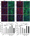
Mechanical stretch protects desmosomes from anti-Dsg3 Ab-induced instability. Confluent keratinocytes were subjected to combinations of AK23 mAb treatment with and without cyclic and static stretch. Cells were then stained and imaged to investigate how expression and distribution of selected proteins related to desmosomes (Dsg3, IF) changed with different treatments. The studied conditions were a) untreated control; b) cyclic stretch (10% strain, 1 Hz); c) static stretch (10% strain); d) AK23 mAb (2 μg mL−1); e) AK23 mAb (2 μg mL−1) with cyclic stretch; and f) AK23 mAb (2 μg mL−1) with static stretch; g) Average intensity of Dsg3 based on five images for each test condition over five separate experiments; all values are mean ± SEM (n = 5); h) A total of 10 lines were drawn across the cell–cell contact on each fluorescence images of Dsg3 under control, CS, SS, AK23+CS, and AK23+SS, and the peak intensity of each line normalized against the baseline intensity at the cytoplasm from each group were plotted as relative intensity (Rel. Dsg3 Intensity). Images are representative of more than five experiments for each condition; all values are normalized against control (control set to 1). all values are mean ± SEM (n = 10). * p < 0.05, **p < 0.01, ***p < 0.005, ns p > 0.05. Scale bar: 10 μm.
Figure 4.
Figure 4.
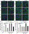
Mechanical stretch protects AJs from anti-Dsg3 Ab-induced instability. Confluent keratinocytes were subjected to combinations of AK23 mAb treatment with and without cyclic and static stretch. Cells were then stained and imaged to investigate how expression and distribution of selected proteins related to AJs (E-cad, F-actin) changed with different treatments. The studied conditions were a) untreated control; b) cyclic stretch (10% strain, 1 Hz); c) static stretch (10% strain); d) AK23 mAb (2 μg mL−1); e) AK23 mAb (2 μg mL−1) with cyclic stretch; and f) AK23 mAb (2 μg mL−1) with static stretch; g) Average intensity of E-cad based on five images for each test condition over five separate experiments; all values are mean ± SEM (n = 5); h) A total of 10 lines were drawn across the cell-cell contact on each fluorescence images of E-cad under control, CS, SS, AK23+CS, and AK23+SS, and the peak intensity of each line normalized against the baseline intensity at the cytoplasm from each group were plotted as relative intensity (Rel. E-cad Intensity); all values are mean ± SEM (n = 10). Images are representative of over five experiments for each condition; all values are normalized against control (control set to 1). *p < 0.05, **p < 0.01, ***p < 0.005, ns p > 0.05. Scale bar: 10 μm.
Figure 5.
Figure 5.
Mechanical stretch increases RhoA expression and localization at cell–cell adhesion without and with AK23 mAb treatment. Expression of RhoA was investigated with western blot and immunofluorescence imaging after exposure to both cyclic stretch (10% strain, 1 Hz) and static stretch (10% strain) to determine if mechanical stretch influences the RhoA pathway. a) Western blot analysis of RhoA, comparing expression in control to cells treated with cyclic and static stretch; GAPDH is shown as a loading control. Images are representative of over three experiments for each condition; b) Western blot results comparing RhoA expression of control to treatment with AK23 mAb and AK23 mAb with CS and SS. GAPDH is shown as a loading control. c) Quantitative Western blot data from (a) and (b), with all values are normalized against control (control set to 1). All values are mean ± SEM (n = 3). d) Fluorescent images of RhoA expression under corresponding test conditions: no anti-Dsg3 Ab and mechanical stretch (Control), cyclic stretch (CS), static stretch (SS), AK23 mAb (AK23), AK23 mAb and cyclic stretch (AK23+CS), AK23 mAb and static stretch (AK23+SS); Images are representative of over five experiments for each condition; e) Average cell intensity of based on five images for each test condition over five separate experiments; all values are mean ± SEM (n = 5); f) A total of 30 lines were drawn across the cell-cell contact on each fluorescence images under each condition, and the peak intensity of each line normalized against the baseline intensity at the cytoplasm from each group were plotted as relative intensity (Rel. RhoA Intensity); all values are mean ± SEM (n = 10). *p < 0.05, **p < 0.01, ***p < 0.005. Scale bar: 20 μm.
Figure 6.
Figure 6.
Activation of RhoA pathway with CN01 triggers changes in cell adhesion and structure proteins similar to mechanical stretch. Cells were studied under the influence of AK23 mAb with and without RhoA pathway activation via CN01 using immunofluorescence imaging. a) Dsg3 localization to cell-cell junctions triggered by AK23 mAb is increased with exposure to CN01; b) E-cad expression and localization to cell-cell junctions is increased with addition of CN01; c) RhoA expression is increased with exposure to AK23 mAb, and further increased with addition of CN01, while localization largely remains unchanged; Images are representative of over five experiments for each condition; d–f) Average intensity of Dsg3, E-cad, and RhoA, respectively, based on five images for each test condition over five separate experiments; all values are mean ± SEM (n = 5); g–i) A total of 30 lines were drawn across the cell-cell contact on each fluorescence images of Dsg3, E-cad, and RhoA, respectively, under control, AK23, and AK23+CN01 conditions, and the peak intensity of each line normalized against the baseline intensity at the cytoplasm from each group were plotted as relative intensity (Rel. Dsg3, E-cad, and RhoA Intensity); all values are mean ± SEM (n = 10). *p < 0.05, ***p < 0.005, ns: p > 0.05. Scale bar: 20 μm.
Figure 7.
Figure 7.
Protective mechanism of applied external stretch against anti-Dsg3-induced cell disassociation. a) Under normal physiology, IFs in keratinocytes provide resistance (blue arrows) to tension in the actin network (red arrows).[26] b) This equilibrium is interrupted by anti-Dsg3 Ab-induced desmosome disassembly, which reduces the resistance capacity of the IF network and lowers the tension in the AJs and actin network and mechanical stress at the cell–cell adhesion. c) A transient tensional compensation is invoked in AJs via RhoA activation and localization to the cell–cell border, resulting in a temporary increase in tension and mechanical stress. However, a continued assault from anti-Dsg3 Ab ultimately results in the complete cell dissociation. d) An externally applied uniaxial cyclic stretch (white arrows) can activate mechanosensitive pathways that upregulate RhoA, which elevates tension in AJs, thickens cortical actin, and stabilizes the cell–cell adhesion.

Similar articles

Cited by

References

    1. Kasperkiewicz M, Ellebrecht CT, Takahashi H, Yamagami J, Zillikens D, Payne AS, Amagai M, Nat. Rev. Dis. Primers 2017, 3, 17026. - PMC - PubMed
    1. Beutner EH, Lever WF, Witebsky E, Jordon R, Chertock B, J. Am. Med. Assoc 1965, 192, 682. - PubMed
    1. Sajda T, Hazelton J, Patel M, Seiffert-Sinha K, Steinman L, Robinson W, Haab BB, Sinha AA, Proc. Natl. Acad. Sci. USA 2016, 113, 1859. - PMC - PubMed
    1. Sinha AA, Sajda T, Front. Med 2018, 5, 218. - PMC - PubMed
    1. Amagai M, Klaus-Kovtun V, Stanley JR, Cell 1991, 67, 869. - PubMed

Publication types

LinkOut - more resources