. 2021 Jan;5(1):e2000176.
doi: 10.1002/adbi.202000176. Epub 2021 Jan 4.

Enhancement of Wound Healing Efficacy by Increasing the Stability and Skin-Penetrating Property of bFGF Using 30Kc19α-Based Fusion Protein

Affiliations

Enhancement of Wound Healing Efficacy by Increasing the Stability and Skin-Penetrating Property of bFGF Using 30Kc19α-Based Fusion Protein

Haein Lee et al. Adv Biol (Weinh). 2021 Jan.

Abstract

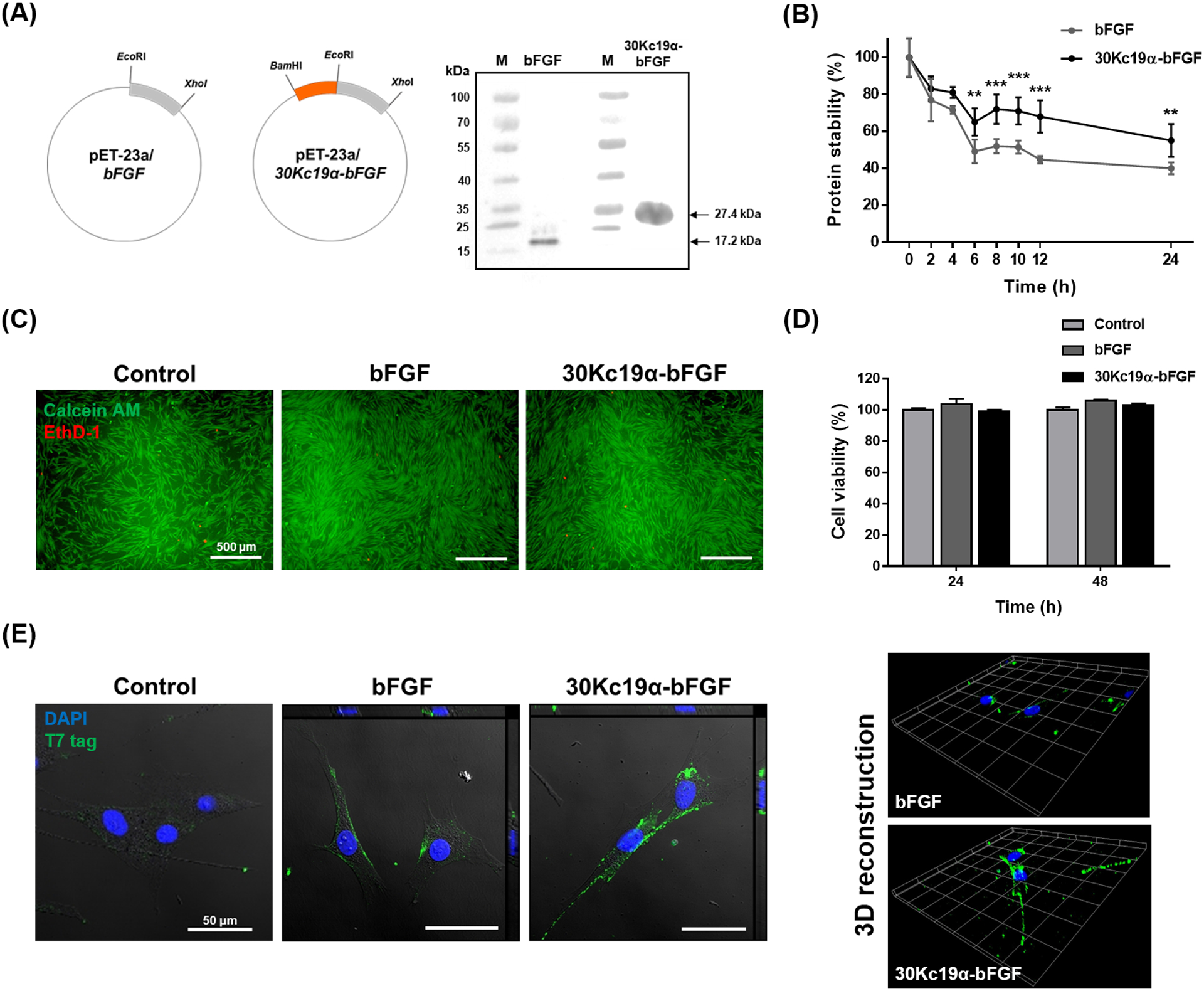
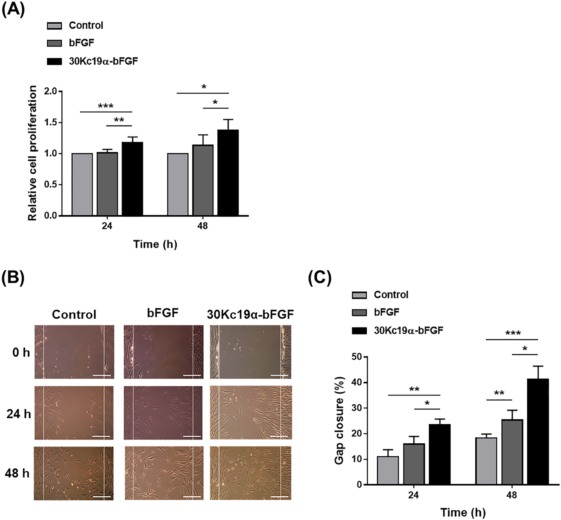
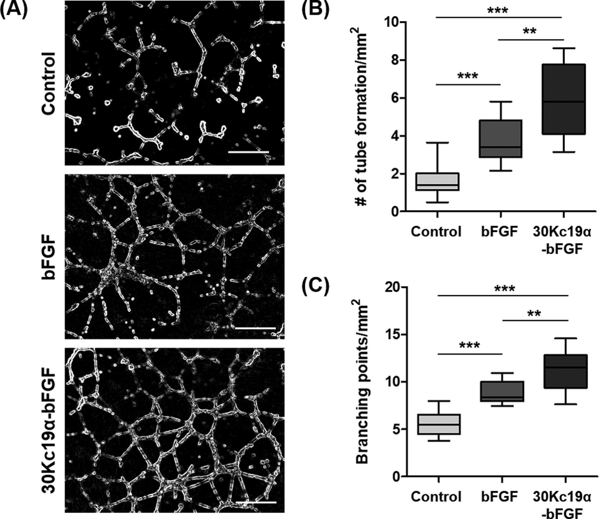
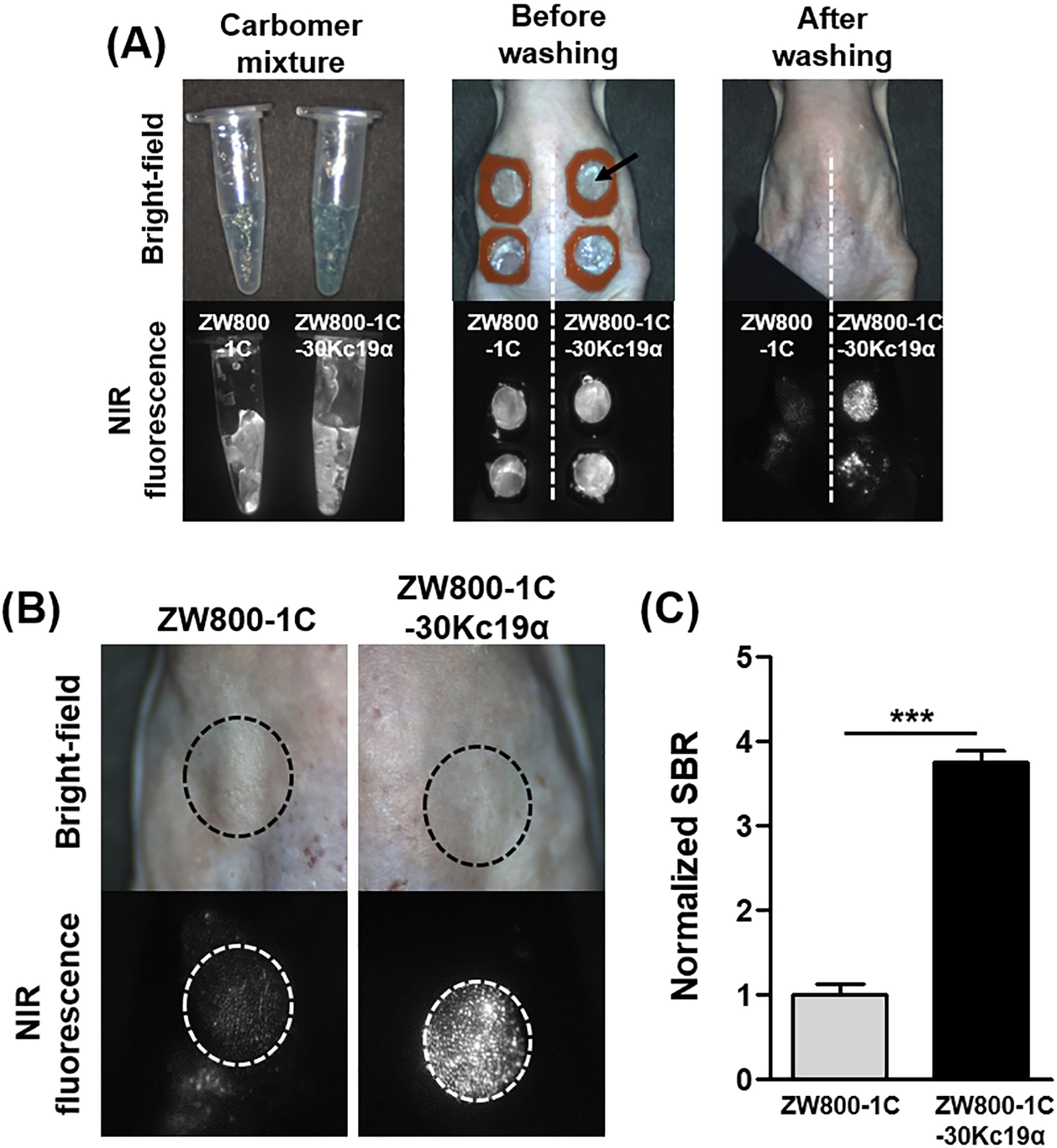
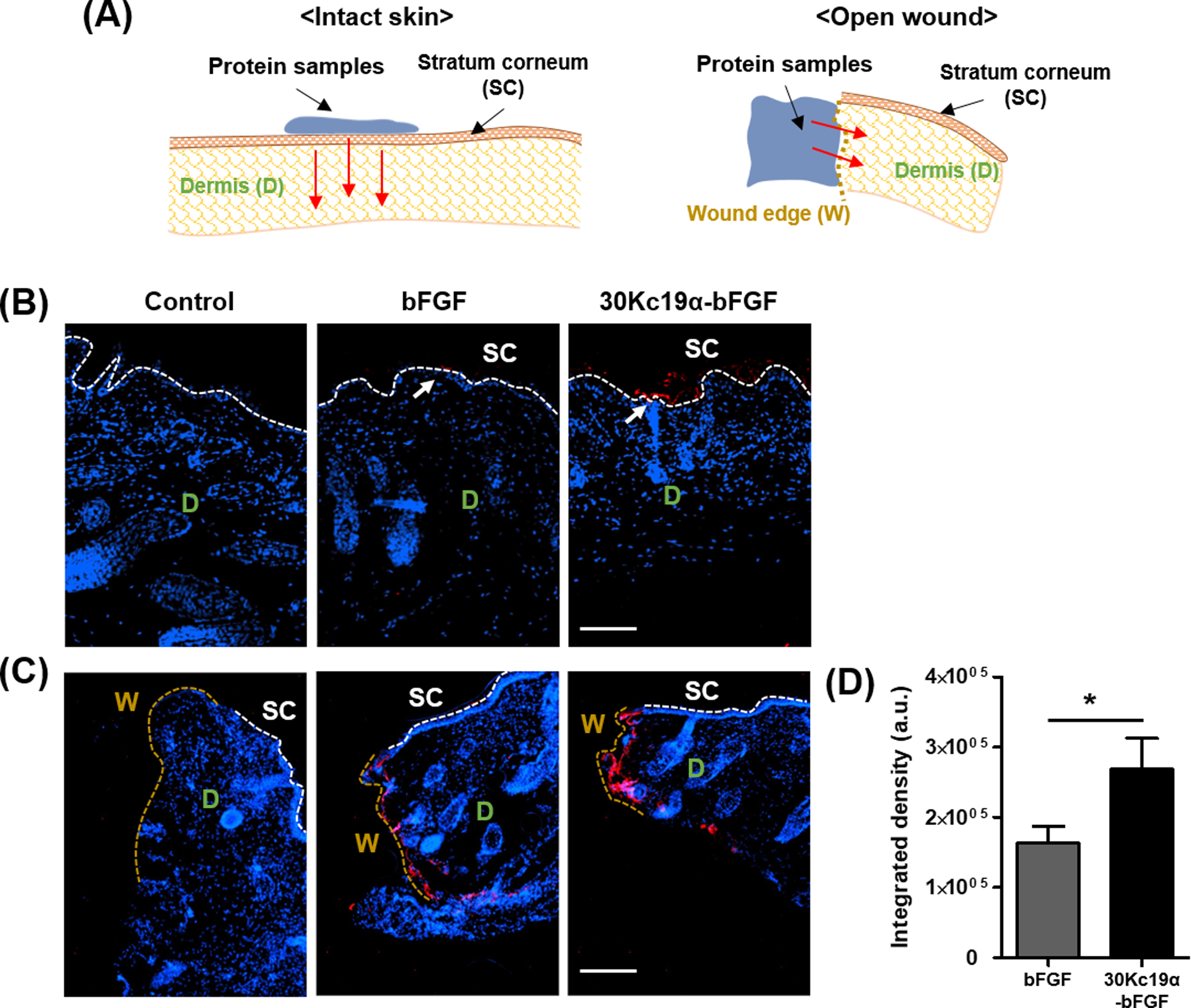
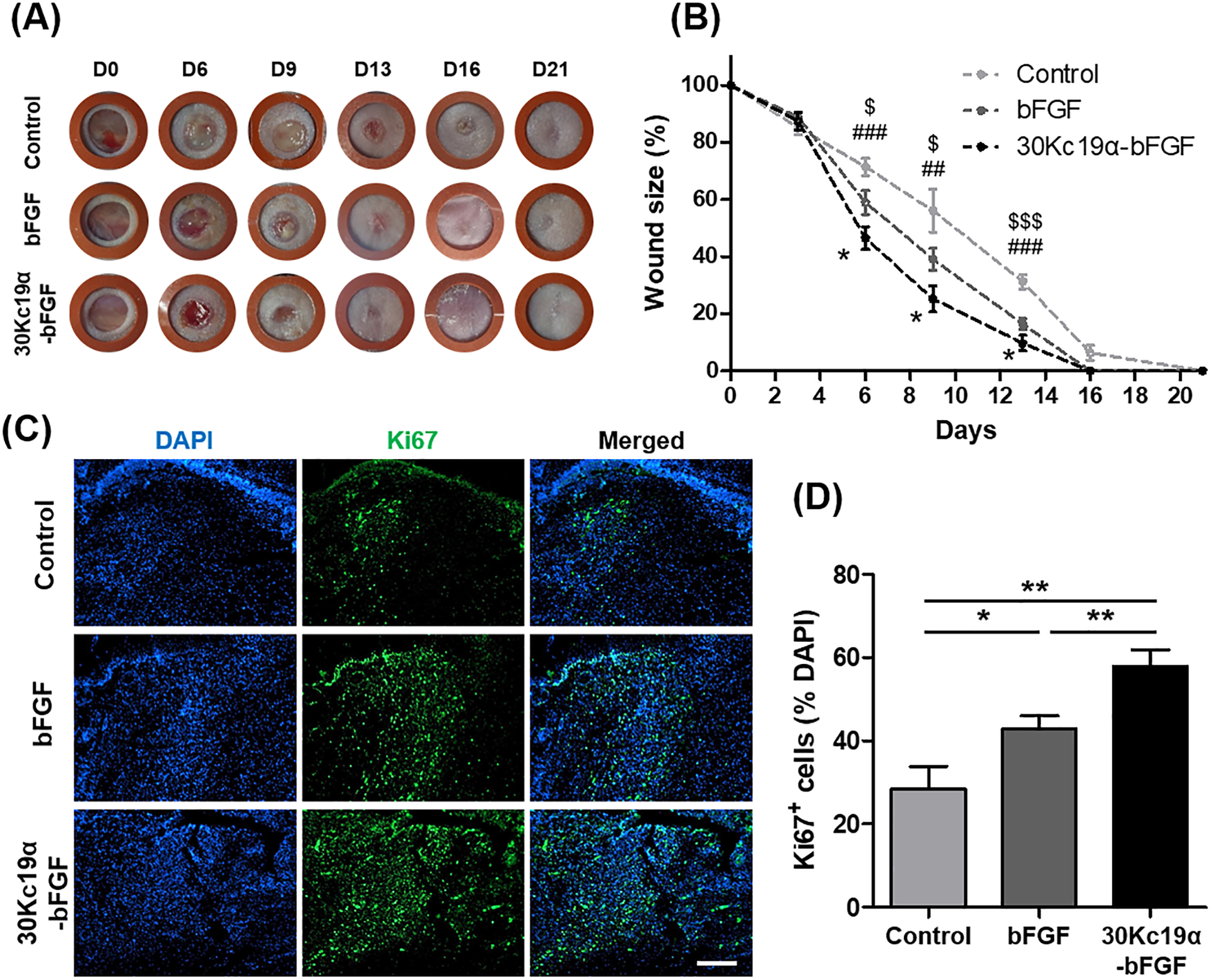
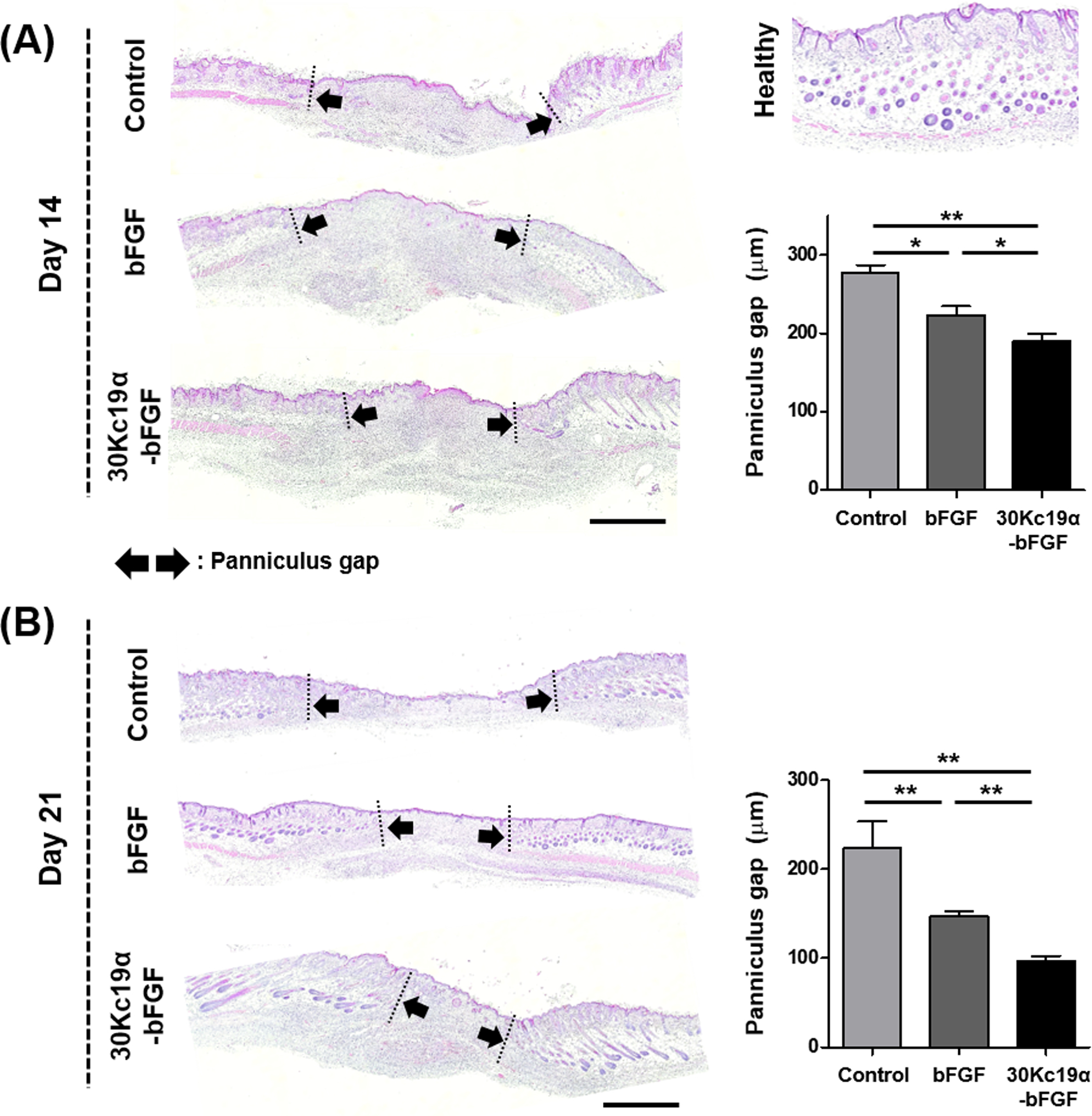
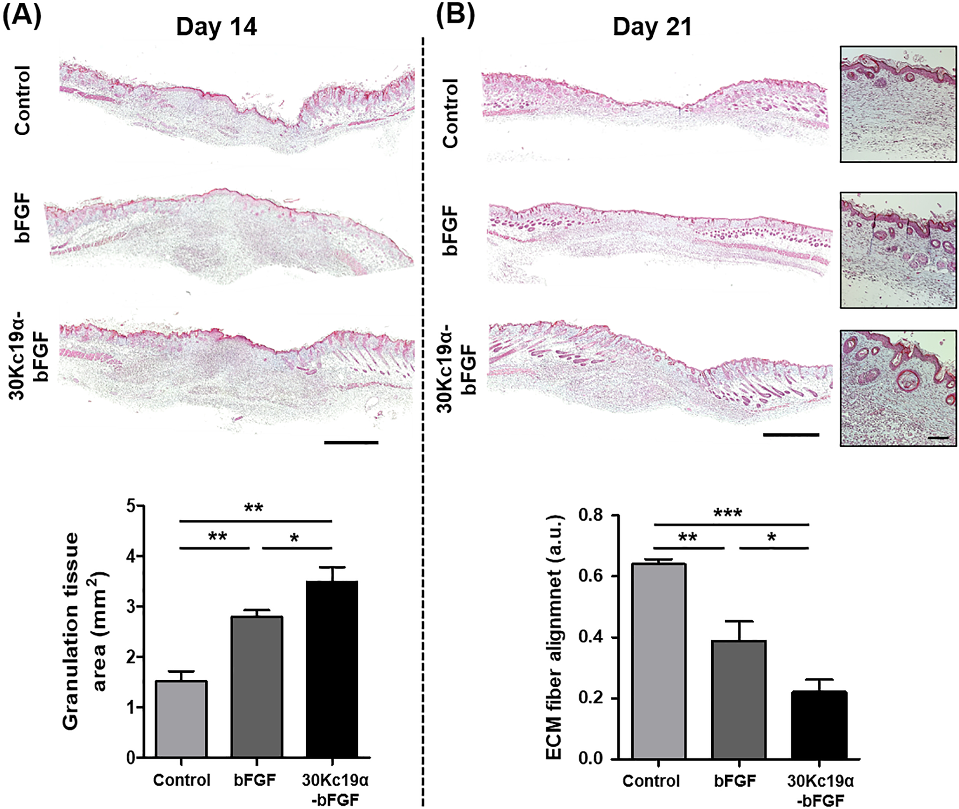
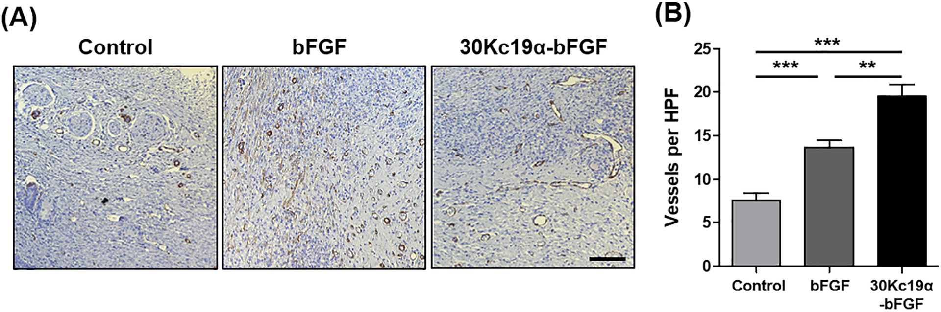
The instability of recombinant basic fibroblast growth factor (bFGF) is a major disadvantage for its therapeutic use and means frequent applications to cells or tissues are required for sustained effects. Originating from silkworm hemolymph, 30Kc19α is a cell-penetrating protein that also has protein stabilization properties. Herein, it is investigated whether fusing 30Kc19α to bFGF can enhance the stability and skin penetration properties of bFGF, which may consequently increase its therapeutic efficacy. The fusion of 30Kc19α to bFGF protein increases protein stability, as confirmed by ELISA. 30Kc19α-bFGF also retains the biological activity of bFGF as it facilitates the migration and proliferation of fibroblasts and angiogenesis of endothelial cells. It is discovered that 30Kc19α can improve the transdermal delivery of a small molecular fluorophore through the skin of hairless mice. Importantly, it increases the accumulation of bFGF and further facilitates its translocation into the skin through follicular routes. Finally, when applied to a skin wound model in vivo, 30Kc19α-bFGF penetrates the dermis layer effectively, which promotes cell proliferation, tissue granulation, angiogenesis, and tissue remodeling. Consequently, the findings suggest that 30Kc19α improves the therapeutic functionalities of bFGF, and would be useful as a protein stabilizer and/or a delivery vehicle in therapeutic applications.

Keywords: basic fibroblast growth factor; cell-penetrating proteins; protein stabilizer; skin wound healing; transdermal delivery.


Conflict of interest statement

Conflict of interest

The authors have declared that no competing interest exists.

Figures

Figure 1.
Figure 1.. The Fusion of 30Kc19α to bFGF and Its Effects on Stability and Cell-Penetration of bFGF.
(A) Plasmid construction of bFGF and 30Kc19α-bFGF and western blot analysis of recombinant proteins. Anti-T7 tag antibody was used as a primary antibody. (B) Protein stability assay using the bFGF ELISA kit. Proteins at 10 nM were incubated at 37°C from 0 to 24 hours. The relative protein stability was obtained by normalizing protein stability at each time point to protein stability at t = 0 (**p < 0.01, ***p < 0.001 compared with bFGF group). (C) Cytotoxicity assay using LIVE/DEAD kit and (D) CCK-8 kit. Human dermal fibroblasts (HDFs) were treated with 4 μM of the proteins for 24 and 48 hours. Live and dead cells from 48 hour treated samples were stained with green and red fluorescence respectively (Scale bar=500 μm). CCK-8 assay was done for both 24 and 48 hour treated samples. Data are expressed as relative cell viability normalized to the control group (n=4). (E) Confocal image with orthogonal projection (left) and 3D recostruction of z-stack images (right) of bFGF- and 30Kc19α-bFGF-treated HDFs. HDFs were treated with 4 μM of the proteins for 1 hour. The proteins were tagged with Alexa Fluro® 488, and the nucleus with DAPI (Scale bar = 50 μm).
Figure 2.
Figure 2.. In Vitro Wound Healing Effect of 30Kc19α-bFGF on HDFs.
(A) Cell proliferation assay using CCK-8 kit. HDFs were treated with 4 μM of the proteins in serum-free conditions for 24 and 48 hours. Data are expressed as relative cell proliferation normalized to the control group (n=8). (B) In vitro wound healing (scratch) assay (Scale bar = 200 μm) and (C) quantification of gap closure in three groups. Gaps were created by scratching the plate using a micropipette tip, and the gap closure was calculated by Equation 1 (n=3) (*p<0.05, **p<0.01, and ***p<0.001).
Figure 3.
Figure 3.. In Vitro Angiogenic Effects of 30Kc19α-bFGF on HUVECs.
(A) Human umbilical vein endothelial cells (HUVECs) formed vasculatures on Matrigel when treated with 4 μM of the proteins for 12 hours (Scale bar=500 μm). Angiogenic effects of 30Kc19α-bFGF were analyzed by measuring (B) the number of tube formation and (C) the count of branching points per unit area based on the microscopic images (n=8–10) (**p<0.01 and ***p<0.001).
Figure 4.
Figure 4.. In Vivo Skin Penetration Ability of 30Kc19α Using NIR Fluorophore.
(A) ZW800–1C-30Kc19α conjugate was prepared and applied to the dorsal skin of hairless mice. Images were obtained with the fluorescence-assisted resection and exploration (FLARE) imaging system. (B) Magnified images after washing showed that the ZW800–1C-30Kc19α could penetrate the SC effectively. (C) The fluorescent signal was quantified based on the magnified images, and the normalized signal-to-background ratio (SBR) in ZW800–1C-30Kc19α was significantly higher than that of ZW800–1C, suggesting that 30Kc19α improves the skin penetration ability of small molecules (n=3) (***p<0.001).
Figure 5.
Figure 5.. In Vivo Transdermal- and Dermal Penetration Ability of 30Kc19α-bFGF.
The transdermal and dermal penetration of 30Kc19α-bFGF on hairless mouse skin was visualized by immunofluorescent staining at 4 hours after application. (A) The experimental scheme showed how the protein samples were applied to the skin and penetrated—the immunofluorescent staining of the proteins in (B) intact skin and (C) open wound cases. 30Kc19α-bFGF exhibited more significant accumulation in the skin tissue than bFGF. (D) The integrated density was measured based on the fluorescent signal in the open wound (n=3–5) (Red arrow: the penetrating direction of proteins) (Scale bar=100 μm).
Figure 6.
Figure 6.. In Vivo Wound Healing Application of 30Kc19α-bFGF.
(A) Photographs of the wound up to 21 days. (B) The wound size-reduction profile was calculated based on the photographs. 30Kc19α-bFGF significantly promoted wound healing compared to bFGF (Statistical significance: * represents 30Kc19α-bFGF to bFGF; # represents 30Kc19α-bFGF to control; and $ represents bFGF to control. *,$p<0.05, ##p<0.01, and ###,$$$p<0.001). (C and D) Proliferative cells in the wound bed were estimated by immunofluorescence staining of Ki67 (green) on day 6, and quantitatively analyzed based on the fluorescent signals. (Scale bar=200 μm) (n=5–8) (*p<0.05 and **p<0.01,).
Figure 7.
Figure 7.. Histological and Qualitative Analysis Based on Hematoxylin and Eosin (H&E) Stain.
Panniculus gap was quantified on day 14 (A) and day 21 (B). It was revealed that 30Kc19α-bFGF accelerated wound regeneration via tissue granulation process. Although the panniculus gap of all groups continuously decreased, it was notably reduced with 30Kc19α-bFGF, whose tissue was being recovered at the fastest rate with a structure similar to healthy tissue (Scale bar=1 mm) (n=3–4) (*p<0.05 and **p<0.01).
Figure 8.
Figure 8.. Histological and Qualitative Analysis Based on Masson’s Trichrome (MTC) Stain.
Tissue granulation and the degree of extracellular matrix (ECM) fiber alignment were analyzed by MTC staining of the skin tissue on day 14 (A) and day 21 (B), respectively. It was confirmed that 30Kc19α-bFGF supported tissue granulation during the proliferative phase of the wound healing process. Also, both collagen deposition and the formation of skin appendages were enhanced in 30Kc19α-bFGF on day 21. The organization of ECM fiber alignment was quantified at the dermis layer, where the value represents its coherence. 30Kc19α-bFGF exhibited better results all round (Scale bar=1 mm and 100 μm in low and high magnified images, respectively) (n=3–4) (*p<0.05, **p<0.01, and ***p<0.001).
Figure 9.
Figure 9.. In Vivo Angiogenic Ability of 30Kc19α-bFGF.
(A) Immunohistochemistry staining of alpha-smooth muscle actin (α-SMA) on day 14 and (B) its quantitative analysis. 30Kc19α-bFGF improved angiogenesis during wound healing compared to control and bFGF and newly formed vessels had enlarged and stretched structures (Scale bar=100 μm) (n=10–15) (**p<0.01 and ***p<0.001).

Similar articles

Cited by

References

    1. Ornitz DM, Itoh N, Wiley Interdiscip. Rev. Dev. Biol 2015, 4, 215. - PMC - PubMed
    1. Behm B, Babilas P, Landthaler M, Schreml S, J. Eur. Acad. Dermatol 2012, 26, 812; - PubMed
    2. Barrientos S, Stojadinovic O, Golinko MS, Brem H, Tomic-Canic M, Wound Repair Regen. 2008, 16, 585. - PubMed
    1. Kanazawa S, Fujiwara T, Matsuzaki S, Shingaki K, Taniguchi M, Miyata S, Tohyama M, Sakai Y, Yano K, Hosokawa K, PLoS One 2010, 5, e12228; - PMC - PubMed
    2. Song YH, Zhu YT, Ding J, Zhou FY, Xue JX, Jung JH, Li ZJ, Gao WY, Mol. Med. Rep 2016, 14, 3336; - PubMed
    3. O’keefe EJ, Chiu ML, Payne RE Jr, J. Investig. Dermatol 1988, 90; - PubMed
    4. Sogabe Y, Abe M, Yokoyama Y, Ishikawa O, Wound Repair Regen. 2006, 14, 457; - PubMed
    5. Przybylski M, J. Wound Care 2009, 18, 516. - PubMed
    1. Montesano R, Vassalli J-D, Baird A, Guillemin R, Orci L, Proc. Natl. Acad. Sci. U.S.A 1986, 83, 7297; - PMC - PubMed
    2. Akita S, Akino K, Hirano A, Adv. Wound Care (New Rochelle) 2013, 2, 44; - PMC - PubMed
    3. Sasaki T, J. Dermatol 1992, 19, 664. - PubMed
    1. Uchi H, Igarashi A, Urabe K, Koga T, Nakayama J, Kawamori R, Tamaki K, Hirakata H, Ohura T, Furue M, Eur. J. Dermatol 2009, 19, 461; - PubMed
    2. Nunes QM, Li Y, Sun C, Kinnunen TK, Fernig DG, PeerJ 2016, 4, e1535; - PMC - PubMed
    3. Akita S, Akino K, Imaizumi T, Hirano A, Wound Repair Regen. 2008, 16, 635. - PubMed

Publication types

Substances

LinkOut - more resources