

Tumor Segmentation in Patients with Head and Neck Cancers Using Deep Learning Based-on Multi-modality PET/CT Images
- PMID: 33724743
- PMCID: PMC7929493
- DOI: 10.1007/978-3-030-67194-5_10
Tumor Segmentation in Patients with Head and Neck Cancers Using Deep Learning Based-on Multi-modality PET/CT Images
Abstract
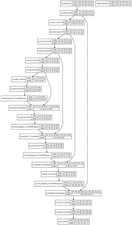
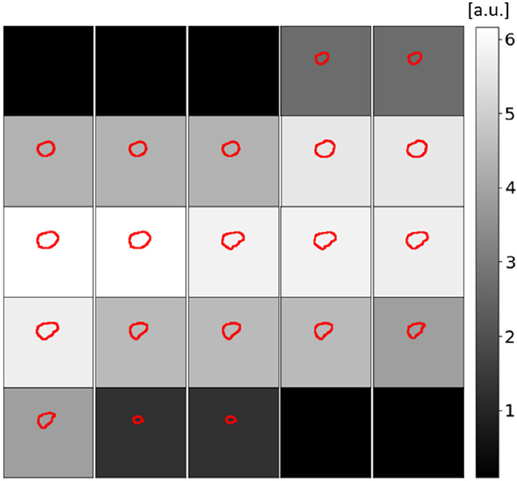
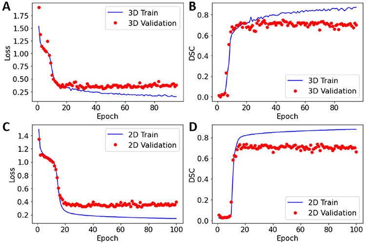
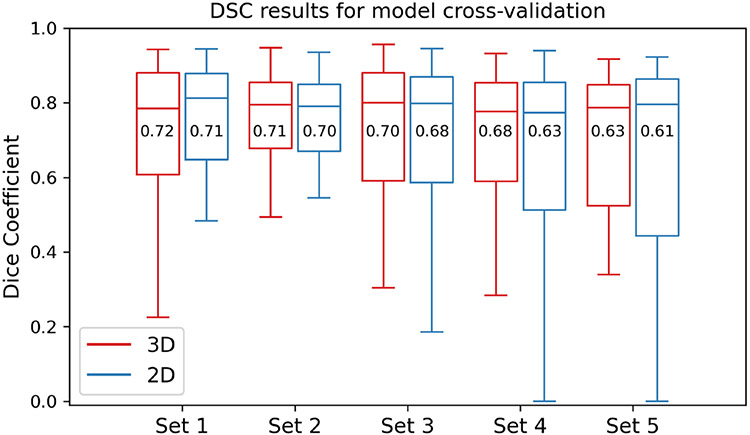
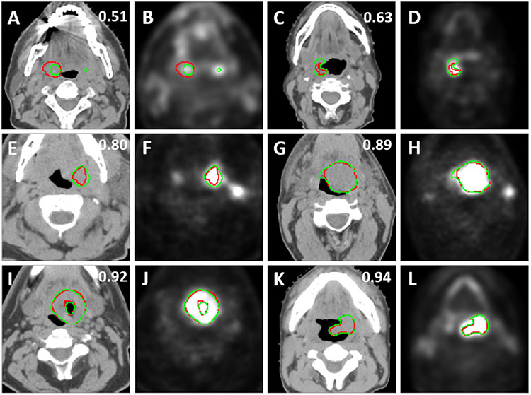
Segmentation of head and neck cancer (HNC) primary tumors onmedical images is an essential, yet labor-intensive, aspect of radiotherapy.PET/CT imaging offers a unique ability to capture metabolic and anatomicinformation, which is invaluable for tumor detection and border definition. Anautomatic segmentation tool that could leverage the dual streams of informationfrom PET and CT imaging simultaneously, could substantially propel HNCradiotherapy workflows forward. Herein, we leverage a multi-institutionalPET/CT dataset of 201 HNC patients, as part of the MICCAI segmentationchallenge, to develop novel deep learning architectures for primary tumor auto-segmentation for HNC patients. We preprocess PET/CT images by normalizingintensities and applying data augmentation to mitigate overfitting. Both 2D and3D convolutional neural networks based on the U-net architecture, which wereoptimized with a model loss function based on a combination of dice similaritycoefficient (DSC) and binary cross entropy, were implemented. The median andmean DSC values comparing the predicted tumor segmentation with the groundtruth achieved by the models through 5-fold cross validation are 0.79 and 0.69for the 3D model, respectively, and 0.79 and 0.67 for the 2D model, respec-tively. These promising results show potential to provide an automatic, accurate,and efficient approach for primary tumor auto-segmentation to improve theclinical practice of HNC treatment.
Keywords: PET; CT; Tumor segmentation; Head and neck cancer; Deep learning; Auto-contouring.
Figures
References
-
- Vorwerk H, et al.: Protection of quality and innovation in radiation oncology: the prospective multicenter trial the German Society of Radiation Oncology (DEGRO-QUIRO study). Strahlentherapie und Onkol. 190, 433–443 (2014) - PubMed
Grants and funding
LinkOut - more resources
Full Text Sources
Other Literature Sources