

Covid-19 Interstitial Pneumonia: Histological and Immunohistochemical Features on Cryobiopsies
- PMID: 33725700
- PMCID: PMC8018216
- DOI: 10.1159/000514822
Covid-19 Interstitial Pneumonia: Histological and Immunohistochemical Features on Cryobiopsies
Abstract
Background: The pathogenetic steps leading to Covid-19 interstitial pneumonia remain to be clarified. Most postmortem studies to date reveal diffuse alveolar damage as the most relevant histologic pattern. Antemortem lung biopsy may however provide more precise data regarding the earlier stages of the disease, providing a basis for novel treatment approaches.
Objectives: To ascertain the morphological and immunohistochemical features of lung samples obtained in patients with moderate Covid-19 pneumonia.
Methods: Transbronchial lung cryobiopsy was carried out in 12 Covid-19 patients within 20 days of symptom onset.
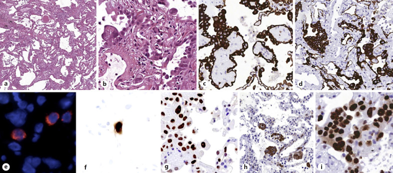
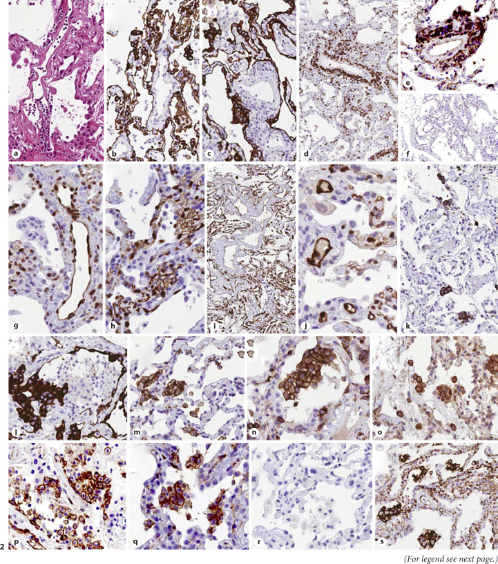
Results: Histopathologic changes included spots of patchy acute lung injury with alveolar type II cell hyperplasia, with no evidence of hyaline membranes. Strong nuclear expression of phosphorylated STAT3 was observed in >50% of AECII. Interalveolar capillaries showed enlarged lumen and were in part arranged in superposed rows. Pulmonary venules were characterized by luminal enlargement, thickened walls, and perivascular CD4+ T-cell infiltration. A strong nuclear expression of phosphorylated STAT3, associated with PD-L1 and IDO expression, was observed in endothelial cells of venules and interstitial capillaries. Alveolar spaces macrophages exhibited a peculiar phenotype (CD68, CD11c, CD14, CD205, CD206, CD123/IL3AR, and PD-L1).
Conclusions: Morphologically distinct features were identified in early stages of Covid-19 pneumonia, with epithelial and endothelial cell abnormalities different from either classical interstitial lung diseases or diffuse alveolar damage. Alveolar type II cell hyperplasia was a prominent event in the majority of cases. Inflammatory cells expressed peculiar phenotypes. No evidence of hyaline membranes and endothelial changes characterized by IDO expression might in part explain the compliance and the characteristic pulmonary vasoplegia observed in less-advanced Covid-19 pneumonia.
Keywords: Acute respiratory distress syndrome; Coronavirus; Covid-19; Cryobiopsy; Indoleamine 2,3-dioxygenase-1; Lung biopsy; SARS-CoV-2; pSTAT-3.
© 2021 S. Karger AG, Basel.
Conflict of interest statement
The authors have no conflicts of interest to declare related to this manuscript.
Figures
Comment on
-
Time Course of Lung Changes at Chest CT during Recovery from Coronavirus Disease 2019 (COVID-19).Radiology. 2020 Jun;295(3):715-721. doi: 10.1148/radiol.2020200370. Epub 2020 Feb 13. Radiology. 2020. PMID: 32053470 Free PMC article.
References
-
- Menter T, Haslbauer JD, Nienhold R, Savic S, Hopfer H, Deigendesch N, et al. Post-mortem examination of COVID19 patients reveals diffuse alveolar damage with severe capillary congestion and variegated findings of lungs and other organs suggesting vascular dysfunction. Histopathology. 2020 - PMC - PubMed
Publication types
MeSH terms
LinkOut - more resources
Full Text Sources
Other Literature Sources
Medical
Research Materials
Miscellaneous