[Congenital pulmonary vein stenosis and bronchopulmonary vascular malformation]
- PMID: 33725717
- PMCID: PMC8351654
- DOI: 10.24875/ACM.20000362
[Congenital pulmonary vein stenosis and bronchopulmonary vascular malformation]
Abstract
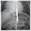
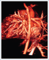
The objective is demonstrate the diagnostic process and evolution of a patient with a diagnosis of congenital pulmonary vein stenosis and broncho-pulmonary vascular malformation. One year old female patient with repeated bronchopneumonia, acrocyanosis, split S2, cardiomegaly, pulmonary hypertension, with a clinical diagnosis of atrial septal defect. The echocardiogram demonstrated left sided vein pulmonary stenosis. The cardiac catheterization demonstrated arterial-venous fistulas apical on the right lung. Magnetic Resonance image and angiography showed an aberrant arterial vessel parallel to the abdominal aorta which flow the right pulmonary lobe. The cardiac tomography angiography reported confluence of right-sided pulmonary veins. A lobectomy is performed. Patient died in post-operative due to massive pulmonary hemorrhaging. This is the first patient mentioned in written literature with pulmonary vein stenosis associated with pulmonary sequestration, with normal venous connection. Echocardiography represents the specific standard study ideal for initial diagnostic for patients with pulmonary vein stenosis.
El objetivo es mostrar el diagnóstico y la evolución de una paciente con estenosis de venas pulmonares y secuestro pulmonar. Se trata de una niña de 1 año de edad, con bronconeumonías de repetición, acrocianosis, 2R intenso, cardiomegalia, hipertensión venocapilar pulmonar, con diagnóstico clínico de comunicación interauricular. El ecocardiograma mostró estenosis de venas pulmonares izquierdas. El cateterismo cardiaco detectó fístulas arteriovenosas en la región apical del pulmón derecho. La imagen de resonancia magnética y la angiografía mostraron un vaso arterial aberrante paralelo a la aorta abdominal y con flujo dirigido al lóbulo pulmonar derecho. La angiotomografía reportó confluencia de las venas pulmonares del lado derecho. Se realizó lobectomía derecha. La paciente falleció en el posoperatorio debido a una hemorragia masiva pulmonar. Esta paciente es la primera descrita en la literatura con estenosis de venas pulmonares congénita asociada a secuestro pulmonar. La ecocardiografía es el estudio diagnóstico ideal inicial en los pacientes con estenosis congénita de venas pulmonares.
Keywords: Pulmonary vein stenosis; Congenital heart disease; Pulmonary heart disease; Pulmonary sequestration.
Conflict of interest statement
Declaramos que no existe ningún conflicto de intereses.
Figures


Similar articles
-
Computed tomographic parenchymal lung findings in premature infants with pulmonary vein stenosis.Pediatr Radiol. 2023 Aug;53(9):1874-1884. doi: 10.1007/s00247-023-05673-y. Epub 2023 Apr 28. Pediatr Radiol. 2023. PMID: 37106091
-
[Echocardiographic diagnosis of infracardiac total anomalous pulmonary venous connection].Beijing Da Xue Xue Bao Yi Xue Ban. 2017 Oct 18;49(5):883-888. Beijing Da Xue Xue Bao Yi Xue Ban. 2017. PMID: 29045974 Chinese.
-
Computed tomography of partial anomalous pulmonary venous connection in adults.J Comput Assist Tomogr. 2003 Sep-Oct;27(5):743-9. doi: 10.1097/00004728-200309000-00011. J Comput Assist Tomogr. 2003. PMID: 14501365
-
Partial anomalous pulmonary venous connection to the right side of the heart.J Thorac Cardiovasc Surg. 1989 Nov;98(5 Pt 2):861-8. J Thorac Cardiovasc Surg. 1989. PMID: 2682021 Review.
-
Computed Tomography Angiography and Magnetic Resonance Angiography of Congenital Anomalies of Pulmonary Veins.J Comput Assist Tomogr. 2019 May/Jun;43(3):399-405. doi: 10.1097/RCT.0000000000000857. J Comput Assist Tomogr. 2019. PMID: 31082945 Review.
References
-
- Holt BD, Moller HJ, Larson S, Johnson CM. Primary pulmonary vein stenosis. Am J Cardiol. 2007;99:568–72. - PubMed
-
- Latson AL, Prieto LR. Congenital and acquired pulmonary vein stenosis. Circulation. 2007;115:103–8. - PubMed
-
- Vick WG, III, Murphy JD, Ludomirsky A, Morrow WR, Morriss JM, -Danford AD, et al. Pulmonary venous systemic ventricular inflow obstruction in patients with congenital heart disease:detection by combined two-dimensional and Doppler echocardiography. J Am Coll Cardiol. 1987;9:580–7. - PubMed
-
- Van Son AJ, Danielson KG, Puga JF, Edwards DW, Driscoll JD. Repair of congenital and acquired pulmonary vein stenosis. Ann Thorac Surg. 1995;60:144–50. - PubMed
-
- Laux D, Rocchisani MA, Boudjemline Y, Gouton M, Bonnet D, Ovaert C. Pulmonary hypertension in the preterm infant with chronic lung disease can be caused by pulmonary bien stenosis:a must-know entity. Pediatr Cardiol. 2016;37:313–21. - PubMed
Publication types
LinkOut - more resources
Full Text Sources
Other Literature Sources

