. 2021 Mar 31;288(1947):20210088.
doi: 10.1098/rspb.2021.0088. Epub 2021 Mar 17.

Drosophila glue protects from predation

Affiliations

Drosophila glue protects from predation

Flora Borne et al. Proc Biol Sci. .

Abstract

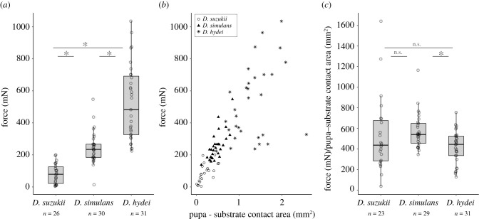
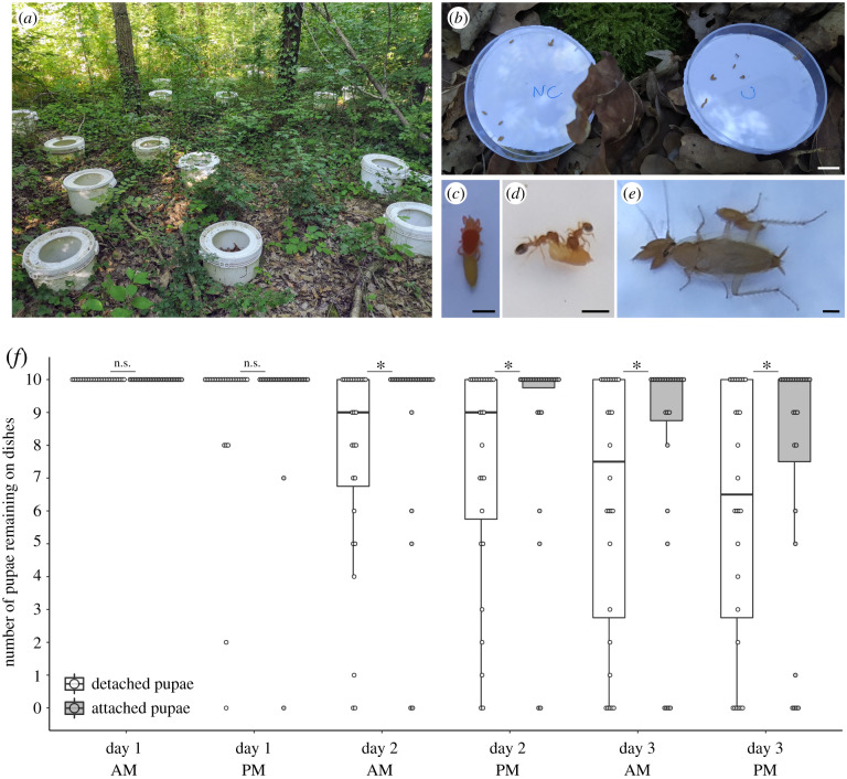
Animals can be permanently attached to a substrate in terrestrial environments at certain stages of their development. Pupa adhesion has evolved multiple times in insects and is thought to maintain the animal in a place where it is not detectable by predators. Here, we investigate whether pupa adhesion in Drosophila can also protect the animal by preventing potential predators from detaching the pupa. We measured the adhesion of Drosophila species sampled from the same area and found that pupa adhesion varies among species, which can be explained by different glue production strategies. Then, we compared attached and manually detached pupae in both field and laboratory assays to investigate the role of pupa adhesion to prevent predation. First, we found that attached pupae remain onsite 30% more than detached pupae in the field after 3 days, probably because they are less predated. Second, we observed that attached pupae are less efficiently predated by ants in the laboratory: they are not carried back to the ant nest and more ants are needed to consume them onsite. Our results show that pupa adhesion can prevent the animal from being taken away by predators and is crucial for Drosophila fly survival.

Keywords: Drosophila; ant; bioadhesive; insect; predation; pupa.


Figures

Figure 1.
Figure 1.
Pupa adhesion varies between species originating from the same location. (a) Adhesion strength of three Drosophila lines collected in Vincennes. Force indicates the force required to detach a pupa naturally attached to a glass slide. Each dot corresponds to a single pupa and n indicates the total number of pupae tested for each species. Ends of the boxes define the first and third quartiles. The black horizontal line represents the median. The vertical line on the top of the box extends to the largest value no further than 1.5 × IQR from the upper hinge of the box. The vertical line on the bottom of the box extends to the smallest value at most 1.5 × IQR of the hinge (IQR, inter-quartile range is the distance between the first and the third quartiles.). Data beyond the end of these lines are ‘outlying’ points. Asterisk indicates significant differences between D. suzukii and D. hydei, D. suzukii and D. simulans and D. simulans and D. hydei (p < 0.05). (b) Relationship between pupa adhesion force and pupa–substrate contact area. Each dot corresponds to a single pupa. Drosophila suzukii pupae are represented as circles, D. simulans as triangles and D. hydei as stars. (c) Adhesion strength corrected by the pupa–substrate contact area. Boxplots and asterisk as in (a). n.s. indicates not significant (p > 0.05).
Figure 2.
Figure 2.
Attached pupae are taken away less frequently than detached pupae in nature. (a) Picture of the half-buried buckets installed in Bois de Vincennes. (b) Picture of two dishes placed in the centre of a bucket and containing attached (C) or detached (NC) pupae. (ce) Predators observed in the dishes during the experiments: a red mite spider (c), two Temnothorax nylanderi ants (d) and a cockroach (e). (f) Boxplot represents the number of pupae present in one dish at the counting time. Pupae were counted twice a day in the morning (AM) and in the afternoon (PM). Each dot represents the count for one dish. White boxes represent dishes with detached pupae and grey boxes dishes with attached pupae. Boxplots are defined as previously (figure 1a). Asterisk indicates significant difference between the number of remaining pupae between the attached and detached conditions (p < 0 0.05, Wilcoxon test). Scale bars: (b) 1 cm, (ce) 1 mm. (Online version in colour.)
Figure 3.
Figure 3.
Attached pupae require more time and more ants to go away. (a) Number of ants in direct contact with the pupa over the duration of the experiment. Each cell represents one experiment with X_Y corresponding to the experiment with the colony X during the trial Y. Grey cells represent experiments using D. suzukii and white cells using D. simulans. Red lines represent the number of ants over time on attached pupa and blue lines on detached pupa. Vertical lines represent the time when the attached pupa (in red) or the detached pupa (in blue) arrives in the nest (dashed line) or is fully consumed outside the nest (full line). (b) Time during which ants are in contact with attached pupa (grey box) and detached pupa (white box) outside the nest (until the pupa is completely consumed outside the nest or enters into the nest). Each dot represents one experiment. (c) Maximum number of ants observed in contact with detached pupa (white box) and attached pupa (grey box) during the duration of the experiment. Each dot represents one experiment. Boxplots are defined as previously (figure 1a). Asterisk indicates significant differences (p < 0.05, Wilcoxon tests).

References

    1. Wahl M. 1997. Living attached: aufwuchs, fouling, epibiosis. Fouling organisms of the Indian Ocean: biology and control technology (eds R Nagabhushanam, M-F Thompson), pp. 31-83. New Delhi, India: Oxford & IBH Publishing Co. Pvt. Ltd.
    1. Fordyce JA, Nice CC. 2002. Variation in butterfly egg adhesion: adaptation to local host plant senescence characteristics?: variation in butterfly egg adhesion. Ecol. Lett. 6, 23-27. (10.1046/j.1461-0248.2003.00389.x) - DOI
    1. Hinton HE. 1981. Biology of insect eggs, p. 3, 1st edn. New York, NY: Pergamon Press.
    1. Voigt D, Gorb S. 2010. Egg attachment of the asparagus beetle Crioceris asparagi to the crystalline waxy surface of Asparagus officinalis. Proc. R. Soc. B 277, 895-903. (10.1098/rspb.2009.1706) - DOI - PMC - PubMed
    1. Heming BS. 2003. Insect development and evolution, pp. 246-247. Cornell University Press; cited 17 December 2020. See https://www.jstor.org/stable/10.7591/j.ctv75d5sv.

Publication types

LinkOut - more resources