. 2021 Mar 16;14(1):157.
doi: 10.1186/s13071-021-04659-9.

A U.S. isolate of Theileria orientalis, Ikeda genotype, is transmitted to cattle by the invasive Asian longhorned tick, Haemaphysalis longicornis

Affiliations

A U.S. isolate of Theileria orientalis, Ikeda genotype, is transmitted to cattle by the invasive Asian longhorned tick, Haemaphysalis longicornis

Kelcey D Dinkel et al. Parasit Vectors. .

Abstract

Background: Theileria orientalis is a tick-borne hemoparasite that causes anemia, ill thrift, and death in cattle globally. The Ikeda strain of T. orientalis is more virulent than other strains, leading to severe clinical signs and death of up to 5% of affected animals. Within the Asia-Pacific region, where it affects 25% of Australian cattle, T. orientalis Ikeda has a significant economic impact on the cattle industry. In 2017, T. orientalis Ikeda was detected in a cattle herd in Albermarle County, Virginia, United States. Months earlier, the U.S. was alerted to the invasion of the Asian longhorned tick, Haemaphysalis longicornis, throughout the eastern U.S. Abundant H. longicornis ticks were identified on cattle in the T. orientalis-affected herd in VA, and a subset of ticks from the environment were PCR-positive for T. orientalis Ikeda. A strain of T. orientalis from a previous U.S. outbreak was not transmissible by H. longicornis; however, H. longicornis is the primary tick vector of T. orientalis Ikeda in other regions of the world. Thus, the objective of this study was to determine whether invasive H. longicornis ticks in the U.S. are competent vectors of T. orientalis Ikeda.

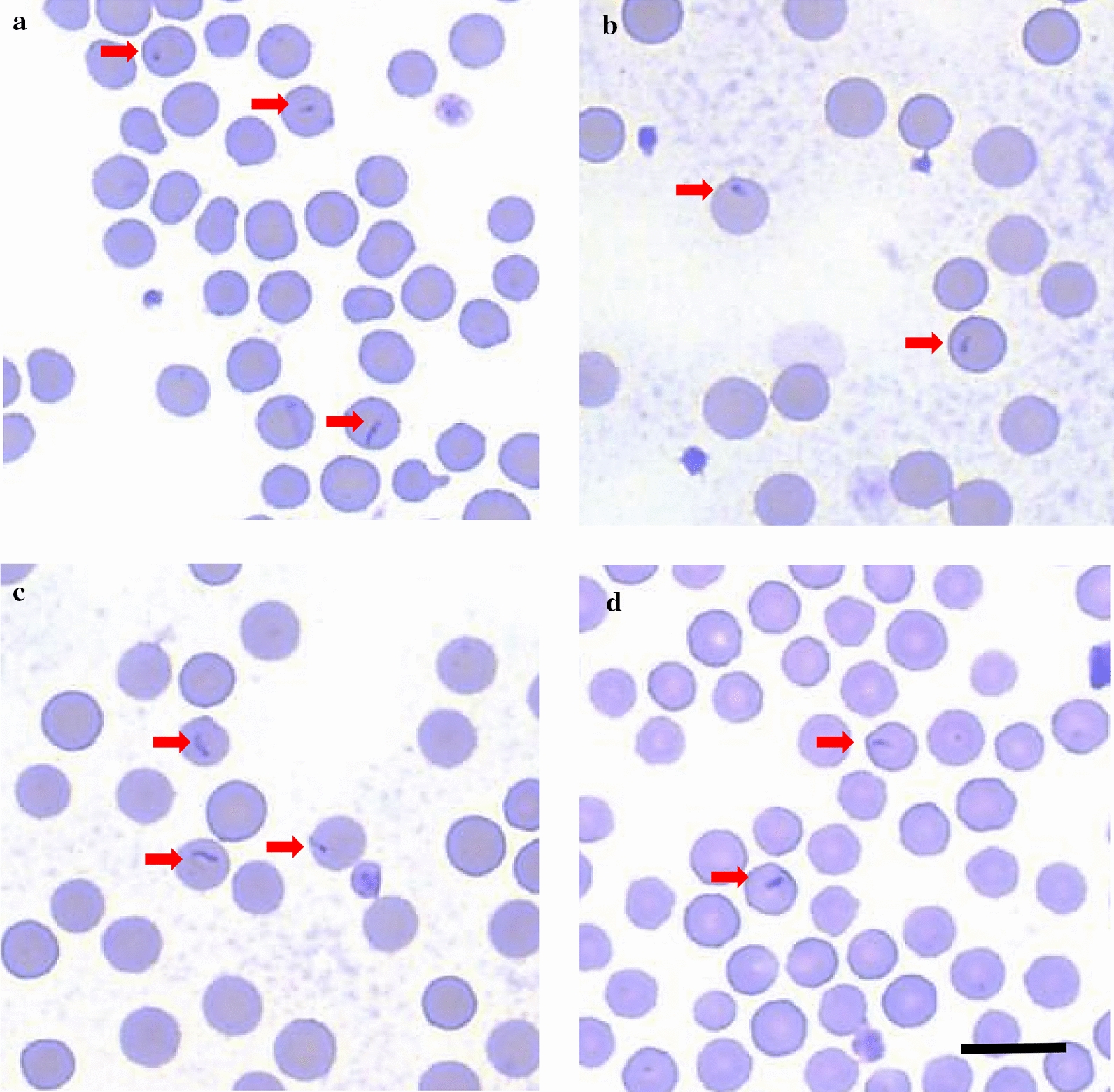
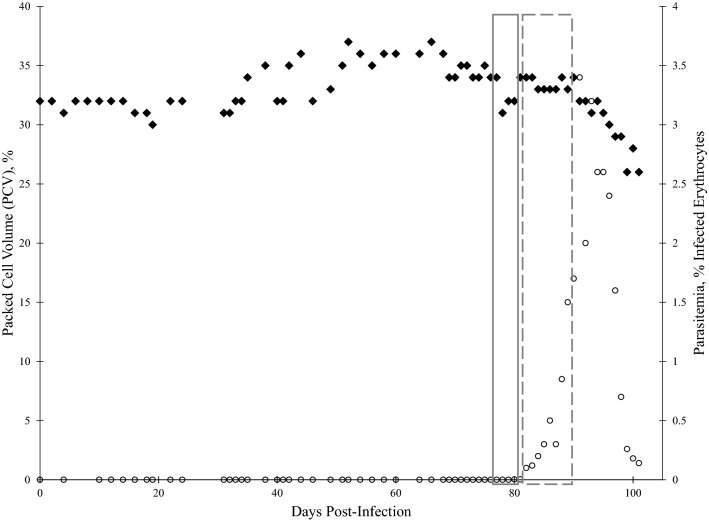
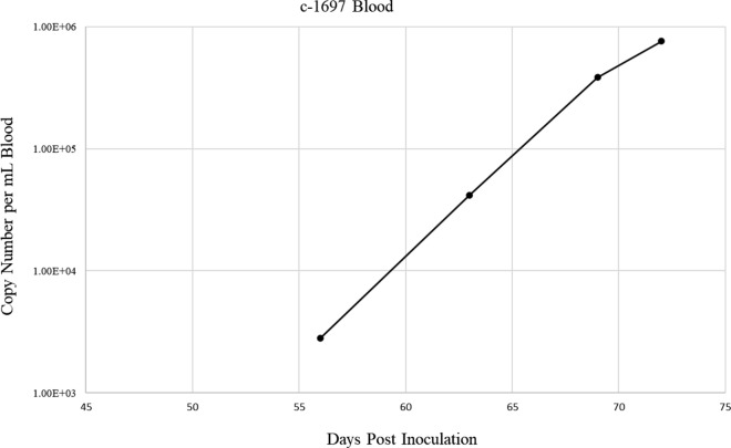
Methods: Nymphal H. longicornis ticks were fed on a splenectomized calf infected with the VA-U.S.-T. orientalis Ikeda strain. After molting, a subset of adult ticks from this cohort were dissected, and salivary glands assayed for T. orientalis Ikeda via qPCR. The remaining adult ticks from the group were allowed to feed on three calves. Calves were subsequently monitored for T. orientalis Ikeda infection via blood smear cytology and PCR.

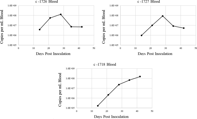
Results: After acquisition feeding on a VA-U.S.-T. orientalis Ikeda-infected calf as nymphs, a subset of molted adult tick salivary glands tested positive by qPCR for T. orientalis Ikeda. Adult ticks from the same cohort successfully transmitted T. orientalis Ikeda to 3/3 naïve calves, each of which developed parasitemia reaching 0.4-0.9%.

Conclusions: Our findings demonstrate that U.S. H. longicornis ticks are competent vectors of the VA-U.S.-T. orientalis Ikeda strain. This data provides important information for the U.S. cattle industry regarding the potential spread of this parasite and the necessity of enhanced surveillance and control measures.

Keywords: Asian longhorned tick; Cattle; Haemaphysalis longicornis; Ikeda genotype; Theileria orientalis; Transmission.


Conflict of interest statement

The authors declare that they have no competing interests.

Figures

Fig. 1
Fig. 1
Representative blood smears from calves 1697, 1718, 1726, and 1727 following T. orientalis Ikeda-infection. In each calf, few erythrocytes contain 1–2.5 µm × 0.5 µm, tear-drop shaped, intracellular piroplasms (arrows). No other evidence of anemia, erythrocyte destruction, or cellular regeneration is present. a calf 1697, b calf 1718, c calf 1726, d calf 1727. Scale bar: 10 µm
Fig. 2
Fig. 2
Packed cell volume (PCV) and peripheral blood percent parasitemia of erythrocytes (PPE) of calf 1697 following T. orientalis Ikeda blood stabilate inoculation. PCV and Diff-quick-stained blood smears were evaluated following intravenous inoculation with T. orientalis-Ikeda-infected blood stabilate. PCV (♦ left y-axis) and PPE (○ right y-axis) are depicted as percentages, and each value is derived from analysis of a single blood sample taken on the indicated day post-inoculation. Grey boxes indicate timeline of nymphal tick acquisition feeds. Solid box: Batch #1, Dashed box: Batch #2
Fig. 3
Fig. 3
Quantitative PCR of T. orientalis mpsp in blood samples from acquisition-fed calf 1697. EDTA-anticoagulated peripheral blood samples were collected during course of infection and evaluated for T. orientalis by mpsp qPCR. Data points represent the copy number per mL of blood collected on the indicated day post inoculation
Fig. 4
Fig. 4
Quantitative PCR of T. orientalis mpsp in salivary gland samples from acquisition-fed adult H. longicornis ticks. Salivary glands dissected from a subset of adult ticks from Batch #1 and Batch #2 were stimulation fed on calves 1718, 1726, and 1727. Only positive ticks are depicted in the graph. Columns represent the copy number per salivary gland pair dissected from a single tick. *Single positive salivary gland pair from batch #2
Fig. 5
Fig. 5
Quantitative PCR analysis of T. orientalis mpsp gene fragment in blood samples from transmission-fed calves 1718, 1726, and 1727. Peripheral blood samples were evaluated as described in Fig. 3
Fig. 6
Fig. 6
Packed cell volume (PCV) and peripheral blood percent parasitemia of erythrocytes (PPE) in calves 1718, 1726, and 1727 following transmission feed by T. orientalis Ikeda-infected H. longicornis adult ticks. PCV and Diff-quick-stained blood smears were evaluated following tick attachment and feeding. a calf 1718, b calf 1726, c calf 1727. For A-C, PCV (left y-axis) and PPE (right y-axis) are depicted as percentages, and each value is derived from analysis of a single blood sample taken on the indicated day post-inoculation. ♦ PCV, ○ PPE

Similar articles

Cited by

References

    1. Uilenberg G, Perie NM, Spanjer AA, Franssen FF. Theileria orientalis, a cosmopolitan blood parasite of cattle: demonstration of the schizont stage. Res Vet Sci. 1985;38(3):352–60. doi: 10.1016/S0034-5288(18)31808-3. - DOI - PubMed
    1. Sivakumar T, Hayashida K, Sugimoto C, Yokoyama N. Evolution and genetic diversity of Theileria. Infect Genet Evol. 2014;27:250–63. doi: 10.1016/j.meegid.2014.07.013. - DOI - PubMed
    1. Watts JG, Playford MC, Hickey KL. Theileria orientalis: a review. N Z Vet J. 2016;64(1):3–9. doi: 10.1080/00480169.2015.1064792. - DOI - PubMed
    1. Kim S, Yu DH, Chae JB, Choi KS, Kim HC, Park BK, et al. Pathogenic genotype of major piroplasm surface protein associated with anemia in Theileria orientalis infection in cattle. Acta Vet Scand. 2017;59(1):51. doi: 10.1186/s13028-017-0318-8. - DOI - PMC - PubMed
    1. Eamens GJ, Gonsalves JR, Jenkins C, Collins D, Bailey G. Theileria orientalis MPSP types in Australian cattle herds associated with outbreaks of clinical disease and their association with clinical pathology findings. Vet Parasitol. 2013;191(3–4):209–17. doi: 10.1016/j.vetpar.2012.09.007. - DOI - PubMed

LinkOut - more resources