. 2021 Mar 16;12(1):3.
doi: 10.1186/s13227-021-00174-1.

Post-metamorphic skeletal growth in the sea urchin Paracentrotus lividus and implications for body plan evolution

Affiliations

Post-metamorphic skeletal growth in the sea urchin Paracentrotus lividus and implications for body plan evolution

Jeffrey R Thompson et al. Evodevo. .

Abstract

Background: Understanding the molecular and cellular processes that underpin animal development are crucial for understanding the diversity of body plans found on the planet today. Because of their abundance in the fossil record, and tractability as a model system in the lab, skeletons provide an ideal experimental model to understand the origins of animal diversity. We herein use molecular and cellular markers to understand the growth and development of the juvenile sea urchin (echinoid) skeleton.

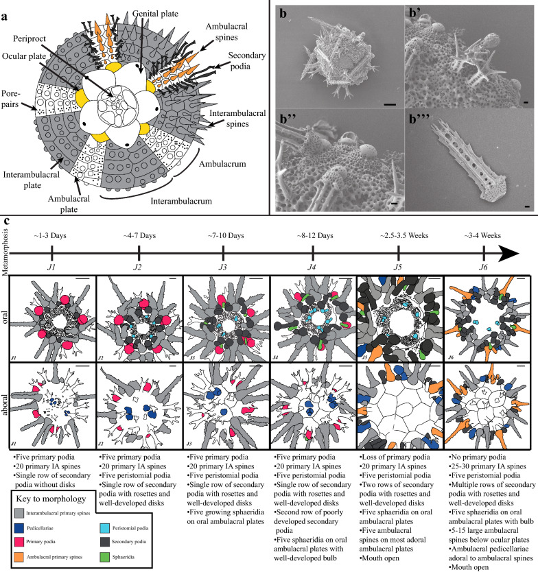
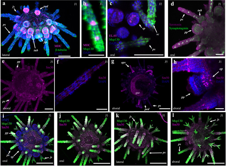
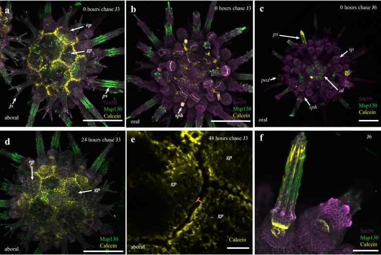
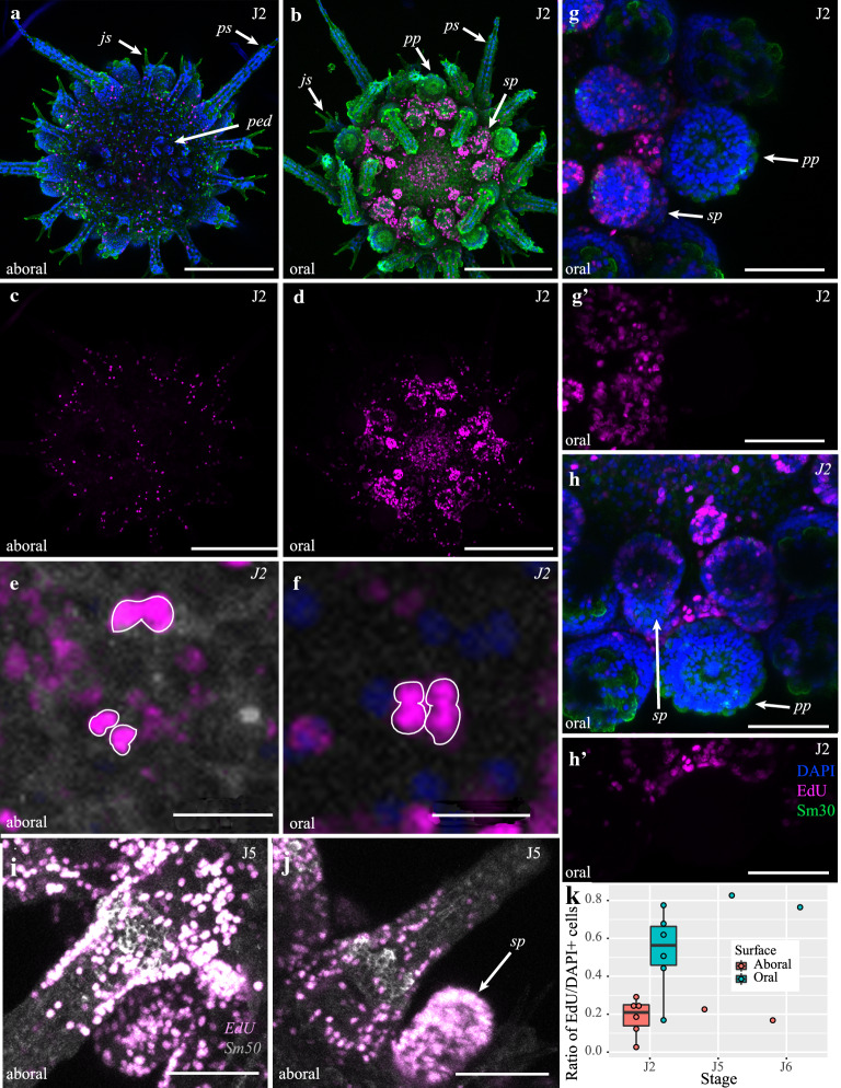
Results: We developed a detailed staging scheme based off of the first ~ 4 weeks of post-metamorphic life of the regular echinoid Paracentrotus lividus. We paired this scheme with immunohistochemical staining for neuronal, muscular, and skeletal tissues, and fluorescent assays of skeletal growth and cell proliferation to understand the molecular and cellular mechanisms underlying skeletal growth and development of the sea urchin body plan.

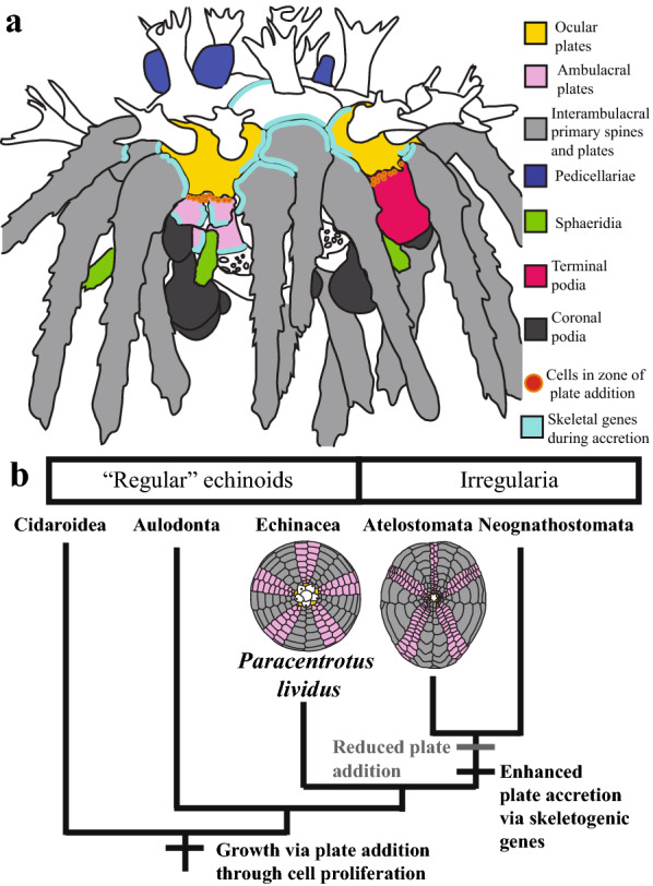
Conclusions: Our experiments highlight the role of skeletogenic proteins in accretionary skeletal growth and cell proliferation in the addition of new metameric tissues. Furthermore, this work provides a framework for understanding the developmental evolution of sea urchin body plans on macroevolutionary timescales.

Keywords: Development; Echinoid; Skeleton.


Conflict of interest statement

We have no competing interests.

Figures

Fig. 1
Fig. 1
a Diagram modified from [46] showing morphology of adult sea urchin test from adoral view. b SEM image showing aboral view of test with juvenile and primary spines Scale bar is 100 μm. b’ Close-up of b showing juvenile spines, ambulacral spine, and sphaeridia. b’’ Close-up of b showing the details of the interambulacra. Primary interambulacral spines articulate with the bosses of primary tubercles on interambulacral plates. b’’ Isolated primary spine. Scale bars in (b’b’’) 10 μm. c Staging scheme showing example drawings and descriptions of each stage and the addition of morphological structures through the first 4 weeks of development. Drawings in c are based off of traces of fixed and stained individuals. Structures are color coded as in the figure legend. IA, interambulacral. Scale bars in c are 100 μm
Fig. 2
Fig. 2
Molecular characterization of juvenile P. lividus cell and tissue types. a Staining with antibodies against Msp130 and β-tubulin reveals the distribution of musculature. Details in text. b Staining against Msp130 and MHC shows MHC+ cells within the spines. c Staining against ELAV and Msp130 shows nerves in the secondary podia, and skeletogenic cells in the spines. d Staining for synaptotagmin and serotonin reveals the extent of the nervous system and serotonergic neurons. eh Immunostaining using antibodies against the skeletogenic proteins Sm30 and Sm50 stains skeletal tissues, with stronger staining in more recently deposited biomineral. il Localization of the skeletogenic proteins Msp130 (green) and Sm50 (purple) in a J1 P. lividus juvenile. Description of stainings given in the main text. js, juvenile spine; ps primary interambulacral spine; ped, pedicellariae; aps, ambulacral primary spine; pp, primary podia; sp, secondary podia; tub, tubercle; gut, gut; op, ocular plate, gp, genital plate; ap, anal plate; sph, sphaeridia; al, Aristotle’s lantern. Scale bars in a, c, e are 100 μm; d is 50 μm; b, f, h are 25 μm, and g, and il are 200 μm
Fig. 3
Fig. 3
Skeletogenesis in juvenile P. lividus. a Localization of Sm50 (purple), Msp130 (green) and incorporation of calcein (yellow) in aboral surface of J3 individual at 0 h chase. b Oral surface of a. c Sm50, Msp130 and calcein in J6 animal at 0 h chase. d Aboral surface of 24 h chase J3 individual. e Zoom of calcein incorporation in margins of aboral plates in 48 h chase J3 individual. Orange bars indicate gap between calcein-marked skeleton due to subsequent accretion. f Close-up of the aboral surface showing incorporation of calcein into growing spines and tubercle. Abbreviations as in Fig. 2 and scale bars in ad are 200 μm, e is 25 μm, and f is 50 μm
Fig. 4
Fig. 4
Growth via cellular proliferation in. P. lividus. ah’ Proliferation on aboral (a, c) and oral (b, d, e, f, gh’) surfaces in J2 animals. e, f Close-ups of proliferating cell doublets in 24-h post-chase animal (e) and quadruplets in 48 h post-chase animal (f). gh’ Zoom showing proliferative zone associated with plate addition. ij Cell proliferation associated with the addition of new ambulacral spines and secondary podia in J5 animal. k Graph showing differential cell proliferation on oral and aboral surfaces. Abbreviations as in Fig. 2. Scale bars in ad 200 μm, ef 10 μm, ij 100 μm, and gh 50 μm
Fig. 5
Fig. 5
a Summary diagram of cell proliferation and expression of skeletal genes during plate accretion and addition in juvenile test growth. b Simplified phylogenetic tree of crown group echinoids showing the transitions in sea urchin growth modes in regular and irregular echinoids, and their hypothesized molecular and cellular foundations. Tree is based on [43] and [47]

References

    1. Levin M, Anavy L, Cole AG, Winter E, Mostov N, Khair S, et al. The mid-developmental transition and the evolution of animal body plans. Nature. 2016;531:637–641. doi: 10.1038/nature16994. - DOI - PMC - PubMed
    1. Davidson EH, Erwin DH. Gene regulatory networks and the evolution of animal body plans. Science. 2006;311(5762):796–800. doi: 10.1126/science.1113832. - DOI - PubMed
    1. Murdock DJ. The ‘biomineralization toolkit’and the origin of animal skeletons. Biolo Rev. 2020;95:1372–1392. doi: 10.1111/brv.12614. - DOI - PubMed
    1. Smith AB. Stereom microstructure of the echinoid test. Spec Pap Palaeontol. 1980;25:1–85.
    1. Hopkins MJ, Smith AB. Dynamic evolutionary change in post-Paleozoic echinoids and the importance of scale when interpreting changes in rates of evolution. Proc Natl Acad Sci. 2015;112(12):3758–3763. doi: 10.1073/pnas.1418153112. - DOI - PMC - PubMed

LinkOut - more resources