. 2021 Mar 16;9(1):44.
doi: 10.1186/s40478-021-01146-1.

Astrocytes respond to a neurotoxic Aβ fragment with state-dependent Ca2+ alteration and multiphasic transmitter release

Affiliations

Astrocytes respond to a neurotoxic Aβ fragment with state-dependent Ca2+ alteration and multiphasic transmitter release

Cuong Pham et al. Acta Neuropathol Commun. .

Abstract

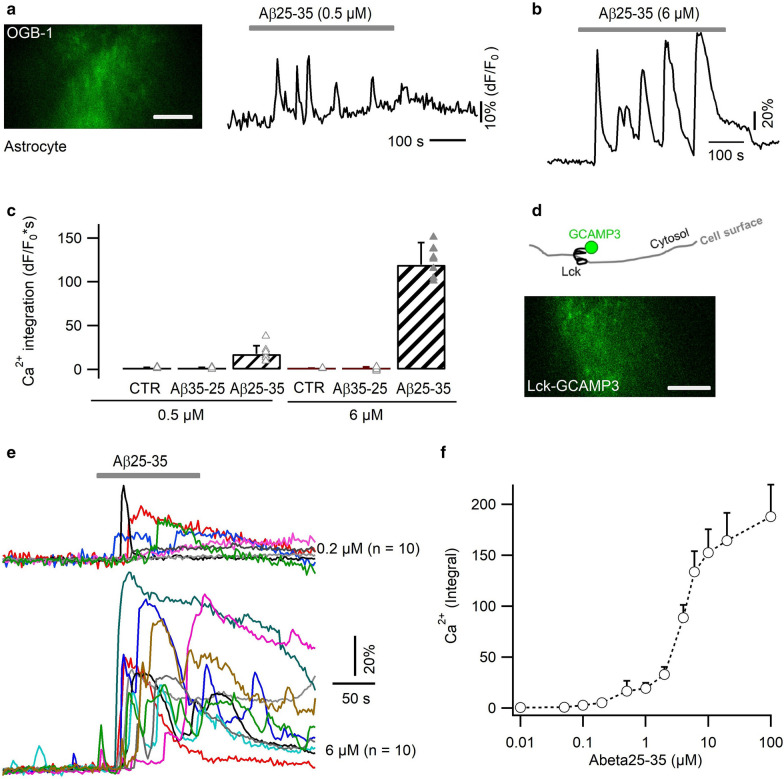
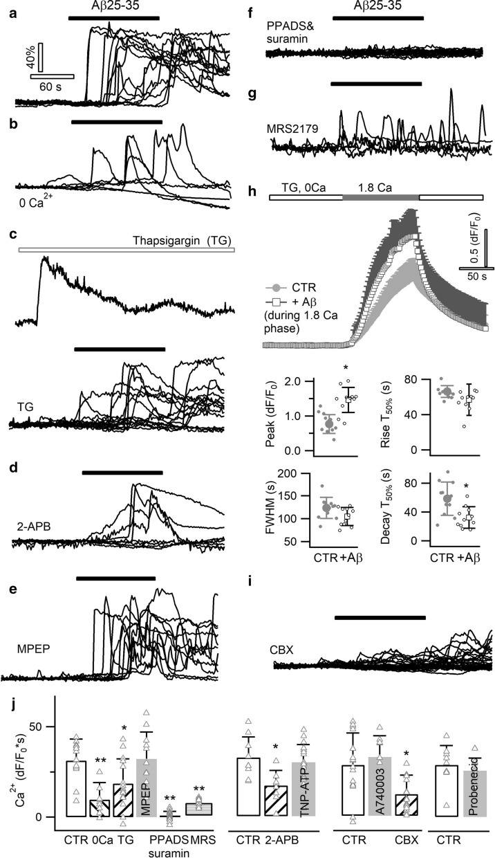
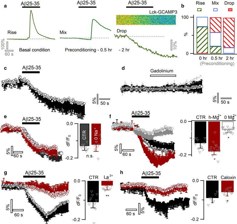
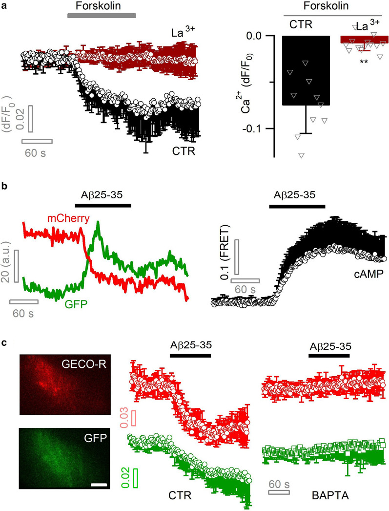
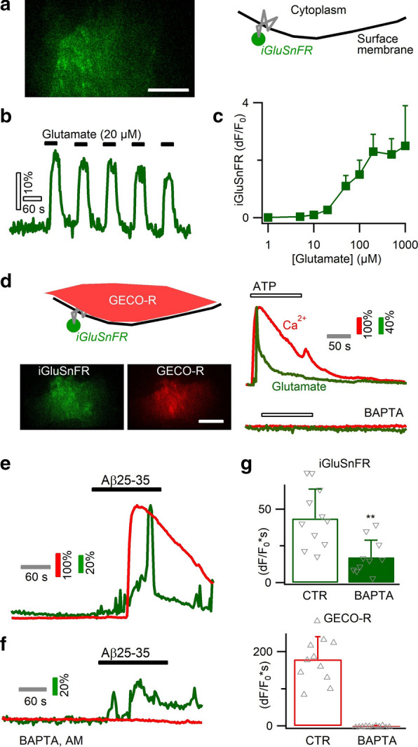
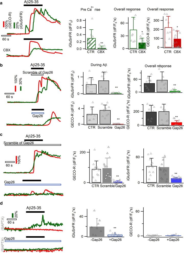
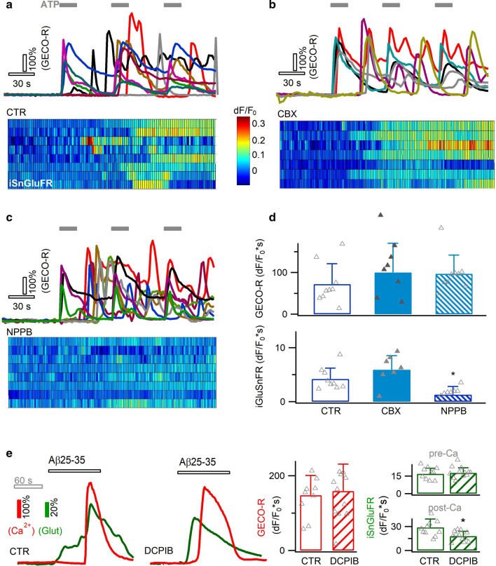
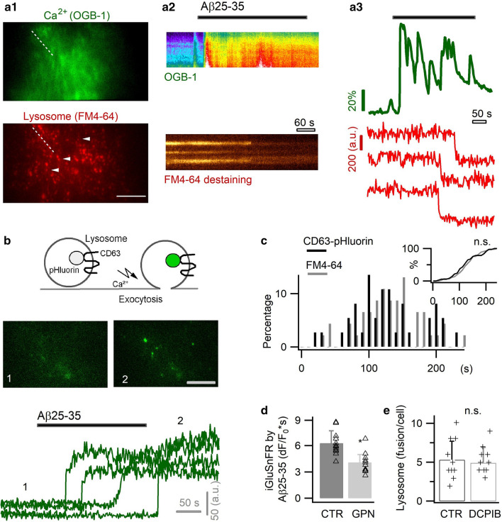
Excessive amounts of amyloid β (Aβ) peptide have been suggested to dysregulate synaptic transmission in Alzheimer's disease (AD). As a major type of glial cell in the mammalian brain, astrocytes regulate neuronal function and undergo activity alterations upon Aβ exposure. Yet the mechanistic steps underlying astrocytic responses to Aβ peptide remain to be elucidated. Here by fluorescence imaging of signaling pathways, we dissected astrocytic responses to Aβ25-35 peptide, a neurotoxic Aβ fragment present in AD patients. In native health astrocytes, Aβ25-35 evoked Ca2+ elevations via purinergic receptors, being also dependent on the opening of connexin (CX) hemichannels. Aβ25-35, however, induced a Ca2+ diminution in Aβ-preconditioned astrocytes as a result of the potentiation of the plasma membrane Ca2+ ATPase (PMCA). The PMCA and CX protein expression was observed with immunostaining in the brain tissue of hAPPJ20 AD mouse model. We also observed both Ca2+-independent and Ca2+-dependent glutamate release upon astrocytic Aβ exposure, with the former mediated by CX hemichannel and the latter by both anion channels and lysosome exocytosis. Our results suggest that Aβ peptide causes state-dependent responses in astrocytes, in association with a multiphasic release of signaling molecules. This study therefore helps to understand astrocyte engagement in AD-related amyloidopathy.

Keywords: ATP; Alzheimer’s disease; Glutamate; Hemichannel; Lysosome.


Conflict of interest statement

The authors declare that they have no competing interests.

Figures

Fig. 1
Fig. 1
Neurotoxic Aβ25–35 peptide triggered irregular Ca2+ rises in primary astrocytes. a, b Aβ-triggered Ca2+ transients in primary cultures of mouse cortical astrocytes, imaged with the chemical Ca2+ indicator OGB-1 AM. Subplasmalemmal Ca2+-dependent fluorescence changes were selectively imaged by TIRFM. c Dose–response of the Aβ25–35 effect. The strength of Ca2+ signals was evaluated by their temporal integral over the same recording period (n = 8–13 astrocytes per condition). d Lck-GCaMP3 was expressed on the inner side of the astrocytic plasma membrane. Below, representative TIRFM image. e Astrocytic Ca2+ signals evoked by 0.2 µM and 6 µM Aβ25–35, respectively. Each trace denotes the response from a single cell. f Dose-responses curve of astrocyte Ca2+ response to Aβ peptide (n = 7–12 per condition). Scale bars, 10 µm
Fig. 2
Fig. 2
A purinergic pathway underlies Aβ-evoked Ca2+ signal. Representative responses evoked by Aβ25–35 in astrocytes loaded with the chemical Ca2+ indicator OGB-1 AM, in control condition (a), in Ca2+-free extracellular solution (b), following thapsigargin (TG, 0.5 µM) depletion of ER Ca2+ store (c, top trace reflecting the Ca2+ leak signal upon TG application), in the presence of the IP3 receptor antagonist 2-APB (d, 200 µM), and of the mGluR5 antagonist MPEP (e, 50 µM). Each trace represents the response of a single astrocyte. f Aβ-evoked Ca2+ responses were fully abolished by blocking purinergic P2 receptors with the combination of wide-spectrum antagonists PPADS (100 µM) and suramin (50 µM). g The P2Y1 antagonist MRS2179 (5 µM) attenuated Aβ-induced Ca2+ responses. h Aβ25–35 enhanced Ca2+ influx via store-operated channels (SOCs). SOCs were activated by fully depleting the ER store with TG in Ca2+-free solution. SOC-mediated Ca2+ influx was induced by re-supplying Ca2+ in the extracellular solution. Ca2+ influx was significantly increased in the presence of Aβ25–35 (n = 11 cells per condition). i Effect of blocking connexin hemichannels with CBX (50 µM). j Aβ-evoked astrocyte Ca2+ responses in different conditions. Ca2+ signal strength was derived from the temporal integral of individual normalized traces (dF/F0*s). Wide-spectrum P2X receptor antagonist TNP-ATP, P2X7 antagonist A740003 and pannexin blocker probenecid were applied at 10 µM, 20 µM and 500 µM, respectively. Control experiments were performed for a defined set of experiments as shown (n = 9–20 cells per condition)
Fig. 3
Fig. 3
Aβ25–35 inhibits Ca2+ levels in preconditioned astrocytes by potentiating PMCA Ca2+ extrusion. a Effect of preconditioning on the acute responses of Aβ25–35 (6 µM) in cultured mouse cortical astrocytes. Left, Ca2+ rise was triggered in intact astrocytes (‘Rise’ type response). Middle, after a short term incubation (i.e., preconditioning) of astrocytes with submicromolar Aβ25–35 (0.5 h, 0.5 µM), acute application of 6 µM Aβ25–35 caused a basal line drop mixed with Ca2+ rise (‘Mix’ type response). Right, following a ~ 2 h preconditioning in 0.5 µM Aβ25–35, astrocytes exhibited only a drop in the intracellular Ca2+ level (‘Drop’ type response). b The percentage of three types of astrocytes that either displayed a Ca2+ rise (‘Rise’), an initial diminution followed by rise (‘mix’), or only a drop in the basal Ca2+ levels (Drop’; n = 19–7 cells per condition). c Average of Aβ-induced Ca2+ diminution in Aβ25–35-preconditioned (2 h) astrocytes (n = 11). d Blocking spontaneous Ca2+ influx by gadolinium (100 µM) failed to mimic Aβ-evoked Ca2+ diminution (n = 8). e Inactivating NCX by Na+-free solution showed no effect (n = 9 per condition). f Aβ-induced Ca2+ diminution affected by ambient Mg2+ concentration, implying the recruitment of an ATP-dependent pathway. Astrocytes were incubated in defined solutions 1 h prior to imaging (n = 11–17 per condition). g, h Inhibiting PMCA by La3+ (50 µM) or Caloxin 3A1 (500 µM) counterbalanced the Aβ-evoked astrocytic Ca2+ diminution (n = 6–11 per condition)
Fig. 4
Fig. 4
Aβ potentiates astrocytic PMCA via cAMP signaling. a Elevation of astrocytic cAMP level increased PMCA-mediated Ca2+ extrusion. cAMP was elevated by forskolin (100 µM) and sub-cellular Ca2+ level imaged with Lck-GCaMP3 and TIRFM. Ca2+ diminution was blocked by the PMCA blocker La3+ (50 µM; n = 9–13 cells per condition). b Intracellular cAMP level was monitored with the FRET sensor GFPnd-EPAC(dDEP)-mCherry and dual-color TIRFM. Left, representative dual-color recording of fluorescence change for EGFP and mCherry of the FRET sensor. Right, averaged astrocytic cAMP rise induced by Aβ25–35 (6 µM; n = 6). c PMCA-mediated astrocytic Ca2+ diminution was coupled with H+ influx. Dual-color TIRFM recorded concomitant cytosolic Ca2+ and pH diminution, by expressing the red genetically encoded Ca2+ sensor GECO-R and the pH-sensitive GFP protein in same astrocytes. Both effects were abolished by chelating astrocyte cytoplasmic Ca2+ with BAPTA AM (100 µM; n = 7–9 cells per condition). Scale bar, 5 µm
Fig. 5
Fig. 5
Biphasic astrocytic glutamate release occurring in both a Ca2+-dependent and -independent manner. a Imaging astrocytic glutamate release with green fluorescent sensor iGluSnFR expressed on the outer face of cell membrane. b CTR: repetitive fluorescence signals upon glutamate exposure. c Dose–response curve for astrocyte-expressed iGluSnR (n = 6–11 cells per concentration). d Dual-color imaging of astrocytic Ca2+ by the red sensor GECO-R and glutamate release by iGluSnFR. Right, ATP application evoked Ca2+ rise and glutamate release, which were both suppressed by BAPTA chelation of intracellular Ca2+. e Aβ25–35 triggered a biphasic glutamate release, which started prior to Ca2+ rise and was further increased during Ca2+ elevation. f, g Chelating astrocytic Ca2+ with BAPTA partially reduced Aβ-induced glutamate release, confirming its occurrence in both Ca2+-independent and -dependent manner (n = 10–11 cells per condition). Scale bars, 5 µm
Fig. 6
Fig. 6
Involvement of CX hemichannels in Ca2+-independent glutamate release. a Glutamate release prior to the Ca2+ elevation (top, CTR) was reduced by the CX hemichannel blocker CBX (100 µM; present throughout the recording; n = 8–13 cells per condition). b During the Aβ application phase, Ca2+-independent glutamate release was blocked by another CX hemichannel blocker Gap26 peptide (200 µM). The inactive scramble peptide of Gap26 showed no effect (n = 8–10 cells per condition). c A more pronounced inhibition effect of Gap26 was observed when applying it throughout the entire imaging period (i.e., pre-, during- and post-Aβ; n = 12–14 per condition). d Chelating astrocytic Ca2+ with BAPTA AM isolated Ca2+-independent astrocytic glutamate release, which was inhibited by CX hemichannel blocker Gap26 (n = 8–10 per condition)
Fig. 7
Fig. 7
Contribution of anion channels to Ca2+-dependent glutamate release. a Aβ-evoked astrocytic Ca2+ rises were due to purinergic receptor activation. To examine Ca2+-dependent glutamtae release, we applied ATP (30 µM) to trigger astrocytic Ca2+ and glutamate release. bd Ca2+-dependent glutamate release was unaffected by inhibition of CX hemichannel (CBX, 100 µM), but reduced by blocking anion channels with NPPB (100 µM) (n = 7–9 cells per condition). e During the response to Aβ25–35 (6 µM), inhibiting anion channels with DCPIB (50 µM) influenced the glutamate release during Ca2+ elevation phase (n = 10–12 cells per condition)
Fig. 8
Fig. 8
Aβ25–35 triggered astrocytic lysosome exocytosis. a Astrocytes co-labeled with the green fluorescent Ca2+ indicator OGB-1 AM and the red-fluorescent lysosomal marker FM4-64 (a1). Application of Aβ25–35 (6 µM) evoked Ca2+ elevation followed by asynchronous exocytosis of lysosomes, as reflected by FM dye destaining (a2, a3). b Aβ-evoked lysosomal exocytosis imaged with CD63-pHluorin. c Temporal distribution of lysosomal exocytosis obtained with FM dye and CD63-pHluorin (n = 51–62 lysosomes from five cells per condition). Inset, cumulative histogram showing the temporal coincidence for the two lysosomal markers (p = 0.7). d Permeabilization of lysosomes by GPN affected the Aβ25–35-induced glutamate release (iGluSnFR, dF/F0*s; n = 12 cells per condition; recording protocol is as Fig. 7e). e The presence of anion channel blocker DCPIB did not affect astrocyte lysosome release rate as measured by FM4-64 destaining (n = 10 cells per condition). Scale bars, 10 µm for a, 5 µm for b

Similar articles

Cited by

References

    1. Abramov AY, Canevari L, Duchen MR. Changes in intracellular calcium and glutathione in astrocytes as the primary mechanism of amyloid neurotoxicity. J Neurosci. 2003;23:5088–5095. doi: 10.1523/JNEUROSCI.23-12-05088.2003. - DOI - PMC - PubMed
    1. Abramov AY, Ionov M, Pavlov E, Duchen MR. Membrane cholesterol content plays a key role in the neurotoxicity of beta-amyloid: implications for Alzheimer's disease. Aging Cell. 2011;10:595–603. doi: 10.1111/j.1474-9726.2011.00685.x. - DOI - PubMed
    1. Alberdi E, Wyssenbach A, Alberdi M, Sanchez-Gomez MV, Cavaliere F, Rodriguez JJ, Verkhratsky A, Matute C. Ca(2+)-dependent endoplasmic reticulum stress correlates with astrogliosis in oligomeric amyloid beta-treated astrocytes and in a model of Alzheimer's disease. Aging Cell. 2013;12:292–302. doi: 10.1111/acel.12054. - DOI - PubMed
    1. Allaman I, Gavillet M, Belanger M, Laroche T, Viertl D, Lashuel HA, Magistretti PJ. Amyloid-beta aggregates cause alterations of astrocytic metabolic phenotype: impact on neuronal viability. J Neurosci. 2010;30:3326–3338. doi: 10.1523/JNEUROSCI.5098-09.2010. - DOI - PMC - PubMed
    1. Araque A, Carmignoto G, Haydon PG, Oliet SH, Robitaille R, Volterra A. Gliotransmitters travel in time and space. Neuron. 2014;81:728–739. doi: 10.1016/j.neuron.2014.02.007. - DOI - PMC - PubMed

Publication types

MeSH terms