Review
. 2021 Apr;69(4):812-823.
doi: 10.4103/ijo.IJO_1917_20.

Imaging the pediatric retina: An overview

Affiliations
Review

Imaging the pediatric retina: An overview

Komal Agarwal et al. Indian J Ophthalmol. 2021 Apr.

Abstract

Recent decade has seen a shift in the causes of childhood blinding diseases from anterior segment to retinal disease in both developed and developing countries. The common retinal disorders are retinopathy of prematurity and vitreoretinal infections in neonates, congenital anomalies in infants, and vascular retinopathies including type 1 diabetes, tumors, and inherited retinal diseases in children (up to 12 years). Retinal imaging helps in diagnosis, management, follow up and prognostication in all these disorders. These imaging modalities include fundus photography, fluorescein angiography, ultrasonography, retinal vascular and structural studies, and electrodiagnosis. Over the decades there has been tremendous advances both in design (compact, multifunctional, tele-consult capable) and technology (wide- and ultra-wide field and noninvasive retinal angiography). These new advances have application in most of the pediatric retinal diseases though at most times the designs of new devices have remained confined to use in adults. Poor patient cooperation and insufficient attention span in children demand careful crafting of the devices. The newer attempts of hand-held retinal diagnostic devices are welcome additions in this direction. While much has been done, there is still much to do in the coming years. One of the compelling and immediate needs is the pediatric version of optical coherence tomography angiography. These needs and demands would increase many folds in future. A sound policy could be the simultaneous development of adult and pediatric version of all ophthalmic diagnostic devices, coupled with capacity building of trained medical personnel.

Keywords: Newborn eye screening; pediatric retina; pediatric retinal diseases; retinal imaging.


Conflict of interest statement

None

Figures

Figure 1
Figure 1
A 2- year old female, born preterm at 32 weeks, presented with bilateral leucocoria. She was referred with a diagnosis of Stage V retinopathy of prematurity. (a) An immersion ultrasonography of left eye shows a mass lesion in the posterior half of the globe. (b-d) Transverse scans of superior, nasal and temporal half showing a mass lesion with intraretinal calcification. She was diagnosed with retinoblastoma and was treated for the same
Figure 2
Figure 2
Wide field fundus photograph of right eye of a preterm neonate showing active neovascularization in zone 1. (a) Picture taken on Retcam with about 130 degrees of field, superior and inferior edges appear cropped. (b) Picture of the same eye taken on “Neo” with about 120 degrees of field in a circular capture
Figure 3
Figure 3
(a) Ultra-wide field fundus photograph of a 28 weeks GA neonate with APROP (b) Follow up image of the same eye 2 weeks post intravitreal avastin showing regression of plus disease. (c) Ultra-wide field fundus photograph of a 4 year old boy with submacular exudates and peripheral telangiectasia in nasal quadrant, He was diagnosed as Coat‘s retinopathy and focal laser was done to the telangiectatic vessels. Inferior view of the fundus is limited due to artefacts due to eye lashes. (d) Follow up image of the same eye showing regressed macular exudates and telangiectatic vessels post focal laser
Figure 4
Figure 4
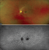
A 14- year old female presented with bilateral gradual decrease in vision. The best corrected visual acuity was 20/200. (a) Color fundus image shows a central area of RPE atrophic changes with fleck like changes around fovea. (b) Autofluorescence shows central dark hypofluorescence corresponding to the area of RPE atrophy and multiple hyper- and hypo-fluorescent fleck like deposits around fovea, more prominent than the color fundus photo. She was diagnosed with Stargardt‘s disease
Figure 5
Figure 5
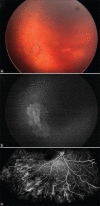
Half zone APROP. (a) Left eye color fundus image shows limited details of vascular retina. (b) Fundus fluorescein angiography (FFA) in the same patient shows vascular retina extent and neovascularization status, not clearly apparent on fundus examination. (c) Wide field FFA shows peripheral avascular retina and neovascular leakage in right eye of 8- year old child presenting with recurrent vitreous hemorrhage
Figure 6
Figure 6
A 6- year old girl presented with reduction in vision in both eyes; best corrected vision was 20/60 in both eyes. She was able to read 20 of 23 plates in Isihara color vision chart. (a and b) The fundus was unremarkable; (c and d) very subtle photo receptor irregularity on OCT. (e and f) A full field electroretinogram showed normal scotopic and photopic responses while mfERG showed hypovoltaged subnormal responses from central macula. This confirmed the diagnosis of occult macular dystrophy

Similar articles

Cited by

References

    1. Position Paper- WHO archives- World Health Organization. [Last accessed 2020 Jun 03]. Available from: www.archives.who.int .
    1. Wadhwani M, Vashist P, Singh SS, Gupta V, Gupta N, Saxena R. Prevalence and causes of childhood blindness in India: A systematic review. Indian J Ophthalmol. 2020;68:311–5. - PMC - PubMed
    1. Gilbert C, Foster A. Childhood blindness in the context of VISION 2020--The right to sight. Bull World Health Organ. 2001;79:227–32. - PMC - PubMed
    1. Agarwal K, Balakrishnan D, Rani PK, Jalali S. Changing patterns of early childhood blinding conditions presenting to a tertiary eye center: The epidemic of retinopathy of prematurity in India. Indian J Ophthalmol. 2019;67:816–8. - PMC - PubMed
    1. Solebo AL, Teoh L, Rahi J. Epidemiology of blindness in children. Arch Dis Child. 2017;102:853–7. Erratum in: Arch Dis Child 2017;102:995. - PubMed