Review
. 2021 Apr;69(4):824-835.
doi: 10.4103/ijo.IJO_1403_20.

Ultra-wide field retinal imaging: A wider clinical perspective

Affiliations
Review

Ultra-wide field retinal imaging: A wider clinical perspective

Vinod Kumar et al. Indian J Ophthalmol. 2021 Apr.

Abstract

The peripheral retina is affected in a variety of retinal disorders. Traditional fundus cameras capture only a part of the fundus even when montaging techniques are used. Ultra-wide field imaging enables us to delve into the retinal periphery in greater detail. It not only facilitates assessing color images of the fundus, but also fluorescein angiography, indocyanine green angiography, fundus autofluorescence, and red and green free images. In this review, a literature search using the keywords "ultra-widefield imaging", "widefield imaging", and "peripheral retinal imaging" in English and non-English languages was done and the relevant articles were included. Ultra-wide field imaging has made new observations in the normal population as well as in eyes with retinal disorders including vascular diseases, degenerative diseases, uveitis, age-related macular degeneration, retinal and choroidal tumors and hereditary retinal dystrophies. This review aims to describe the utility of ultra-wide field imaging in various retinal disorders.

Keywords: Retinal imaging; UWF Indocyanine angiography; UWF autofluorescence; UWF fluorescein angiography; Ultra-widefield; retinal disorders.


Conflict of interest statement

None

Figures

Figure 1
Figure 1
UWF-FFA of normal peripheral retina showing absence of capillary details, ground glass hyperfluorescence (arrow, a) and peripheral microaneurysms (arrow, b). In the setting of high myopia, UWF-FFA shows peripheral avascular retina (white arrow, c)
Figure 2
Figure 2
UWF pseudocolour image showing rhegmatogenous retinal detachment with superior 180° giant retinal tear (arrow, a). Postoperative UWF image of the same eye with giant retinal tear showing gas-filled eye and attached retina (b). Old rhegmatogenous retinal detachment showing demarcation lines and intraretinal cyst (arrow, c) and subretinal bands (arrow, d). UWF image of an eye after scleral buckling showing infero-temporal cryo scar (arrow, e); re-detachment with extensive PVR changes following vitreoretinal surgery with silicone oil (f)
Figure 3
Figure 3
UWF-FFA in diabetic retinopathy showing predominantly posterior pole involvement (a) and panretinal involvement (b) with microaneurysm and vascular changes extending beyond posterior pole, NVEs are also seen along supero-temporal vascular arcade (b). UWF-FFA of proliferative diabetic retinopathy showing large NVD and multiple NVEs with extensive capillary non-perfusion areas (c) and almost total capillary drop out (d)
Figure 4
Figure 4
UWF pseudocolour image (a) of CRVO along with inferotemporal BRVO showing dilated tortuous veins, 360° resolving hemorrhages and prominent inferotemporal area of intra-retinal hemorrhages. UWF-FFA in CRVO (b) and BRVO (c) showing panretinal and sectoral vessel wall staining (arrows) and capillary non-perfusion (CNP) areas extending up to retinal periphery. UWF-FFA in old BRVO (d) showing peripheral laser spots, large areas of uncovered CNP and collateral formation (red arrow) at junction of vascularized and non-vascularized retina
Figure 5
Figure 5
UWF pseudocolor image of Coat‘s disease showing peripheral avascular area and retinal neovascularization (a) UWF- FFA showing bulb like dilatations in addition (b). UWF-FFA of another case showing wide-spread involvement in Coat‘s disease (c). UWF image in familial exudative vitreoretinopathy (FEVR) showing retinal fold, peripheral tractional retinal detachment and disc-macula drag (d). UWF-FFA in FEVR shows temporal avascular retina, temporal straightening of vessels and super-numerary vascular branching in addition to early (e) and frank neovascularization (f)
Figure 6
Figure 6
UWF pseudocolor image of VKH syndrome showing multiple neurosensory detachments (a). UWF-FFA showed multiple pin point leaks (b) which pool in the late phases (c) extending up to the periphery. UWF image of cytomegalovirus retinitis showing peripheral involvement (d); Multifocal choroiditis (e) showing areas of active as well as healed patches. Ocular Toxocariasis showing peripheral granuloma with falciform fold extending from the lesion to posterior pole (f)
Figure 7
Figure 7
UWF-FFA of active retinal vasculitis which can be generalized (a) or localised (b). UWF-FFA of healed retinal vasculitis with extensive capillary non-perfusion areas (CNP) in the retinal periphery and neovascularization (c). Fibro-vascular proliferation (d) in the background of large CNPs is evident in a case of healed retinal vasculitis on UWF-FFA
Figure 8
Figure 8
UWF pseudocolor image showing peripheral drusen (a) which are more evident on UWF-FAF (b). UWF pseudocolour image shows retinal lesion in the periphery with subretinal and sub retinal pigment epithelium hemorrhage (c). UWF- FFA showed peripheral leakage along with blocked hypofluorescence confirming peripheral exudative hemorrhagic chorioretinopathy (d)
Figure 9
Figure 9
UWF pseudocolour image of Leber congenital amaurosis showing chorioretinal degeneration, macular coloboma and peripheral bone-spicule like pigmentation (a). UWF-FAF shows peripheral involvement in a case of Stargardt disease (b). UWF pseudocolour image of pigmented paravenous retinochoroidal atrophy (c) showing perivascular pigmentary changes extending into periphery, which are better characterized on FAF (d). UWF-FFA of gyrate dystrophy (e) and choroideremia (f)
Figure 10
Figure 10
UWF pseudocolour image (a) of choroidal hemangioma at posterior pole. UWF-FAF shows large hypoautofluorescent areas extending to inferior retina due to chronic fluid exudation and retinal pigment epithelium loss (b). UWF–FFA in Von-Hippel-Lindau shows multiple retinal capillary hemangioblastomas and inferior large lesion with feeder vessels (c). UWF imaging (d) of choroidal osteoma shows irregular, yellow-white, juxtrapapillary lesion which had central hypoautofluorescence (signifying decalcification) with surrounding hyperautofluorescence on UWF-FAF (e)

Comment in

  • Wide-field imaging - An update.
    Alone A, Chandra K, Chhablani J. Alone A, et al. Indian J Ophthalmol. 2021 Apr;69(4):788-789. doi: 10.4103/ijo.IJO_2726_20. Indian J Ophthalmol. 2021. PMID: 33727434 Free PMC article. No abstract available.

Similar articles

Cited by

References

    1. Diabetic retinopathy study. Report Number 6. Design, methods, and baseline results. Report Number 7. A modification of the Airlie House classification of diabetic retinopathy. Prepared by the Diabetic Retinopathy. Invest Ophthalmol Vis Sci. 1981;21:1–226. - PubMed
    1. Diabetic Retinopathy Clinical Research Network. Peripheral diabetic retinopathy (DR) lesions on ultrawide-field fundus images and risk of DR worsening over time. DRCRnet. DRCR Retina Network-Public Site. Available from: https://public.jaeb.org/drcrnet .
    1. Choudhry N, Duker JS, Freund KB, Kiss S, Querques G, Rosen R, et al. Classification and guidelines for widefield imaging: Recommendations from the International Widefield Imaging Study Group. Ophthalmol Retina. 2019;3:843–9. - PubMed
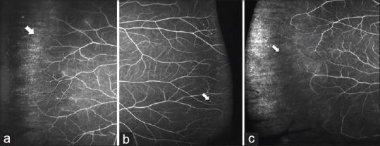
    1. Shah AR, Abbey AM, Yonekawa Y, Khandan S, Wolfe JD, Trese MT, et al. Widefield fluorescein angiography in patients without peripheral disease: A Study of Normal Peripheral Findings. Retina (Philadelphia, Pa) 2016;36:1087–92. - PubMed
    1. Lu J, Mai G, Luo Y, Li M, Cao D, Wang X, et al. Appearance of far peripheral retina in normal eyes by ultra-widefield fluorescein angiography. Am J Ophthalmol. 2017;173:84–90. - PubMed