

On the morphological relations of the Achilles tendon and plantar fascia via the calcaneus: a cadaveric study
- PMID: 33727610
- PMCID: PMC7966405
- DOI: 10.1038/s41598-021-85251-0
On the morphological relations of the Achilles tendon and plantar fascia via the calcaneus: a cadaveric study
Abstract
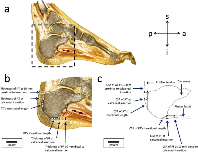
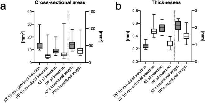
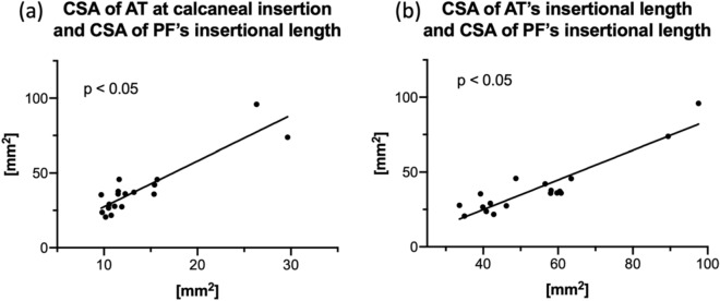
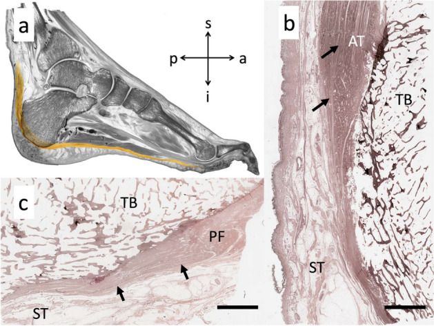
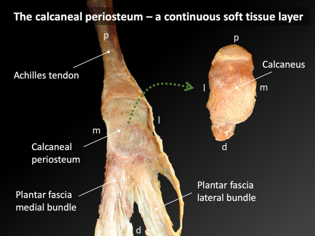
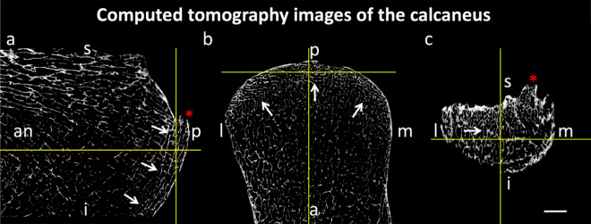
Current treatments of plantar fasciitis are based on the premise that the Achilles tendon (AT) and plantar fascia (PF) are mechanically directly linked, which is an area of debate. The aim of this study was to assess the morphological relationship between the AT and PF. Nineteen cadaveric feet were x-ray imaged, serially sectioned and plastinated for digital image analyses. Measurements of the AT and PF thicknesses and cross-sectional areas (CSA) were performed at their calcaneal insertion. The fiber continuity was histologically assessed in representative subsamples. Strong correlations exist between the CSA of the AT and PF at calcaneal insertion and the CSA of PF's insertional length (r = 0.80), and between the CSAs of AT's and PF's insertional lengths. Further correlations were observed between AT and PF thicknesses (r = 0.62). This close morphological relationship could, however, not be confirmed through x-ray nor complete fiber continuity in histology. This study provides evidence for a morphometric relationship between the AT and PF, which suggests the presence of a functional relationship between these two structures following the biological key idea that the structure determines the function. The observed morphological correlations substantiate the existing mechanical link between the AT and PF via the posterior calcaneus and might explain why calf stretches are a successful treatment option for plantar heel pain.
Conflict of interest statement
The authors declare no competing interests.
Figures
Similar articles
-
A Radiographic Study of Biomechanical Relationship between the Achilles Tendon and Plantar Fascia.Biomed Res Int. 2020 Feb 18;2020:5319640. doi: 10.1155/2020/5319640. eCollection 2020. Biomed Res Int. 2020. PMID: 32149113 Free PMC article.
-
Plantar fascia anatomy and its relationship with Achilles tendon and paratenon.J Anat. 2013 Dec;223(6):665-76. doi: 10.1111/joa.12111. Epub 2013 Sep 12. J Anat. 2013. PMID: 24028383 Free PMC article.
-
The Calcaneal Crescent in Patients With and Without Plantar Fasciitis: An Ankle MRI Study.AJR Am J Roentgenol. 2018 Nov;211(5):1075-1082. doi: 10.2214/AJR.17.19399. Epub 2018 Aug 30. AJR Am J Roentgenol. 2018. PMID: 30160979
-
The plantar calcaneal spur: a review of anatomy, histology, etiology and key associations.J Anat. 2017 Jun;230(6):743-751. doi: 10.1111/joa.12607. Epub 2017 Mar 29. J Anat. 2017. PMID: 28369929 Free PMC article. Review.
-
Diagnosis of heel pain.Am Fam Physician. 2011 Oct 15;84(8):909-16. Am Fam Physician. 2011. PMID: 22010770 Review.
Cited by
-
Relationships between Plantar Pressure Distribution and Rearfoot Alignment in the Taiwanese College Athletes with Plantar Fasciopathy during Static Standing and Walking.Int J Environ Res Public Health. 2021 Dec 8;18(24):12942. doi: 10.3390/ijerph182412942. Int J Environ Res Public Health. 2021. PMID: 34948551 Free PMC article.
-
Why heel spurs are traction spurs after all.Sci Rep. 2021 Jun 24;11(1):13291. doi: 10.1038/s41598-021-92664-4. Sci Rep. 2021. PMID: 34168232 Free PMC article.
-
Does the Calcaneus Serve as Hypomochlion within the Lower Limb by a Myofascial Connection?-A Systematic Review.Life (Basel). 2021 Jul 26;11(8):745. doi: 10.3390/life11080745. Life (Basel). 2021. PMID: 34440492 Free PMC article. Review.
References
Publication types
MeSH terms
LinkOut - more resources
Full Text Sources
Other Literature Sources
