

Attenuation of Aggregatibacter actinomycetemcomitans virulence using curcumin-decorated nanophytosomes-mediated photo-sonoantimicrobial chemotherapy
- PMID: 33727630
- PMCID: PMC7966776
- DOI: 10.1038/s41598-021-85437-6
Attenuation of Aggregatibacter actinomycetemcomitans virulence using curcumin-decorated nanophytosomes-mediated photo-sonoantimicrobial chemotherapy
Abstract
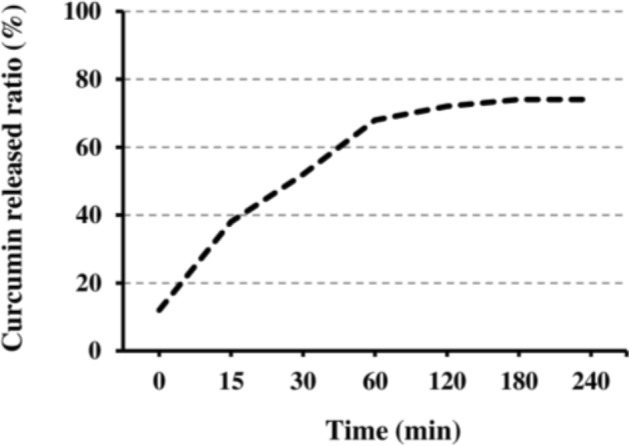
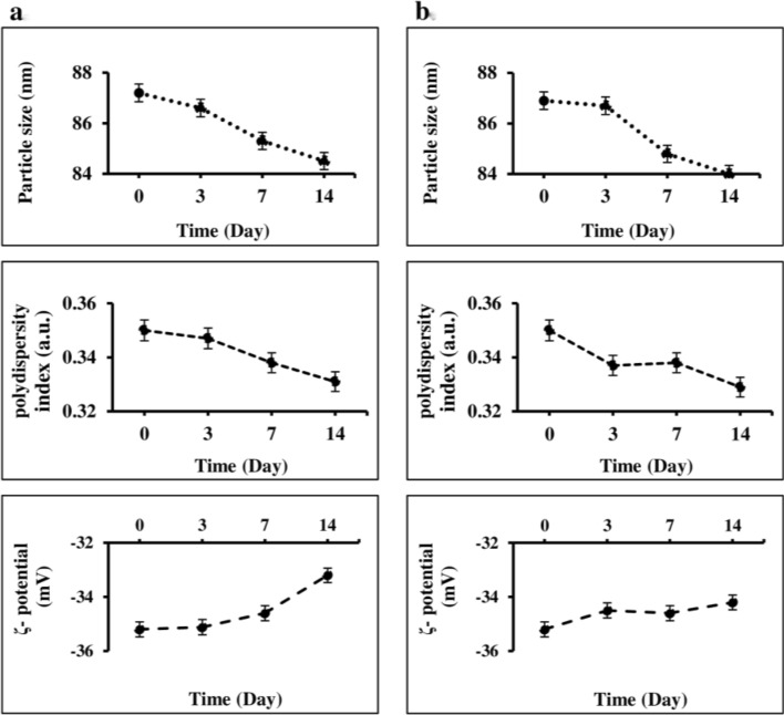
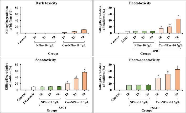
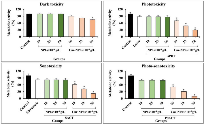
This study aimed to focus on the simultaneous use of antimicrobial photodynamic therapy (aPDT) and sonodynamic antimicrobial chemotherapy (SACT), which is called photo-sonodynamic antimicrobial chemotherapy (PSACT) to attenuate the virulence of Aggregatibacter actinomycetemcomitans. Following the synthesis of Curcumin-decorated nanophytosomes (Cur-NPhs) as a novel photo-sonosensitizer, its particle size, polydispersity, ζ-potential surface morphology, physical stability, drug release, and entrapment efficiency were determined. In the Cur-NPhs-PSACT, the antimicrobial activities of Cur-NPhs against A. actinomycetemcomitans were investigated using cell viability, biofilm killing/degradation, metabolic activity, expression of quorum-sensing-associated qseB and qseC genes, and biofilm-associated rcpA gene under blue laser irradiation plus ultrasonic waves. Characterization tests showed the presence of a sphere-shaped vesicle and the self-closed structure of Cur-NPhs, resulting in a high drug-loading content and encapsulation efficiency. However, the antimicrobial effect of Cur-NPhs-PSACT was dose-dependent, PSACT using the high concentrations of Cur-NPhs (50 × 10-4 g/L) showed significant reductions (P < 0.05) in cell viability (13.6 log10 CFU/mL), biofilm killing/degradation (65%), metabolic activity (89.6%,), and mRNA levels of virulence determinant genes (qseB; 9.8-fold, qseC; 10.2-fold, and recA; 10.2-fold). This study concludes that the Cur-NPhs-PSACT had antimicrobial activities against A. actinomycetemcomitans by downregulating the expression of virulence genes, and may attenuate this bacterium that decreases periodontal disease severity in patients.
Conflict of interest statement
The authors declare no competing interests.
Figures


Similar articles
-
Photo-sonodynamic antimicrobial chemotherapy via chitosan nanoparticles-indocyanine green against polymicrobial periopathogenic biofilms: Ex vivo study on dental implants.Photodiagnosis Photodyn Ther. 2020 Sep;31:101834. doi: 10.1016/j.pdpdt.2020.101834. Epub 2020 May 25. Photodiagnosis Photodyn Ther. 2020. PMID: 32464265
-
Evaluation of Antimicrobial Effects of Photo-sonodynamic Antimicrobial Chemotherapy Based on Nano-micelle Curcumin on Virulence Gene Expression Patterns in Acinetobacter baumannii.Infect Disord Drug Targets. 2022;22(3):e201221199163. doi: 10.2174/1871526522666211220121725. Infect Disord Drug Targets. 2022. PMID: 34931970
-
Photoexcitation triggering via semiconductor Graphene Quantum Dots by photochemical doping with Curcumin versus perio-pathogens mixed biofilms.Photodiagnosis Photodyn Ther. 2019 Dec;28:125-131. doi: 10.1016/j.pdpdt.2019.08.025. Epub 2019 Aug 31. Photodiagnosis Photodyn Ther. 2019. PMID: 31479805
-
Effect of curcumin-mediated photodynamic therapy on Streptococcus mutans and Candida albicans: A systematic review of in vitro studies.Photodiagnosis Photodyn Ther. 2019 Sep;27:455-461. doi: 10.1016/j.pdpdt.2019.07.010. Epub 2019 Jul 25. Photodiagnosis Photodyn Ther. 2019. PMID: 31352059
-
Sonodynamic antimicrobial chemotherapy: An emerging alternative strategy for microbial inactivation.Ultrason Sonochem. 2021 Jul;75:105591. doi: 10.1016/j.ultsonch.2021.105591. Epub 2021 May 20. Ultrason Sonochem. 2021. PMID: 34082219 Free PMC article. Review.
Cited by
-
Photodynamic therapy on mRNA levels in bacteria.Lasers Med Sci. 2024 Aug 31;39(1):229. doi: 10.1007/s10103-024-04179-9. Lasers Med Sci. 2024. PMID: 39214913 Review.
-
Host mRNA Analysis of Periodontal Disease Patients Positive for Porphyromonas gingivalis, Aggregatibacter actinomycetemcomitans and Tannerella forsythia.Int J Mol Sci. 2022 Aug 31;23(17):9915. doi: 10.3390/ijms23179915. Int J Mol Sci. 2022. PMID: 36077312 Free PMC article. Review.
-
Effects on Colonization Factors and Mechanisms Involved in Antimicrobial Sonophotodynamic Inactivation Mediated by Curcumin.Pharmaceutics. 2023 Sep 30;15(10):2407. doi: 10.3390/pharmaceutics15102407. Pharmaceutics. 2023. PMID: 37896167 Free PMC article.
-
Resolvin E1's Antimicrobial Potential Against Aggregatibacter Actinomycetemcomitans.Front Oral Health. 2022 Apr 27;3:875047. doi: 10.3389/froh.2022.875047. eCollection 2022. Front Oral Health. 2022. PMID: 35571980 Free PMC article.
-
Aggregatibacter actinomycetemcomitans: From the Oral Cavity to the Heart Valves.Microorganisms. 2024 Jul 17;12(7):1451. doi: 10.3390/microorganisms12071451. Microorganisms. 2024. PMID: 39065217 Free PMC article. Review.
References
Publication types
MeSH terms
Substances
LinkOut - more resources
Full Text Sources
Other Literature Sources
