

Cell wall N-glycan of Candida albicans ameliorates early hyper- and late hypo-immunoreactivity in sepsis
- PMID: 33727664
- PMCID: PMC7966402
- DOI: 10.1038/s42003-021-01870-3
Cell wall N-glycan of Candida albicans ameliorates early hyper- and late hypo-immunoreactivity in sepsis
Abstract
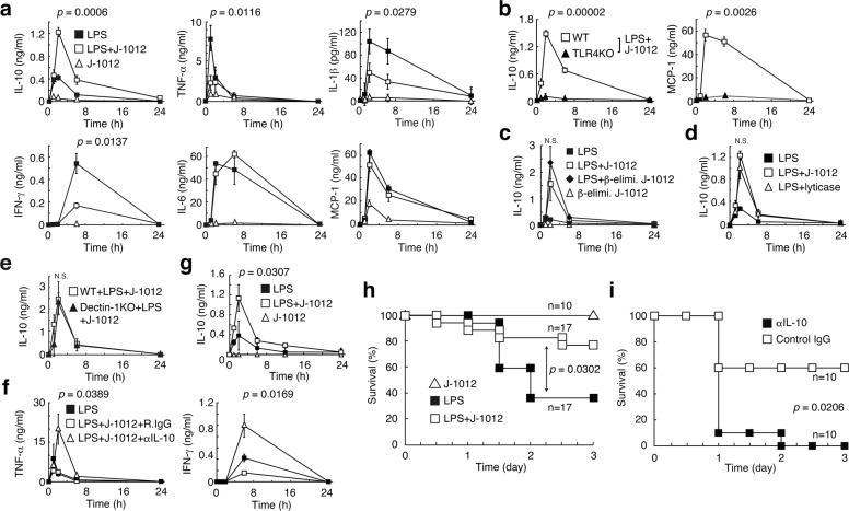
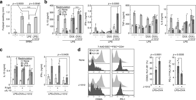
Severe infection often causes a septic cytokine storm followed by immune exhaustion/paralysis. Not surprisingly, many pathogens are equipped with various anti-inflammatory mechanisms. Such mechanisms might be leveraged clinically to control septic cytokine storms. Here we show that N-glycan from pathogenic C. albicans ameliorates mouse sepsis through immunosuppressive cytokine IL-10. In a sepsis model using lipopolysaccharide (LPS), injection of the N-glycan upregulated serum IL-10, and suppressed pro-inflammatory IL-1β, TNF-α and IFN-γ. The N-glycan also improved the survival of mice challenged by LPS. Analyses of structurally defined N-glycans from several yeast strains revealed that the mannose core is key to the upregulation of IL-10. Knocking out the C-type lectin Dectin-2 abrogated the N-glycan-mediated IL-10 augmentation. Furthermore, C. albicans N-glycan ameliorated immune exhaustion/immune paralysis after acute inflammation. Our results suggest a strategy where the immunosuppressive mechanism of one pathogen can be applied to attenuate a severe inflammation/cytokine storm caused by another pathogen.
Conflict of interest statement
The authors declare no competing interests.
Figures


Similar articles
-
Mnn10 Maintains Pathogenicity in Candida albicans by Extending α-1,6-Mannose Backbone to Evade Host Dectin-1 Mediated Antifungal Immunity.PLoS Pathog. 2016 May 4;12(5):e1005617. doi: 10.1371/journal.ppat.1005617. eCollection 2016 May. PLoS Pathog. 2016. PMID: 27144456 Free PMC article.
-
Intestinal colonization by Candida albicans alters inflammatory responses in Bruton's tyrosine kinase-deficient mice.PLoS One. 2014 Nov 7;9(11):e112472. doi: 10.1371/journal.pone.0112472. eCollection 2014. PLoS One. 2014. PMID: 25379804 Free PMC article.
-
Dectin-2-mediated signaling triggered by the cell wall polysaccharides of Cryptococcus neoformans.Microbiol Immunol. 2019 Dec;63(12):500-512. doi: 10.1111/1348-0421.12746. Epub 2019 Nov 22. Microbiol Immunol. 2019. PMID: 31544981
-
Candida albicans phospholipomannan: a sweet spot for controlling host response/inflammation.Semin Immunopathol. 2015 Mar;37(2):123-30. doi: 10.1007/s00281-014-0461-5. Epub 2014 Nov 14. Semin Immunopathol. 2015. PMID: 25394861 Review.
-
Role of IFN-gamma in immune responses to Candida albicans infections.Front Biosci (Landmark Ed). 2014 Jun 1;19(8):1279-90. doi: 10.2741/4281. Front Biosci (Landmark Ed). 2014. PMID: 24896350 Review.
Cited by
-
The F1Fo-ATP synthase α subunit of Candida albicans induces inflammatory responses by controlling amino acid catabolism.Virulence. 2023 Dec;14(1):2190645. doi: 10.1080/21505594.2023.2190645. Virulence. 2023. PMID: 36914568 Free PMC article.
-
Pathogenesis and virulence of Candida albicans.Virulence. 2022 Dec;13(1):89-121. doi: 10.1080/21505594.2021.2019950. Virulence. 2022. PMID: 34964702 Free PMC article. Review.
-
Immunological hyporesponsiveness in tuberculosis: The role of mycobacterial glycolipids.Front Immunol. 2022 Dec 2;13:1035122. doi: 10.3389/fimmu.2022.1035122. eCollection 2022. Front Immunol. 2022. PMID: 36544778 Free PMC article. Review.
-
An immune-adrenergic pathway induces lethal levels of platelet-activating factor in mice.Commun Biol. 2024 Jun 29;7(1):782. doi: 10.1038/s42003-024-06498-7. Commun Biol. 2024. PMID: 38951147 Free PMC article.
-
Translational and Clinical Significance of DAMPs, PAMPs, and PRRs in Trauma-induced Inflammation.Arch Clin Biomed Res. 2022;6(5):673-685. doi: 10.26502/acbr.50170279. Epub 2022 Aug 26. Arch Clin Biomed Res. 2022. PMID: 36147548 Free PMC article.
References
-
- Chousterman BG, Swirski FK, Weber GF. Cytokine storm and sepsis disease pathogenesis. Semin. Immunopathol. 2017;39:517–528. - PubMed
Publication types
MeSH terms
Substances
LinkOut - more resources
Full Text Sources
Other Literature Sources
Medical
