. 2021 Apr;6(4):467-478.
doi: 10.1038/s41564-021-00884-1. Epub 2021 Mar 16.

ISG15-dependent activation of the sensor MDA5 is antagonized by the SARS-CoV-2 papain-like protease to evade host innate immunity

Affiliations

ISG15-dependent activation of the sensor MDA5 is antagonized by the SARS-CoV-2 papain-like protease to evade host innate immunity

GuanQun Liu et al. Nat Microbiol. 2021 Apr.

Abstract

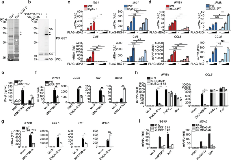
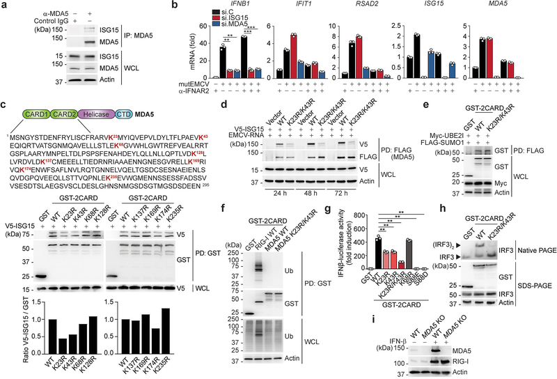
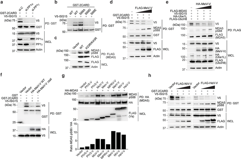
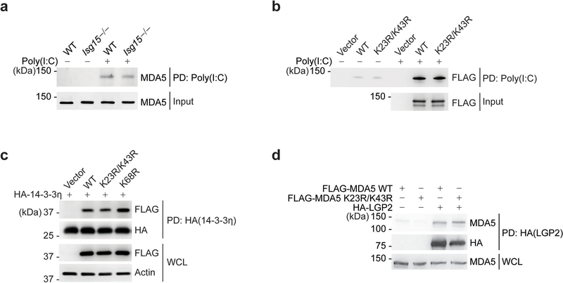
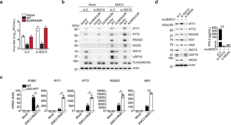
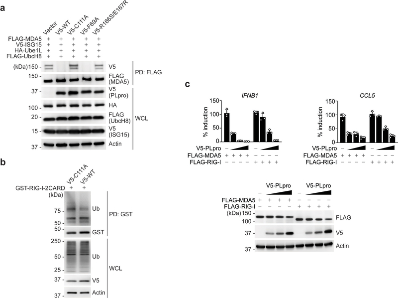
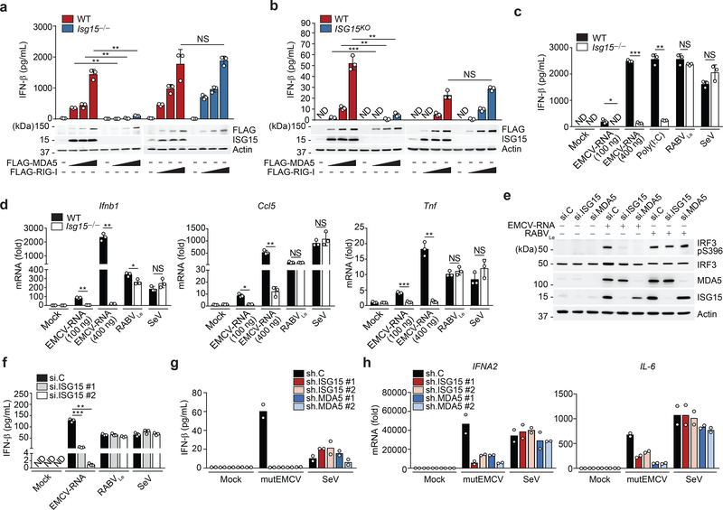
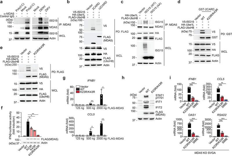
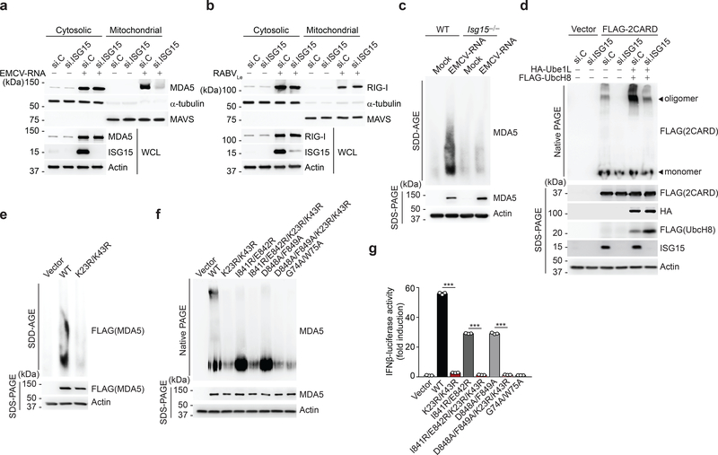
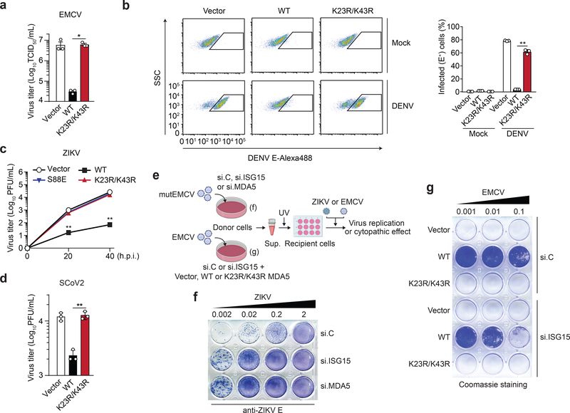
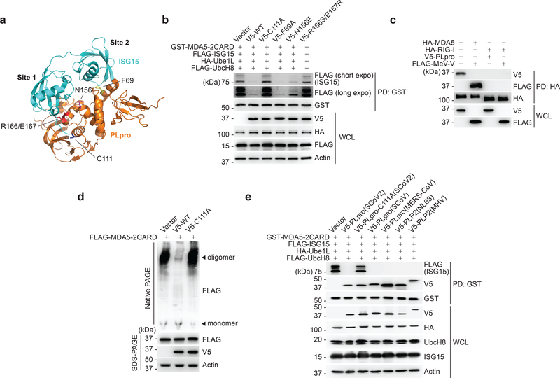
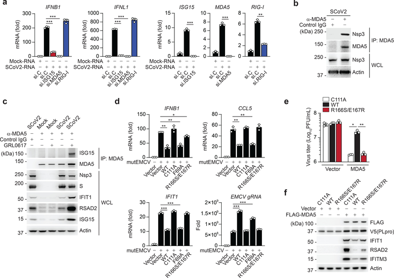
Activation of the RIG-I-like receptors, retinoic-acid inducible gene I (RIG-I) and melanoma differentiation-associated protein 5 (MDA5), establishes an antiviral state by upregulating interferon (IFN)-stimulated genes (ISGs). Among these is ISG15, the mechanistic roles of which in innate immunity still remain enigmatic. In the present study, we report that ISG15 conjugation is essential for antiviral IFN responses mediated by the viral RNA sensor MDA5. ISGylation of the caspase activation and recruitment domains of MDA5 promotes its oligomerization and thereby triggers activation of innate immunity against a range of viruses, including coronaviruses, flaviviruses and picornaviruses. The ISG15-dependent activation of MDA5 is antagonized through direct de-ISGylation mediated by the papain-like protease of SARS-CoV-2, a recently emerged coronavirus that has caused the COVID-19 pandemic. Our work demonstrates a crucial role for ISG15 in the MDA5-mediated antiviral response, and also identifies a key immune evasion mechanism of SARS-CoV-2, which may be targeted for the development of new antivirals and vaccines to combat COVID-19.


Conflict of interest statement

Competing interests

The authors declare no competing interests.

Figures

Extended Data Fig. 1
Extended Data Fig. 1. ISG15 is required for MDA5, but not RIG-I, mediated signal transduction.
(a) Silver-stained affinity-purified GST and GST-MDA5–2CARD from transiently transfected HEK293T cells. Asterisks denote the GST and GST-MDA5–2CARD (aa 1–295) proteins. Arrows indicate the bands that identified ISG15 by MS analysis. (b) GST-MDA5–2CARD ISGylation in transiently transfected HEK293T cells with or without co-expressed V5-ISG15, determined by GST pulldown (PD) and immunoblot (IB) with anti-V5 and anti-GST. Whole cell lysates (WCL) were probed by IB with anti-V5. (c, d) qRT-PCR analysis of IFNB1 and CCL5 transcripts in WT and Isg15−/− MEFs (c) or WT and ISG15 KO HeLa cells (d) that were transfected with empty vector or increasing amounts of FLAG-MDA5 or FLAG-RIG-I for 40 h. (e) ELISA of IFN-β in the supernatants of WT or ISG15 KO HeLa cells that were mock-transfected or transfected with EMCV-RNA (0.4 μg/mL) or RABVLe (1 pmol/mL) for 24 h. (f) qRT-PCR analysis of IFNB1, CCL5, TNF, and MDA5 mRNA in WT and ISG15 KO HeLa cells that were mock-transfected or transfected with EMCV-RNA (0.4 μg/mL) for 24 h. (g) qRT-PCR analysis of IFNB1, CCL5, TNF, and MDA5 mRNA in WT and ISG15 KO HAP-1 cells that were stimulated as in (f). (h) qPCR analysis of IFNB1 and CCL5 mRNA in NHLFs that were transfected with the indicated siRNAs for 30 h and then mock-stimulated or transfected with EMCV-RNA (0.4 μg/mL) or RABVLe (1 pmol/mL), or infected with SeV (10 HAU/mL) for 16 h. (i) qRT-PCR analysis of ISG15 and MDA5 mRNA in PBMCs that were transduced for 40 h with the indicated shRNA lentiviral particles and then infected with mutEMCV (MOI 10) or SeV (200 HAU/mL) for 8 h Data are representative of at least two independent experiments (mean ± s.d. of n = 3 biological replicates in c, d, e, f, g, and h; mean of n = 2 biological replicates in i). *p < 0.05, **p < 0.01, ***p < 0.001 (two-tailed unpaired t-test). NS, not significant; ND, not detected.
Extended Data Fig. 2
Extended Data Fig. 2. ISGylation at K23 and K43 is essential for MDA5 activation.
(a) ISGylation of endogenous MDA5 in uninfected NHLFs, determined by IP with anti-MDA5 (or IgG isotype control) and IB with anti-ISG15. (b) qRT-PCR analysis of the indicated transcripts in NHLFs that were transfected for 30 h with the indicated siRNAs and then infected with mutEMCV (MOI 0.005) for 16 h in the absence or presence of anti-IFNAR2 (2 μg/mL). (c) Upper panels: Amino acid sequence of the MDA5 CARDs. Lysine (K) residues are highlighted in red. CTD, C-terminal domain. Middle panels: ISGylation of GST-MDA5–2CARD WT or mutants in HEK293T cells that co-expressed V5-ISG15, determined by GST-PD and IB with anti-V5. Lower panels: Densitometric analysis of the ISGylation levels of GST-MDA5–2CARD WT and mutants, normalized to protein levels in GST-PD. (d) ISGylation of FLAG-MDA5 WT and K23R/K43R in transfected MDA5 KO HEK293 cells that co-expressed V5-ISG15 and were stimulated with EMCV-RNA (0.4 μg/mL) for the indicated times, determined by FLAG-PD and IB with anti-V5. (e) SUMOylation of GST-MDA5–2CARD WT and K23R/K43R in transfected HEK293T cells that co-expressed Myc-UBE2I and FLAG-SUMO1, determined by FLAG-PD and IB with anti-GST. (f) Ubiquitination of GST-RIG-I-2CARD WT, or GST-MDA5–2CARD WT and K23R/K43R in HEK293T cells, determined by GST-PD and IB with anti-Ub. (g) IFN-β-luciferase activity in HEK293T cells transfected for 40 h with GST, or GST-MDA5–2CARD (GST-2CARD) WT or mutants. (h) Endogenous IRF3 dimerization in HEK293T cells transfected with GST, or GST-MDA5–2CARD WT or K23R/K43R for 24 h, determined by Native PAGE and IB with anti-IRF3. WCLs were analyzed by SDS-PAGE and IB with anti-GST, anti-IRF3, and anti-Actin. (i) Validation of MDA5 gene editing in SVGAs. MDA5 protein abundance in WT control and MDA5 KO SVGAs treated with IFN-β (1,000 U/mL) for 16 h, or left untreated, assessed by IB with anti-MDA5. Immunoblotting for RIG-I and Actin served as control. Data are representative of at least two independent experiments with similar results (mean ± s.d. of n = 3 biological replicates in b and g). **p < 0.01, ***p < 0.001 (two-tailed unpaired t-test).
Extended Data Fig. 3
Extended Data Fig. 3. Dephosphorylation of MDA5 induces ISGylation.
(a) ISGylation of GST-MDA5–2CARD in HEK293T cells transfected with V5-ISG15 and the indicated siRNAs for 48 h, assessed by GST-PD and IB with anti-V5 and anti-GST. WCLs were probed by IB with anti-V5, anti-PP1α, anti-PP1γ, and anti-Actin. (b) ISGylation of GST-MDA5–2CARD WT or S88A, S88D and S88E mutants in transiently transfected HEK293T cells that also co-expressed V5-ISG15, determined by GST-PD and IB with anti-V5 and anti-GST forty hours after transfection. (c) Phosphorylation of FLAG-MDA5 WT or K23R/K43R mutant in HEK293T cells, determined by FLAG-PD and IB with anti-pS88-MDA5 and anti-FLAG. WCLs were probed by IB with anti-FLAG and anti-Actin. (d) ISGylation and phosphorylation of GST-MDA5–2CARD in HEK293T cells transfected with V5-ISG15 and either empty vector or increasing amounts of FLAG-MeV-V for 24 h, determined by GST-PD and IB with anti-pS88-MDA5, anti-V5, and anti-GST. (e) ISGylation and phosphorylation of FLAG-tagged MDA5 in transfected HEK293T cells that co-expressed V5-ISG15, HA-Ube1L, and FLAG-UbcH8 as well as either empty vector or increasing amounts of HA-MeV-V for 24 h, determined by FLAG-PD and IB with anti-pS88-MDA5, anti-V5, and anti-FLAG. WCLs were probed by IB with the indicated antibodies. (f) ISGylation of GST-MDA5–2CARD, or GST (negative control), in HEK293T cells transiently transfected with V5-ISG15 and either empty vector or HA-tagged MeV-V WT or Δtail for 48 h, determined by GST-PD and IB with anti-V5 and anti-GST. (g) Upper panel: Phosphorylation of HA-MDA5 in transfected HEK293T cells that co-expressed empty vector or the indicated FLAG-tagged paramyxoviral V proteins, assessed by HA-PD and IB with anti-pS88-MDA5 and anti-HA. WCLs were probed by IB with anti-FLAG. Lower panel: Densitometric analysis of the levels of MDA5 S88 phosphorylation, normalized to protein levels in HA-PD. (h) ISGylation and phosphorylation of GST-MDA5–2CARD in transfected HEK293T cells that co-expressed V5-ISG15 and either empty vector or increasing amounts of FLAG-NiV-V or FLAG-HeV-V for 24 h, determined by GST PD and IB with anti-V5, anti-pS88-MDA5, and anti-GST. WCLs were probed by IB with anti-FLAG, anti-V5, and anti-Actin. Data are representative of at least two independent experiments with similar results.
Extended Data Fig. 4
Extended Data Fig. 4. CARD ISGylation does not affect the ability of MDA5 to bind RNA, 14–3-3η, or LGP2.
(a) In vitro RNA-binding ability of endogenous MDA5 from WT or Isg15−/− MEFs that were stimulated with IFN-β (1,000 U/mL) for 24 h, assessed by biotin-HMW-poly(I:C)-PD and IB with anti-MDA5. Equal input MDA5 protein amounts were confirmed by IB with anti-MDA5. (b) In vitro RNA-binding ability of FLAG-MDA5 WT and K23R/K43R from transiently transfected HEK293T cells, assessed by biotin-HMW-poly(I:C)-PD and IB with anti-FLAG. Equal input FLAG-MDA5 protein amounts were confirmed by IB with anti-FLAG. (c) Binding of FLAG-tagged MDA5–2CARD WT or mutants to HA-14–3-3η in transiently transfected HEK293T cells, determined by HA-PD and IB with anti-FLAG and anti-HA. WCLs were probed by IB with anti-FLAG and anti-Actin. (d) Binding of FLAG-tagged MDA5 WT and K23R/K43R to HA-tagged LGP2 in transiently transfected HEK293T cells, determined by HA-PD and IB with anti-MDA5 and anti-HA. WCLs were probed by IB with anti-MDA5. Data are representative of at least two independent experiments with similar results.
Extended Data Fig. 5
Extended Data Fig. 5. Aberrant ISG upregulation in ISG15-deficient cells upon MDA5 stimulation.
(a) EMCV titers in the supernatant of RIG-I KO HEK293 cells that were transfected for 24 h with nontargeting control siRNA (si.C) or ISG15-specific siRNA (si.ISG15) and then transfected with either empty vector or FLAG-tagged MDA5 WT or K23R/K43R for 24 h prior to infection with EMCV (MOI 0.001) for 16 h, determined by plaque assay. (b) Protein abundance of the indicated ISGs and USP18 in mock-infected or EMCV-infected (MOI 0.001 for 16 h) RIG-I KO HEK293 cells that were transfected with the indicated siRNAs, and 24 h later, transfected with empty vector or FLAG-MDA5 WT or K23/K43R for 24 h, determined by IB with the indicated antibodies. WCLs were further immunoblotted with anti-FLAG and anti-Actin. (c) qRT-PCR analysis of IFNB1 and ISG transcripts in WT and ISG15 KO HeLa cells that were mock-treated or transfected with EMCV-RNA (0.4 μg/mL) for 16 h. (d) Left panel: Protein abundance of the indicated ISGs and USP18 in NHLFs that were transfected for 40 h with the indicated siRNAs and then infected with mutEMCV (MOI 0.1) for 16 h, determined by IB with the indicated antibodies. Right panel: ELISA of IFN-β from supernatants of NHLFs from the same experiment (left panel). Data are representative of at least two independent experiments with similar results (mean of n = 2 biological replicates in a; mean ± s.d. of n = 3 biological replicates in c and d). **p < 0.01, ***p < 0.001 (two-tailed unpaired t-test). ND, not detected.
Extended Data Fig. 6
Extended Data Fig. 6. SCoV2 PLpro does not affect RIG-I ubiquitination and preferentially antagonizes the MDA5 pathway.
(a) ISGylation of FLAG-MDA5 in HEK293T cells that were co-transfected for 20 h with vector or V5-tagged SCoV2 PLpro WT or mutants, along with V5-ISG15, HA-Ube1L, and FLAG-UbcH8, determined by FLAG-PD and IB with anti-V5 and anti-FLAG. WCLs were probed by IB with anti-V5, anti-HA, anti-FLAG, and anti-Actin. (b) Ubiquitination of GST-RIG-I-2CARD in HEK293T cells that were transfected with V5-tagged SCoV2 PLpro WT or C111A for 24 h, determined by GST-PD and IB with anti-Ub and anti-GST. WCLs were probed by IB with anti-Ub, anti-V5, and anti-Actin. (c) Upper panels: qPCR analysis of IFNB1 and CCL5 transcripts in HeLa cells that were transfected with FLAG-MDA5 or FLAG-RIG-I together with either empty vector (Vec) or increasing amounts of V5-tagged SCoV2 PLpro (10 ng, 25 ng, and 50 ng) for 24 h. Data are presented as percentage of induction relative to the values for cells transfected with the respective RLR (i.e. FLAG-MDA5 or FLAG-RIG-I) and vector, set to 100%. Lower panels: WCLs from the same experiment were probed by IB with the indicated antibodies. Data are representative of at least two independent experiments with similar results (mean ± s.d. of n = 3 biological replicates in c).
Fig. 1 |
Fig. 1 |. ISGylation is required for MDA5-, but not RIG-I, signaling.
a, b, ELISA of IFN-β from supernatants of MEFs (WT or Isg15−/−) (a) and HeLa cells (WT or ISG15 KO) (b) transiently transfected with increasing amounts of FLAG-tagged MDA5 or RIG-I for 40 h. Whole cell lysates (WCLs) were probed by immunoblotting (IB) with anti-ISG15, anti-FLAG, and anti-Actin (loading control). c, ELISA of IFN-β from supernatants of WT or Isg15−/− MEFs that were mock-stimulated or transfected with EMCV-RNA (0.1 or 0.4 μg/mL), HMW-poly (I:C) (0.5 μg/mL), or RABVLe (1 pmol/mL), or infected with SeV (10 HAU/mL) for 24 h. d, Quantitative RT-PCR (qRT-PCR) analysis of IFNB1 and CCL5 mRNA in WT and Isg15−/− MEFs stimulated as in (c). e, IRF3 phosphorylation in the WCLs of NHLFs that were transfected with the indicated siRNAs for 30 h and then mock-stimulated or transfected with EMCV-RNA (0.4 μg/mL) or RABVLe (1 pmol/mL) for 6 h, assessed by IB with anti-pS396-IRF3 and anti-IRF3. f, ELISA of IFN-β from supernatants of NHLFs that were transfected with the indicated siRNAs for 30 h and then mock-stimulated or transfected with EMCV-RNA (0.4 μg/mL) or RABVLe (1 pmol/mL), or infected with SeV (10 HAU/mL) for 16 h. g, ELISA of IFN-β from the supernatants of PBMCs that were transduced for 40 h with the indicated shRNAs and then infected with mutEMCV (MOI 10) or SeV (200 HAU/mL) for 8 h. h, qRT-PCR analysis of IFNA2 and IL-6 mRNA in PBMCs that were transduced and infected as in (g). Data are representative of at least two independent experiments with similar results (mean ± s.d. of n = 3 biological replicates in a, b, c, d, f and mean of n = 2 biological replicates in g and h). *p < 0.05, **p < 0.01, ***p < 0.001 (two-tailed unpaired t-test). ND, not detected; NS, not significant.
Fig. 2 |
Fig. 2 |. MDA5 activation requires ISGylation at K23 and K43.
a, Endogenous MDA5 ISGylation in NHLFs that were mock-treated, transfected with HMW-poly (I:C) (0.1 μg/mL) for 40 h (left), or infected with DENV or ZIKV (MOI 1 for each) for 48 h (right), determined by immunoprecipitation (IP) with anti-MDA5 (or an IgG isotype control) and IB with anti-ISG15. b, ISGylation of FLAG-tagged MDA5–2CARD and MDA5ΔCARD in transiently transfected HEK293T cells that also expressed V5-ISG15, HA-Ube1L, and FLAG-UbcH8, assessed by FLAG pulldown (PD) and IB with anti-V5 at 40 h post-transfection. c, Endogenous MDA5 ISGylation in ISG15 KO HeLa cells stably reconstituted with vector, WT ISG15 or ISG15-AA and co-transfected with HA-Ube1L and FLAG-UbcH8 after IFN-β treatment (1,000 U/mL) for 24 h, determined by IP with anti-MDA5 and IB with anti-ISG15. d, ISGylation of GST-MDA5–2CARD WT and K23R/K43R in HEK293T cells that were co-transfected with V5-ISG15, HA-Ube1L, and FLAG-UbcH8 for 24 h, determined by GST-PD and IB with anti-V5. e, ISGylation of FLAG-MDA5 WT and K23R/K43R in HEK293T cells that were co-transfected with V5-ISG15, HA-Ube1L, and FLAG-UbcH8, determined by FLAG-PD and IB with anti-V5. f, IFN-β-luciferase reporter activity in HEK293T cells that were transfected for 40 h with vector, or FLAG-MDA5 WT or mutants. Luciferase values are presented as fold induction relative to the values for vector-transfected cells, set to 1. WCLs were probed by IB with anti-FLAG and anti-Actin. g, qRT-PCR analysis of IFNB1 and CCL5 mRNA in HEK293T cells that were transiently transfected with either vector, or increasing amounts of FLAG-MDA5 WT or K23R/K43R. h, STAT1 phosphorylation and ISG (IFIT1 and 2) protein abundance in the WCLs of HEK293T cells that were transiently transfected with vector or FLAG-MDA5 WT or K23R/K43R, determined by IB. i, qRT-PCR analysis of the indicated antiviral genes in MDA5 KO SVGAs that were reconstituted with either empty vector or FLAG-tagged MDA5 WT, K23R/K43R or S88E. Data are representative of at least two independent experiments with similar results (mean ± s.d. of n = 3 biological replicates in f, g, and i). *p < 0.05, **p < 0.01, ***p < 0.001 (two-tailed unpaired t-test). NS, not significant.
Fig. 3 |
Fig. 3 |. CARD ISGylation is essential for formation of higher-order MDA5 assemblies.
a,b, Cytosol-mitochondria fractionation of WCLs from NHLFs that were transfected for 30 h with non-targeting control siRNA (si.C) or ISG15-specific siRNA (si.ISG15) and then mock-treated or transfected with EMCV-RNA (0.4 μg/mL) (a) or RABVLe (1 pmol/mL) (b) for 16 h. IB was performed with anti-MDA5 (a), anti-RIG-I (b), anti-ISG15 and anti-Actin (a, b). α-Tubulin and MAVS served as purity markers for the cytosolic and mitochondrial fraction, respectively (a, b). c, Endogenous MDA5 oligomerization in WT and Isg15−/− MEFs that were transfected with EMCV-RNA (0.5 μg/mL) for 16 h, assessed by SDD-AGE and IB with anti-MDA5. WCLs were further analyzed by SDS-PAGE and probed by IB with anti-MDA5 and anti-Actin. d, Oligomerization of FLAG-MDA5–2CARD in HEK293T cells that were transfected with the indicated siRNAs together with or without HA-Ube1L and FLAG-UbcH8 for 48 h, determined by native PAGE and IB with anti-FLAG. WCLs were further analyzed by SDS-PAGE and probed by IB with anti-FLAG, anti-HA, anti-ISG15, and anti-Actin. e, Oligomerization of FLAG-MDA5 WT and K23R/K43R in transiently transfected MDA5 KO HEK293 cells, assessed by SDD-AGE and IB with anti-FLAG. WCLs were further analyzed by SDS-PAGE and IB with anti-FLAG and anti-Actin. f, Oligomerization of FLAG-tagged MDA5 WT and mutants in transiently transfected MDA5 KO HEK293 cells, assessed by native PAGE and IB with anti-MDA5. WCLs were further analyzed by SDS-PAGE and probed by IB with anti-MDA5 and anti-Actin. g, IFN-β-luciferase reporter activity in MDA5 KO HEK293 cells that were transfected for 24 h with either empty vector, or FLAG-tagged MDA5 WT or mutants. Luciferase activity is presented as fold induction relative to the values for vector-transfected cells, set to 1. Data are representative of at least two independent experiments with similar results (mean ± s.d. of n = 3 biological replicates in f). ***p < 0.001 (two-tailed unpaired t-test).
Fig. 4 |
Fig. 4 |. ISGylation is required for viral restriction by MDA5.
a, EMCV titers in the supernatant of HEK293T cells that were transfected for 40 h with either vector, or FLAG-MDA5 WT or K23/K43R and then infected with EMCV (MOI 0.001) for 24 h, determined by TCID50 assay. b, Percentage of DENV-infected MDA5 KO HEK293 cells that were transfected for 24 h with either vector or FLAG-MDA5 WT or K23R/K43R and then mock-treated or infected with DENV (MOI 5) for 48 h, assessed by FACS using anti-flavivirus E (4G2). SSC, side scatter. c, ZIKV titers in the supernatant of MDA5 KO SVGAs that were transfected for 30 h with vector or FLAG-tagged MDA5 WT, K23R/K43R, or S88E and then infected with ZIKV (MOI 0.1) for the indicated times, determined by plaque assay. d, SCoV2 titers in the supernatant of HEK293T-hACE2 cells that were transfected for 24 h with either empty vector, or FLAG-MDA5 WT or K23/K43R and then infected with SCoV2 (MOI 0.5) for 24 h, determined by plaque assay. e, Schematic of the experimental approach to ‘decouple’ the role of ISG15 in MDA5-mediated IFN induction from its role in dampening IFNAR signaling. f, NHLF ‘donor’ cells were transfected for 40 h with the indicated siRNAs and then infected with mutEMCV (MOI 0.1) for 16 h. Cell supernatants were UV-inactivated and transferred onto Vero ‘recipient’ cells. 24 h later, cells were infected with ZIKV (MOI 0.002 to 2) for 72 h, and ZIKV-positive cells determined by immunostaining with anti-flavivirus E (4G2) and TrueBlue peroxidase substrate. g, RIG-I KO HEK293 ‘donor’ cells were transfected with si.C or si.ISG15 together with either vector or FLAG- MDA5 WT or K23R/K43R for 24 h, followed by EMCV infection (MOI 0.001) for 16 h. UV-inactivated cell supernatants were transferred onto Vero ‘recipient’ cells for 24 h, followed by infection with EMCV (MOI 0.001 to 0.1) for 40 h. EMCV-induced cytopathic effects were visualized by Coomassie blue staining. Data are representative of at least two independent experiments with similar results (mean ± s.d. of n = 3 biological replicates in a, b, c). *p < 0.05, **p < 0.01 (two-tailed unpaired t-test).
Fig. 5 |
Fig. 5 |. SCoV2 PLpro binds to and de-ISGylates MDA5–2CARD.
a, Ribbon representation of the crystal structure of the SCoV2 PLpro: ISG15 complex (PDB: 6YVA). Key residues that mediate ‘site 1’ interaction (N156 and R166/E167) or ‘site 2’ interaction (F69) in PLpro, as well as its catalytically-active site (C111), are indicated. b, ISGylation of GST-MDA5–2CARD in HEK293T cells that were co-transfected for 20 h with vector or V5-tagged SCoV2 PLpro WT or mutants, along with FLAG-ISG15, HA-Ube1L, and FLAG-UbcH8, determined by GST-PD and IB with anti-FLAG and anti-GST. WCLs were probed by IB with anti-V5, anti-HA, anti-FLAG, and anti-Actin. (c) Binding of HA-tagged MDA5 or RIG-I to V5-SCoV2-PLpro or FLAG-MeV-V (positive control) in transiently transfected HEK293T cells, determined by HA-PD and IB with anti-V5 or anti-FLAG, and anti-HA. WCLs were probed by IB with anti-V5 and anti-FLAG. d, Oligomerization of FLAG-MDA5–2CARD in HEK293T cells that were co-transfected with vector, or V5-SCoV2 PLpro WT or C111A for 24 h, assessed by Native PAGE and IB with anti-FLAG. WCLs were further analyzed by SDS-PAGE and probed by IB with anti-FLAG, anti-V5 and anti-Actin. e, ISGylation of GST-MDA5–2CARD in HEK293T cells that also expressed FLAG-ISG15, HA-Ube1L and FLAG-UbcH8, and were co-transfected for 40 h with vector or the indicated V5-tagged coronaviral PLpro, determined by GST-PD and IB with anti-FLAG, anti-V5, and anti-GST. Data are representative of at least two independent experiments with similar results.
Fig. 6 |
Fig. 6 |. SCoV2 PLpro inhibits ISG15-mediated MDA5 signaling via its deISGylase activity.
a, qRT-PCR analysis of IFNB1, IFNL1, ISG15, MDA5, and RIG-I transcripts in NHLFs that were transfected with the indicated siRNAs for 40 h and then transfected with mock-RNA or SCoV2-RNA (0.4 μg/mL) for 24 h. b, Binding of SCoV2 Nsp3 to endogenous MDA5 in A549-hACE2 cells that were infected with SCoV2 (MOI 0.5) for 24 h, determined by IP with anti-MDA5 (or an IgG isotype control) followed by IB with anti-PLpro and anti-MDA5. WCLs were probed by IB with anti-PLpro (Nsp3) and anti-Actin. c, Endogenous MDA5 ISGylation in A549-hACE2 cells that were mock-infected or infected with SCoV2 (MOI 0.5) for 40 h in the presence of PLpro inhibitor (GRL-0617; 50 μM) or vehicle control (DMSO), determined by IP with anti-MDA5 (or an IgG isotype control) followed by IB with anti-ISG15 and anti-MDA5. Protein abundance of IFIT1, RSAD2, ISG15 and actin in the WCLs were probed by IB. Efficient virus replication was verified by immunoblotting WCLs with anti-PLpro (Nsp3) and anti-Spike (S). d, qRT-PCR analysis of IFNB1, CCL5, IFIT1 transcript, and EMCV genomic RNA (gRNA) in HeLa cells that were transiently transfected for 24 h with vector, or V5-SCoV2 PLpro WT or mutants and then infected with mutEMCV (MOI 0.5) for 12 h. e, EMCV titers in the supernatant of RIG-I KO HEK293 cells that were transiently transfected for 24 h with vector or FLAG-MDA5 along with V5-SCoV2 PLpro WT, C111A, or R166S/E167R and then infected with EMCV (MOI 0.001) for 16 h, determined by plaque assay. f, Protein abundance of the indicated ISGs in the WCLs from the experiment in (e), determined by IB with the indicated antibodies. Data are representative of at least two independent experiments with similar results (mean ± s.d. of n = 3 biological replicates in a, d, e). *p < 0.05, **p < 0.01, ***p < 0.001 (two-tailed unpaired t-test).

Update of

References

    1. Liu G & Gack MU Distinct and Orchestrated Functions of RNA Sensors in Innate Immunity. Immunity 53, 26–42 (2020). - PMC - PubMed
    1. Wu J & Chen ZJ Innate immune sensing and signaling of cytosolic nucleic acids. Annu Rev Immunol 32, 461–488 (2014). - PubMed
    1. Chow KT, Gale M Jr. & Loo YM RIG-I and Other RNA Sensors in Antiviral Immunity. Annu Rev Immunol 36, 667–694 (2018). - PubMed
    1. Schlee M Master sensors of pathogenic RNA - RIG-I like receptors. Immunobiology 218, 1322–1335 (2013). - PMC - PubMed
    1. Rehwinkel J & Gack MU RIG-I-like receptors: their regulation and roles in RNA sensing. Nat Rev Immunol 20, 537–551 (2020). - PMC - PubMed

Publication types